Reading...
![]()
Play button
![]()
Play button
![]()
Use LEFT and RIGHT arrow keys to navigate between flashcards;
Use UP and DOWN arrow keys to flip the card;
H to show hint;
A reads text to speech;
29 Cards in this Set
- Front
- Back

What structure surrounds the entire eye?
|

Bony orbit
|
|

Identify 9. What does it do?
|

Lacrimal gland. Allows tears to flow.
|
|
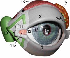
Identify 11
|

Lacrimal canals (canaliculi)
|
|

Identify 10
|

Lacrimal sac
|
|

What structure is located directly inferior to #10?
|

Nasolacrimal duct
|
|

What structures are indicated by #2 and #3?
|

Palpebrae
|
|

What is the corner of the eye by #12 called? How about the opposing corner?
|

Medial canthus. Lateral canthus
|
|

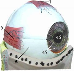
What is indicated by "C"?
|

The caruncle
|
|

What are conjunctiva? What types do humans have, and where?
|

The covering of your eyelids/eyeball. The membrane underneath the eyelid is called the palpebral conjunctiva. The one on the eye itself is called the occular conjunctiva
|
|

What gland lies high and to the left of the label for #30 on this model? What type of gland do they resemble?
|

The cilliary gland. A sweat gland
|
|

What glands lie between the hairs of your eyelashes? What type of glands are they?
|

Bobeum or tarsals. Sebaceous.
|
|

What is a sty?
|
When your bobeum or ciliary glands get inflamed/infected.
|
|

What muscle is indicated by "I"? What is the opposing muscle? (Not shown, but on the bottom of the eye)
|
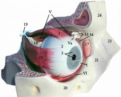
The superior rectus. The inferior rectus.
|
|

What muscle is indicated by "IV"? What is the opposing muscle?
|

Lateral rectus. Medial rectus
|
|

What muscle is indicated by "V"? By "VI"?
|

Superior oblique. Inferior oblique
|
|

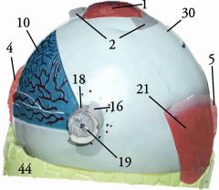
What is the outer layer of your eye called? What are the two parts?
|

Fibrous tunic. Sclera (2) and cornea (clear layer in front of lens, pupil, and iris)
|
|

What is the name of the second layer deep in the outer surface of the eye?
|

The uvea or vascular tunic
|
|

What is the back part of the vascular tunic called?
|
The choroid (10)
|
|

What is indicated by 45?
|

Cilliary bodies
|
|
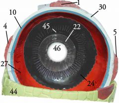
What is indicated by 22?
|

Iris
|
|

What is indicated by 46?
|

Pupil
|
|

What is the pinkish(orange in this model) surface at the back of your eye called?
|

Retina
|
|

What is indicated by #31?
|

Optic disc (formed by the optic nerve and giving us our blind spot)
|
|
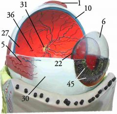
What is indicated by #36?
|

Macula lutea, with the Fovea centralis at it's center
|
|

What is indicated by #22?
|

Lens
|
|

What is the "scalloping" outside of #45 called?
|

Aura serrata
|
|

What is the space between the lens and the retina called? What is the space between the lens and the cornea called?
|

The posterior cavity or segment, containing vitrious humor. The anterior cavity or segment, containing aqueous humor.
|
|

The anterior segment of the eye is divided into two sections - what divides them, and what are the sections called?
|

Divided by the iris into the anterior and posterior chambers.
|
|

Where do the layers shown lie within the eye? Name the layers of the eye as shown, bottom to top.
|
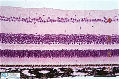
At the top of the screen is the vitrious humor, and at the bottom is the sclera of the eye.
(From bottom): Choroid (1) Photoreceptors (2) Bipolar cells (3) Ganglion cells (4) |