![]()
![]()
![]()
Use LEFT and RIGHT arrow keys to navigate between flashcards;
Use UP and DOWN arrow keys to flip the card;
H to show hint;
A reads text to speech;
51 Cards in this Set
- Front
- Back
|
sensory receptors
|
usually modified neurons or epithelial cells that occur singly or in groups within sensory organs
|
|
|
sensory transduction |
The conversion of a physical or chemical stimulus to a change in the membrane potential of a sensory receptor. a graded change in the membrane potential of a receptor cell is proportional to the strength of the stimulus |
|
|
amplification
|
The strengthening of stimulusenergy during transduction. Forexample, an action potential conducted from the eye to thehuman brain has about 100,000 times as much energy as thefew photons of light that triggered it.
|
|
|
Transmission
|
of receptor potential to the central nergvous system may occur either as a result of the generation of an action potential when the sensory receptor is a sensory neuron, or by the release of neurotransmitter from a receptor cell into a synspse with a sensory neuron, which then may transmit an action potential.
|
|
|
sensory adaptation |
caused by continuous stimulation result in a decline in sensitivity of the receptor cell |
|
|
mechanoreceptors |
sensory receptros that respond to the mechanical energy of pressure, touch, stretch, motion, and sound. Bendin or stretching of the mechanoreceptor cell membrane increases its permeability to sodium and potassium ions, creating a receptor potential. |
|
|
muscle spindles
|
stertch receptors that monitor the length of skeletal muscles
|
|
|
Hair cells |
mechanoreceptors (motion detection) in the ear, when motion produces bending in the cilia or microvilli projecting from a hair cell, ion permeabilities either increase or decrease, and the rate of action potential firing changes. |
|
|
nociceptors
|
also called Pain receptors. naked dendrites in the epidermis called nociceptors, different groups of receptors respond to exceess heat, pressure, or chemicals released by injured cells.
|
|
|
eye cup |
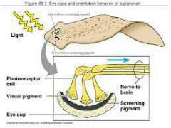
in planarians, detect light intensity and direction, consists of receptor cells within a cup formed from darkly pigmented cells. Brain compared impulses to help the animal navigate a direct path away from a light source. |
|
|
compound eye |
contains up to thousnads of light detectors called ommatidia, each with its own lens. for example insect eyes. |
|
|
single-lens eye |
light is focused through the signle lens onto the retina containing light. for example humen eyes. |
|
|
choroid
|

thin, pigmented inner layer of eye
|
|
|
retina
|

הרשתית innermost layer, containts the photoreceptors cells, the optic nerve attaches to the eye at the optic disk, forming a blind spot on the retina |
|
|
lens |
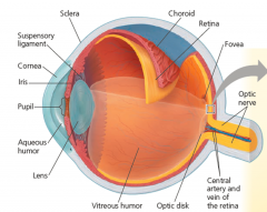
עדשת העין focuses an image onto the retina |
|
|
rod cells and cone cells
|
the photoreceptors in the retina. The relative proportion of each of these receptors correlates with the activity pattern of the animal: Rods are more light sensitive and enable night vision, whereas cones distinguish colors. In the human eye, rods are most concentrated towart the edge of the retina, whereas the center of the visual field, the fovea, is filled with cones.
|
|
|
receptor potential |
An initial response of a receptor cell to a stimulus, consisting of a change in voltage across the receptor membrane. proportional to the stimulus strength. |
|
|
sensory adaptation |
The tendency of sensory neurons to become less sensitive when they are stimulated repeatedly. |
|
|
statocysts |
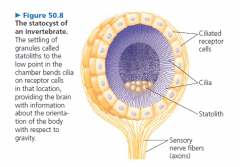
To sense gravity and maintain equilibrium, most invertebratesrely on mechanoreceptors located in organs called statocysts |
|
|
Statolith |

in statocysts, granules formed by grains of sand or other dense materials,sit freely in a chamber lined with ciliated cells. |
|
|
outer ear |

consists of the external pinna and the auditory canal, which collect sound waves and channel them to the tympanic membrane (eardrum), which separates the outer ear from the middle ear. |
|
|
tympanic membrane |

עור התוף. (eardrum) transmits sound waves to bones of the middle ear. |
|
|
middle ear |

three small bones—the malleus (hammer), incus (anvil), and stapes(stirrup)—transmit vibrations to the oval window, which is a membrane beneath the stapes. |
|
|
oval window |

In the vertebrate ear, a membrane-covered gap in the skull bone,through which sound waves pass from the middle ear to the inner ear. |
|
|
Eustachian tube |
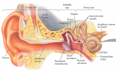
The tube that connects the middle ear to the pharynx. equalizes pressure between the middle ear and the atmosphere |
|
|
inner ear |

consists of fluid-filled chambers, including the semicircular canals, which function in equilibrium, and the coiled cochlea (from the Latin meaning “snail”), a bony chamber that is involved in hearing. |
|
|
Cochlea |
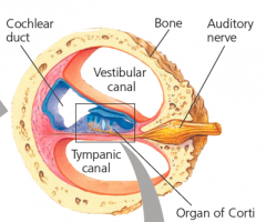
שבלול האוזן. The complex, coiled organ of hearing that contains the organ of Corti. |
|
|
semicircular canals |

A three-part chamber ofthe inner ear that functions in maintaining equilibrium. |
|
|
Organ of Corti |

The actual hearing organ of the vertebrate ear, located in the floor of the cochlear duct in the inner ear; contains the receptor cells (hair cells) of the ear. |
|
|
Vestibular canal\Tympanic canal |

The cochlea has two large canals—an upper vestibular canal and a lower tympanic canal—separated by a smaller cochlear duct. Both canals are filled with fluid. |
|
|
Basilar membrane |

Sound waves make the basilarme mbrane vibrate, which results in bending of the hairs anddepolarization of the hair cells. |
|
|
round window |
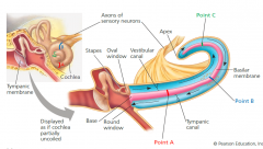
In the mammalian ear, the point of contact where vibrations of the stapes create a traveling series of pressure waves in the fluid of the cochlea. |
|
|
ommatidia |
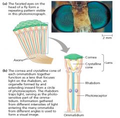
(singular,Ommatidium) A compound eye (like insects eye) consists of up to several thousand light detectors called ommatidia. |
|
|
Pupil |

האישון The opening in the iris, which admits light into the interior of the vertebrate eye. Muscles in the iris regulate its size. |
|
|
Iris |

הקשתית By changing size, the iris regulates the amount of light entering the pupil. |
|
|
retinal |
(a derivative of vitamin A). The light-absorbing molecule in rods and cones of the vertebrate eye. Absorption of light shifts one bond in retinal from a cis to a trans arrangement,converting the molecule from an angled shape to a straight shape. |
|
|
Ganglion cell |

each ganglion cell gathers input from several bipolar cells. |
|
|
Bipolar cell |
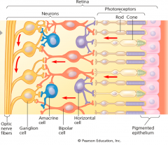
Each bipolar cell receives information from several rods or cones |
|
|
Horizontal cell |

horizontal cells carry signals from one rod or cone to other photoreceptors and to several bipolar cells while doing lateral inhibition. |
|
|
Amacrine cell |

Amacrine cells distribute some information from one bipolar cell to several ganglion cells. Lateral inhibitionis repeated by the interactions of the amacrine cells with the ganglion cells and occurs at all levels of visual processing in the brain. |
|
|
opsin |
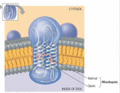
retinal bound to a membrane protein called an opsin. Seven α helices of each opsin molecules pan the disk membrane. The visualpigment of rods, shown here, is called rhodopsin. |
|
|
thin filaments |
Muscle cell contraction relies on the interaction betweenprotein structures called thin and thick filaments. two strands of polymerized actinare coiled around one another; similar actin structurescalled microfilaments function in cell motility. |
|
|
thick filaments |

are staggered arrays of myosin molecules. |
|
|
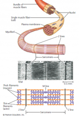
myofibrils |

contain the thin and thick filaments |
|
|
sarcomeres |
The myofibrils in muscle fibers are made up of repeatingsections called sarcomeres, which are the basic contractileunits of skeletal muscle. |
|
|
Tropomyosin |
The regulatory protein thatblocks the myosin-binding sites on actinmolecules. |
|
|
troponin complex |
The regulatory proteins that control the position of tropomyosin on the thin filament. |
|
|
T tubules |

transverse (T) tubules. An infolding of theplasma membrane of skeletal muscle cells. |
|
|
SR |
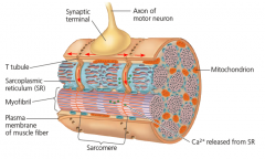
sarcoplasmic reticulum (SR). A specialized endoplasmicreticulum that regulates the calciumconcentration in the cytosol of muscle cells. |
|
|
motor unit |
A single motor neuron and all themuscle fibers it controls. |
|
|
tetanus |
When the rate is so high that the muscle fiber cannot relaxat all between stimuli, the twitches fuse into one smooth,sustained contraction called tetanus. |