![]()
![]()
![]()
Use LEFT and RIGHT arrow keys to navigate between flashcards;
Use UP and DOWN arrow keys to flip the card;
H to show hint;
A reads text to speech;
70 Cards in this Set
- Front
- Back
- 3rd side (hint)
|
The skeletal system pictured: |
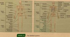
Back (Definition) |
|
|
|
The human movement system |
Combination & interrelation of the nervous, muscular, & skeletal systems |
|
|
|
Nervous system |
A conglomeration of billions of cells specifically designed to provide a communication network within the body. |
|
|
|
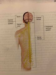
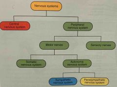
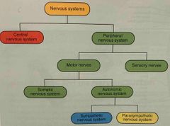
What are the 2 parts of the nervous system? |
The Central nervous system The Peripheral nervous system |
|
|
|
The central nervous system (CNS) |
Is composed of the brain & spinal cord. |
|
|
|
The peripheral nervous system (PNS) |
Contains only nerves & connects the brain & spinal cord (CNS) to the rest of the body. |
|
|
|
What are the 3 primary functions of the nervous system? |
Sensory Integrative Motor |
|
|
|
Sensory function |
The ability of the nervous system to sense changes in either the internal/external environment. |
A stretch placed on a muscle (internal) Change from walking on the sidewalk to walking on sand (external) |
|
|
Integrative function |
The ability of the nervous system to analyze & interpret sensory information to allow for proper decision making, which produces the appropriate response. |
|
|
|
Motor function |
The neuromuscular response to the sensory information. |
Causing a muscle to contract when stretched too far. Changing walking pattern when walking on the sand vs. sidewalk. |
|
|
What is the nervous system responsible for? |
Recruitment of muscles Learned patterns of movement Functioning of every organ in the body |
There are 3 |
|
|
Proprioception |
The cumulative sensory input to the CNS from all mechanoreceptors that sense body position & limb movement. |
When walking/running the feedback we get about the surface/terrain we’re on. |
|
|
Training the body’s proprioceptive abilities will improve |
Balance Coordination Posture And enable the body to adapt to its surroundings without cautiously thinking about what movement is appropriate |
|
|
|
The functional unit of the nervous system is known as the |
Neuron |
|
|
|
What do neurons do? |
Neurons are specialized cells that transmit & coordinate signals (electrically & chemically), providing a communication network within the human body. |
|
|
|
Neurons form the core of the nervous system, what does this include? |
Brain Spinal cord Peripheral ganglia |
There are 3 |
|
|
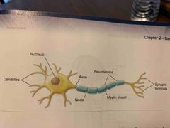
Neurons are composed of 3 main parts, what are they? |
The cell body (Soma) Axon Dendrites |
|
|
|
The cell body (soma) of a neuron contains a nucleus and other organelles including |
Lysosomes Mitochondria Golgi complex |
There are 3 |
|
|
The Axon is a cylindrical rejection from the cell body that |
Transmits nervous impulses to other neurons or effector sites (muscles, organs) providing communication from the brain & spinal cord to other parts of the body. |
|
|
|
Dendrites |
Gather information from other structures & transmit it back into the neuron |
|
|
|
The PNS consists of how many cranial nerves, pairs of spinal nerves & sensory receptors? |
12 cranial nerves 31 pairs of spinal nerves Sensory receptors |
|
|
|
Spinal nerves |
Branch out from the brain & spinal cord |
|
|
|
Sensory afferent neurons respond to |
Touch Sound Light Heat Taste Motion |
|
|
|
What are the 2 further subdivisions of the PNS? |
Somatic nervous system Autonomic nervous system |
|
|
|
The somatic nervous system |
Consists of nerves that serve the outer areas of the body and skeletal muscle, they are largely responsible for the voluntary control movement. |
|
|
|
The autonomic nervous system |
Supplies neural input to the involuntary systems of the body (heart, digestive systems, endocrine glands). |
|
|
|
The autonomic system is further divided into what systems? |
Sympathetic nervous system Parasympathetic nervous system |
|
|
|
Both the sympathetic and parasympathetic nervous system’s serve to |
Increased levels of activation in preparation for activity (sympathetic)/serve to decrease levels of activation during rest and recovery (parasympathetic) |
|
|
|
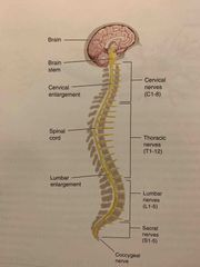
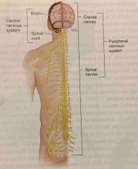
The CNS pictured |
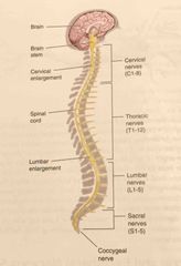
Back (Definition) |
|
|
|
The neuron pictured |
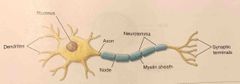
Back (Definition) |
|
|
|
What are the 3 main functional classifications of neurons that are determined by the direction of their nerve impulses? |
Sensory (afferent) neurons Interneurons Motor (efferent) neurons |
|
|
|
Sensory afferent neurons |
Transmit nerve impulses from effector sites (muscles, organs) via receptors to the brain & spinal cord. |
|
|
|
Sensory afferent neurons respond to |
Touch Sound Light And other stimuli |
|
|
|
Interneurons |
Transmit nerve impulses from 1 neuron to another. |
|
|
|
Motor efferent neurons |
Transmit nerve impulses from the brain & spinal cord to effector sites (muscles, glands). |
|
|
|
When a person touches a hot object the 3 neurons work together, what happens? |
This sensory afferent neurons send a signal from the hands of the brain telling the brain that the object is hot. This signal makes us way to the brain by traveling from one neuron to another via interneuron. Signal makes it to the brain sending the appropriate signals to the muscle of the hand and arm via motor neurons to pull away. |
|
|
|
The CNS |
The brain and spinal cord portion of the NS. |
It’s primary function is to coordinate the activity of all parts of the body. |
|
|
The CNS pictured |
Back (Definition) |
|
|
|
The neuron pictured |
Back (Definition) |
|
|
|
What are the 4 categories of receptors? |
Mechanoreceptors Nociceptors Chemoreceptors Photo receptors |
|
|
|
Mechanoreceptors are |
Sensory receptors responsible for sensing distortion in body tissues. Mechanical/outside forces (touch, pressure, stretching, sound waves, motion). |
|
|
|
Nociceptors respond to |
Pain |
|
|
|
ChemoreceptorsRespond to |
Chemical interaction (smell, taste) |
|
|
|
Photo receptors respond to |
Light (vision) |
|
|
|
Mechanoreceptors are |
Sensory receptors responsible for sensing distortion (pressure) in body tissues. |
|
|
|
Mechanoreceptors are located in the muscles tendons ligaments in joint capsules & include |
Muscle spindles Gogi tendon organs Joint receptors |
|
|
|
The PNS system pictured |
Back (Definition) |
|
|
|
The nervous system structure pictured muscle spindles |
Back (Definition) |
|
|
|
Joint receptors |
Receptors surrounding a joint that responsive pressure, acceleration, & deceleration of the joint. |
|
|
|
Do you want receptors act to signal |
Extreme joint positions, helping to prevent injury. |
|
|
|
The PNS system pictured |
Back (Definition) |
|
|
|
The nervous system structure pictured |
Back (Definition) |
|
|
|
Muscle spindles |
Receptors sensitive to change in length of the muscle & the rate of that change. |
|
|
|
When specific muscle is stretched the spindles within that |
Are also stretched, conveying information about its lengths to the CNS via sensory neurons. |
|
|
|
Once the information from muscle spindles reaches the brain it can then. |
Determine the position of various body parts. |
|
|
|
What can help regulate the contraction of muscles via stretch reflex mechanism? |
Muscle spindles |
|
|
|
What is the stretch reflex? |
It is a normal response by the body to a stretch stimulus in the muscle. |
|
|
|
Stretched muscle spindles send impulses to the |
Spinal cord immediately, and a response to contract the muscle is received within 1-2 milliseconds. |
|
|
|
The rapid neural response is designed as a |
Protective mechanism to prevent overstretching & potential muscle damage. |
|
|
|
Joint receptors |
Receptors surrounding a joint that responsive pressure, acceleration, & deceleration of the joint. |
Ruffini endings Pacinian Corpuscles |
|
|
What are the 3 main functional classifications of neurons that are determined by the direction of their nerve process? |
Sensory (afferent) neurons Interneurons Motor (efferent) neurons |
|
|
|
How does the CNS receive sensory input & initiate responses? |
With the nerves of the PNS. |
|
|
|
What are the two main functions of the peripheral nerves? |
Provide a connection for the nervous system to activate different effector sites, muscles (motor function). Relay information from the effector sites back to the brain via sensory receptors, providing constant updates on the relation between the body & the environment. |
|
|
|
Mechanoreceptors transmit impulses through |
sensory nerves enabling us to detect the position far muscles bones joints proprioception. |
|
|
|
Where are Mechanoreceptors located? |
Muscles, tendons, ligaments, joint capsules. include muscle spindles, golgi tendon organs, and joint receptors. |
|
|
|
Golgi tendon organs |
Receptor sensitive to change in the tension of the muscle & the rate of that change. |
|
|
|
Where is the golgi tendon organ located? |
The point where the skeletal muscle fibers insert into the tendons of the skeletal muscle. |
|
|
|
When the Golgi tendon organ is activated what happens? |
The muscle relaxers, preventing the muscle from excessive stress/possibility of injury. |
|
|
|
Where are joint receptors located? |
In & around the joint capsule. |
|
|
|
Giant receptors signal |
Extreme joint positions, to help prevent injury. To initiate a reflexive inhibitory response in the surrounding muscles if there’s too much stress on the joint. |
|