![]()
![]()
![]()
Use LEFT and RIGHT arrow keys to navigate between flashcards;
Use UP and DOWN arrow keys to flip the card;
H to show hint;
A reads text to speech;
187 Cards in this Set
- Front
- Back
|
Nutrient Broth (liquid) is used to observe |
growth patterns |
|
|
In a nutrient broth you inoculate with a |
loop |
|
|
Nutrient Agar Slant is used to observe |
margin growth patterns |
|
|
In a Nutrient Agar Slat you inoculate with a |
Needle |
|
|
Nutrient Agar Plate is used to observe |
colony growth patterns |
|
|
In a Nutrient Agar Plate you inoculate with a |
loop |
|
|
DEFINE undefined media |
General growth media where the amount of carbon and nitrogen are unknown |
|
|
What is the common media used to maintain bacterial cultures? |
Nutrient broth and nutrient agar |
|
|
What sources are necessary for growth media? |
they are formulated from sources that supply carbon and nitrogen in a variety of forms- amino acids, purines, pyrimidines, monosaccharides to polysaccharides, and various lipids. Generally these are provided in digest of plant material (phytone) or animal material (peptone and others). |
|
|
How long and at what temperature must media be autoclaved before it is considered sterile? |
15 min at 121 C |
|
|
BSL-1 |
Organisms do not typically cause disease in healthy individuals and present a minimal threat to the environment and lab personnel. Standard microbiological practices are adequate. These microbes may be handled in the open, and no special containment equipment is required. |
|
|
BSL-2 |
Organisms are commonly encountered in the community and present a moderate environmental and/or health hazard. These organisms are associated with human diseases of varying severity. Individuals may do laboratory work that is not especially prone to splashes or aerosol generation, using standard microbiological practices. |
|
|
BSL-3 |
Organisms are of location or exotic origin and are associated with respiratory transmission and serious or lethal diseases Special ventilation systems are used to prevent aerosol transmission out of the laboratory, and access to the lab is restricted. Specially trained personnel handle microbes in a Class I or II biological safety cabinet (BSC), not on the open bench. |
|
|
BSL-4 |
Organisms have a great potential for lethal infection. Inhalation of infectious aerosols, exposure to infection droplets, and autoinoculation are of primary concern. The lab is isolated from other facilities, and access is strictly controlled. Ventilation and waste management are under rigid control to prevent release of the microbial agents to the environment. Specifically trained personnel perform transfers in Class III BSCs. Class II BSCs may be used as long as personal wear protective pressure, one-piece body suits with a life-support system. |
|
|
Control |
A constant that is unchanging A controlled experiment is one in which all variables except one- the (experimental variable)- are maintained without change. |
|
|
Experimental variable |
The variable in which has changes |
|
|
false positive |
A test that indicates a test is positive but is actually negative |
|
|
false negative |
A test that indicates a test is negative but is actually positive. |
|
|
What Biological Safety Level is associated with the microbes used in this lab? |
BSL-2 |
|
|
List the characteristics of proper student conduct |
To reduce the risk of infection, do not smoke, eat, drink, or bring food or drinks into the laboratory room- even if lab work is not being done at the time. Do not apply cosmetics or handle contact lenses in the laboratory. Wash your hands thoroughly with soap and water after handling living microbes and before leaving the laboratory each day. Do not remove any organisms or chemicals from the laboratory. Lab time is precious, so come to lab prepared for that day's work. Figuring out what to do as you go is likely to produce confusion and accidents. Work carefully and methodically. Do not hurry through any laboratory procedure. |
|
|
List the characteristics of basic laboratory safety |
read page 3 |
|
|
Aseptic Transfer |
Aseptic transfer is the process of adding living microbes to a media without contaminating the media with unwanted microbes. To transfer microbes without contamination all media and tools used in the process must be sterile. |
|
|
Aseptically |
Without contamination of the culture, the sterile medium, or the surroundings. |
|
|
Meniscus |
The curved upper surface of a liquid in a tube. When reading volumes, use the base of the meniscus |
|
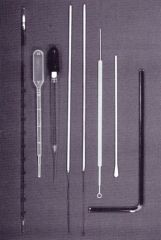
list from left to right |
Serological pipette, disposable transfer pipette,Pasteur pipette, inoculating needle, inoculating loop, disposable inoculatingneedle/loop, cotton swab and glassspreading rod. |
|
|
Epidemiology |
Is the study of the causes, occurrence, transmission, distribution, and prevention of diseases in a population. |
|
|
index case |
the first case of a disease |
|
|
incidence rate |
number of new cases in a time period/size of at-risk population at midpoint of time period. X K |
|
|
prevalence rate |
number of existing cases at a point in time/total population X K |
|
|
How are diseases transmitted? |
ingestion, inhalation, direct skin contact, open wounds, or lesions in the skin, animal bites, direct blood-to-blood contact as in blood transfusions, and sexual contact. sick people, or animals, healthy people or animals carrying the infectious organism or virus, water contaminated with human feces, contaminated objects (fomites) aerosols, or biting insects (vectors) |
|
|
How is incidence rate calculated? |
number of new cases in a time period/size of at-risk population at midpoint of time period. |
|
|
Ubiquity |
found just about everywhere |
|
|
Free-living |
they do not reside on or in a specific plant or animal host and are not knows to cause disease. |
|
|
Host |
A animal or plant on or in which a parasite or commensal organism lives. |
|
|
nonpathogenic |
Does not cause disease |
|
|
commensal |
a commensal organism, such as many bacteria. |
|
|
mutualistic |
relationship that benefits two/both organisms |
|
|
opportunistic |
They are capable of producing a disease state if introduced into a suitable part of the body. |
|
|
pathogens |
A bacterium, virus, or other microorganism that can cause disease. |
|
|
reservoir |
Any area including sites outside that host organisms, where a microbe resides and serves as a potential source of infection |
|
|
pathogenic |
Causes disease |
|
|
mixed culture |
A microbial culture consisting of two or more species. |
|
|
pure culture |
Contains only a single species.
|
|
|
quadrant streak plate |
A type of isolation technique where a bacterial sample is streaked over the surface of an agar medium. During streaking the cell density decreases, eventually leading to individual cells being deposited separate on the agar surface (colonies) or CFU (Colony-forming Unit) |
|
|
colony-forming unit (CFU) |
colony origin
|
|
|
filiform |
Dense and opaque with a smooth edge |
|
|
pigmented |
Color- such as opaque, translucent, shiny, or dull
|
|
|
friable |
crusty
|
|
|
spreading edge |

|
|
|
translucent |
allowing light, but not detailed images, to pass through; semitransparent. |
|
|
transparent |
allowing light to pass through so that objects behind can be distinctly seen
|
|
|
pellicle |
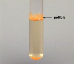
organisms that float on top of the medium and produce a type of surface membrane |
|
|
sediment |
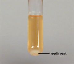
organisms that sink to the bottom of a broth |
|
|
uniform fine turbidity |

cloudy broth with no visible floating particles |
|
|
flocculent |
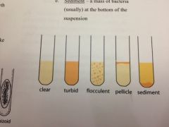
medium has small masses visible in suspension |
|
|
What is the purpose of the four-quadrant streak? |
Designed to separate deposited cells (CFUs) on the agar surface so individual cells (CFUs) grow into isolated colonies. Generally use high cell density samples.
|
|
|
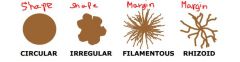
What are the 5 basic categories of colony morphology |
Shape
Margin Elevations Texture Pigment production |
|
|
What is the difference between colony morphology and margin morphology? |

margin morphology is a section inside of colony morphology that may be described:
Entire (smooth, with no irregularities) Undulate (wavy) Lobate (lobed) Filamentous (thread like) Rhizoid (branched like roots) |
|
|
Be able to distinguish between round, irregular, filamentous, and rhizoid colony morphology. |
|
|
|
Be able to distinguish between smoother, rhizoid, irregular, lobate, and filamentous margin morphology. |
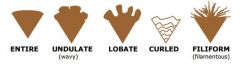
margin morphology is a section inside of colony morphology that may be described:Entire (smooth, with no irregularities)Undulate (wavy) Lobate (lobed) Filamentous (thread like) Rhizoid (branched like roots)
|
|
|
What terminology is used to describe colony elevation? |
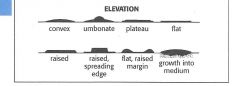
Convex
Umbonate Plateau Flat raised raised, spreading edge flat, raised margin growth into medium |
|
|
How is growth described on an agar slant? |
Filiform- (dense and opaque with a smooth edge)
Pigmented Friable- (crusty) Spreading Edge Translucent Transparent |
|
|
How is growth described in broth? |

Pellicle- organisms that float on top of the medium and produce a type of surface membrane Sediment- organisms that sink to the bottom of a broth Uniform Fine Turbidity- cloudy broth with no visible floating particles Flocculent- medium has small masses visible in suspension |
|
|
Resolution/ resolving power |
ability of the unaided human eye to distinguish two points as separate.
unaided human eye= 0.5 and 1 millimeter |
|
|
parfocal |
able to maintain relative focus when switching objectives |
|
|
Condenser Lens |
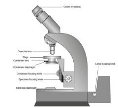
focuses the light through the specimen. The purpose of the condenser s to change the contrast of the specimen viewed. Condensers are more useful at the highest powers where they render a sharper image than those who no condenser adjustment. |
|
|
Course Adjustment Knob |
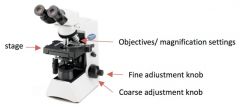
It is used to focus on the specimen; it may move either the stage or the upper part of the microscope (in a relative up and down motion). Always focus with the coarse knob first. 4x and 10x only |
|
|
Fine focus adjustment knob |

Found on the coarse focus adjustment knob, used to bring the specimen into sharp focus under low power lenses and for all focusing under high power lenses. |
|
|
Lamp |

Provides the light to view the specimen. It may have an intensity knob to vary the brightness |
|
|
Ocular Lenses/ eyepiece |
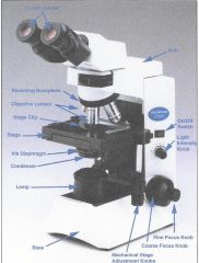
Magnifies the image usually 10x |
|
|
Objective lenses |
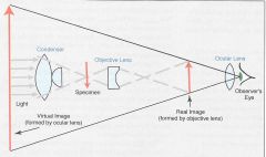
gathers light from the specimen, magnifies the image and projects it into the body tube. The revolving nose piece "clicks" the particular objective lens into place. There are 3 or 4 objective lenses on a microscope. They usually consist of 4x, 10x, 40x, and 100x powers. |
|
|
Stage clips |

Holds specimen slides in place
|
|
|
Stage |

It’s where the sample or specimen is placed for examination.
can be moved up and down left and right |
|
|
Total Magnification |
Magnification by the Objective Lens X Magnification by the Ocular Lens |
|
|
Working distance |
The amount of space between the very tip of the objective lens and the object you are viewing.
|
|
|
Field of view |
The amount of area you can see at any given magnification. |
|
|
Depth of Field (Focus) |
The amount of an object in clear focus at any given magnification |
|
|
Size of a prokaryotic cell |
0.1-0.5 um |
|
|
Size of an eukaryotic cell |
10-100um
|
|
|
Contrast |
Contrast is when you have two different refractive indexes
Contrast is the ratio between the dark and the light Typically, most microscopes use absorption contrast, that is the specimen is subjected to stains in order to be seen |
|
|
Morphology |
The study of the form of things (microbes on this class) |
|
|
cocci (coccus) |

sphere |
|
|
Bacilli (bacillus) |

rods |
|
|
Spirilla (spirillum) |

spirals |
|
|
Vibrios |
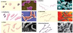
slightly curved rods |
|
|
coccobacillus |
short rods |
|
|
spirochetes |
flexible spirals |
|
|
pleomorphism |
more then 1 shape in a sample |
|
|
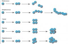
diplococcus |
two daughter cells attached, even after a coccus divides
|
|
|
diplobacilli |
2 bacilli attached together
|
|
|
streptococcus |
3+ coccus formation
|
|
|
streptobacilli |
3+ bacilli formation
|
|
|
staphylococcus |
a cluster of cells happens if the division planes of a coccus are irregular
|
|
|
chromogen |
The colored molecule in a stain
|
|
|
acidic stains |
Nigrosen, Congo Red , eosin stain |
|
|
basic stains |
Basic stains at + charged and stain the inside of the cell. Crystal Violet, Methylene Blue, Safranin
|
|
|
heat-fixed emulsion |
Heat-fixing kills the bacteria, makes them adhere to the slide, and coagulates cytoplasmic protein to make them more visible. it also distorts the cell to some extent. |
|
|
artifact |
things that appear on you slide that do not have to do with the specimen that you are looking at. |
|
|
What limits the size of bacteria? |
In general the reasons for cell size limits are due to the mechanisms needed for cell survival and how cells' requirements are met by the structures that form and are contained within cells. Nucleo-cytoplasmic ratio Fragility of cell membrane Mechanical structures necessary to hold the cell together (and the contents of the cell in place) |
|
|
how does a basic stain work? |
it dyes the inside of the cell so we can view the cell morphology, size, and arrangement |
|
|
What features of the cell attract a basic stain? |
negative changes on the surface of bacteria cells
|
|
|
list 3 common basic stains |
Crystal Violet, Methylene Blue, Safranin
|
|
|
What is the chemical property of the chromogen (dye) used in a negative stain? |
acidic and negative charge
|
|
|
Titrate
|
adding a little at a time
|
|
|
Differential stains |
allow a microbiologist to detect differences between organisms or differences between parts of the same organism |
|
|
structural stains |
capsule, spore, and flagella stain
|
|
|
decolorization |
removing the color from a stained slide
|
|
|
primary stain |
the initial stain of a staining process
|
|
|
mordant |
secures stain to bacteria, enhances CV, and forms CV iodine complex
|
|
|
Crystal Violet-iodine complex |
makes it so that the dye cannot be removed easily. Formed after iodine is added to CV during a gram stain |
|
|
counterstain |
the secondary stain. Used for identifying purposes at the end of a staining process.
|
|
|
what is the importance of a differential stain? |
It can help us find information about other features not seen by simple stain.
|
|
|
what is the most common differential stain in bacteriology? |
Gram Stain
|
|
|
what are examples of other differential stains? |
acid fast= differential capsule= special flagella= special Spore= special |
|
|
how many basic stains are used in the gram staining procedure |
2
Primary Stain= CV Counter Stain = Safranin |
|
|
what is the primary stain of the gram staining procedures? |
CV
|
|
|
what is the purpose of iodine in the gram staining procedure? Which cells are decolorized? Which are not decolorized? |
secures stain to bacteria, enhances CV, and forms CV iodine complex Gram- decolorized Gram+ colorized |
|
|
What color are gram + cells after the gram staining procedure?
Gram-? |
Purple
Red |
|
|
What structural features give rise to the difference in the gram + and gram - cells and the resulting color results? |
The thick layer of peptidoglycan and cross-linking in Gram + trap the CV I complex more effectively, making the Gram + wall less susceptible to decolorization.
The Gram- cell walls have a high level of lipid content (because of the outer membrane) and a thinner peptidoglycan. The alcohol/acetone in the decolorizer extracts the lipids, making Gram - wall more porous and incapable of retaining the CV-I complex, thereby decolorizing it. |
|
|
How can over-decolorizing or under-decolorizing affect the gram staining results? |
over-decolorizing= reddish gram+ cells
under-decolorizing= purple gram- cells false positive |
|
|
What is the best culture "age" to use for gram staining? |
24 hours or less
|
|
|
What is the KOH (potassium hydroxide) test? |
It is a secondary test for Gram identification. This test is only valid if performed on 24-48 hour culture. The KOH dissolves the cell wlls of Gram- bacteria, but does not affect Gram + cells. The Gram - bacteria will lyse and cellular contents, including DNA, spill out of the cell. DNA is very viscous, and with a large enough cell mass, the DNA strands can be seen sticking to a loop when touched. G+ cells are not lysed, no free DNA, no viscosity will be observed.
|
|
|
Acid Fast Stain is used to test__________ |
For the presence of mycolic acid in the cell walls of acid fast organisms. Mycobacterium Tuberculosis Leprosy Nocardia (N.brasiliensis, N.asteroides) Cryptosporidium Isospora ZN and K method |
|
|
Acid-Fast Bacilli |
An AFB smear is used as a rapid test to detect mycobacteria that may be causing an infection such as tuberculosis. |
|
|
What type of molecule is found in the cell wall of acid-fast bacteria? What properties of that component give rise to the acid-fast nature of the cell? |
Mycolic Acid Mycolic Acid is a waxy (lipid) substance which gives the cell wall high affinity for the primary stain (carbolfuchsin- lipid soluble) and resistance to the decolorization of acid alcohol. |
|
|
Why is it used? |
To test for Acid-fast bacteria that can cause TB and Leprosy
|
|
|
What color do acid-fast cells appear after the acid-fast procedure? Non acid-fast cells? |
Red Green (Brillian Green) or Blue (Methylene Blue) Counterstains |
|
|
What genus of bacteria can be detected using the acid-fast procedure? |
Mycobacterium
Nocardia Cryptosporidium Isospora |
|
|
What disease can be diagnosed using the acid-fast procedure? |
Mycobacterium Tuberculosis
Leprosy N.brasiliensis, N.asteroides Cryptosporidium Isospora |
|
|
capsule |
are composed of mucoid polysaccharides or polypeptides that repel most stains.
|
|
|
is capsul type of staining procedure a negative, positive, or both? Why? |
Both, because Congo Red is a negative stain while Maneval's stain is both a negative and positive mixture.
Maneval's solution (a mixture of acetic acid and acid fuchsin) is added to the slide. The acetic acid lowers the pH in the sample and causes the Congo red to change from a red color to blue. The acid fuchsin (a basic dye) interacts with the bacterial cell, staining the cell bright red. |
|
|
What is the medical importance of a capsule? |
capsule production increases virulence in some microbes because it protects them from phagocytosis
Capsules have several functions and often have multiple functions in a particular organism. Like fimbriae, capsules, slime layers, and glycocalyx often mediate adherence of cells to surfaces. Capsules also protect bacterial cells from engulfment by predatory protozoa or white blood cells (phagocytes), or from attack by antimicrobial agents of plant or animal origin. |
|
|
What stain/s are used for a capsule stain? |
Congo Red and Maneval's stain
|
|
|
What color is the capsule after staining? |
clear
|
|
|
endospore |
Is a dormant form of the bacterium that allows it to survive poor environment conditions.
|
|
|
keratin |
protein that is also a tough outer layer on spores
|
|
|
vegetative cells |
a cell of a bacterium or unicellular alga that is actively growing rather than forming spores.
|
|
|
spore cell |
cell from which a spore develops
|
|
|
sporulate |
produce or form a spore or spores.
|
|
|
germinate |
begin to grow and put out shoots after a period of dormancy
|
|
|
What stain/s is are used for endospore stain? |
Malachite Green (Primary stain)
Safranin (Counter Stain) |
|
|
what is the function of an endospore? |
ensure the survival of a bacterium through periods of environmental stress.
|
|
|
what is the endospore make of? |
protein keratin
|
|
|
what color is the endospore and veg cells after staining? |
Endospore= green
Veg= red |
|
|
what are the possible locations of endospores within the cell? |
Middle (central), end (terminal), and between the middle and the end (subterminal)
|
|
|
what genera produce spores? |
Bacillus and Clostridium
|
|
|
what is the purpose of the steam? |
So the stain can be forced into the spore.
|
|
|
which culture would have more spores? 24 hr or 73 hour culture? |
72 hour |
|
|
What ingredients make up a nutrient broth? |
beef extract peptone distilled or deionized water |
|
|
What ingredients make up nutrient agar? |
Beef extract peptone agar distilled or deionized water |
|
|
Label your media with your |
Name, date, medium, and inoculum |
|
|
When you want to get bacteria out of a tube, do you insert the loop into the tube or do move the tube to the loop? |
Move the tube to the loop. |
|
|
Do you hold the tube right ride up or at an angle? |
at an angle |
|
|
Point Prevalence (Prevalence Rate) |
Number of existing cases at a point in time/ total population x k |
|
|
dark-field microscopy |
a special condenser is used so only the light reflected off the specimen enters the objective. The appearance is of a brightly lit specimen against a dark background, and ofte with better resolution then that of the bright-field microscope. |
|
|
Phase Contrast Microscope |
uses special optical components to exploit subtle differences in the refractive indices of water and cytoplasmic components to produce contract. As a result, the specimen appears as various shades of "darks" against a bright background. |
|
|
Fluorescence Microscopy |
uses a fluorescent dye that emits florescence when illuminated with ultraviolet radiation. |
|
|
always keep petri dishes media side down or up? |
up |
|
|
what type of gloves are you allowed to use in class? |
Nitrile (best) or latex |
|
|
What type of gloves are you not allowed to wear in class? |
Vinal |
|
|
let the loop cool for |
20 seconds |
|
|
characteristic of plates |
Shape Color: pigment Margin Elevation Texture |
|
|
Characteristic of broths |
Turbidity Flocculence Pellicle Sediment |
|
|
Characteristics of Slants |
Filiform:line friable:crusty spreading edge transparent pigments |
|
|
The number on a pipette indicated the |
total volume and its smallest calibrated increments |
|
|
acid fast stain |
|
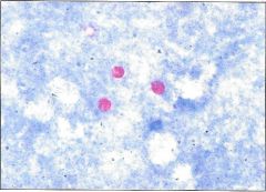
|
acid fast stain |
|
|
acid fast stain |
|
|
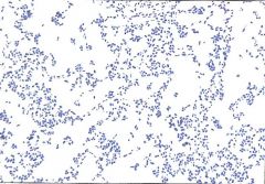
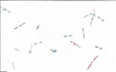
coccobacillus |
|
|
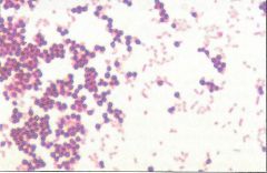
coccus shapes |
|
|
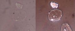
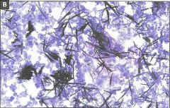
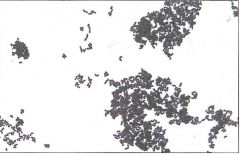
crystal violet crystals |
|
|
crystal violet crystals |
|
|
diplococcus arrangement |
|
|
endospore stain |
|
|
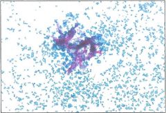
gram stain |
|
|
over decolorized gram stain |
|
|
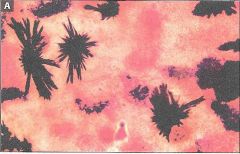
pleomorphism |
|
|
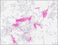
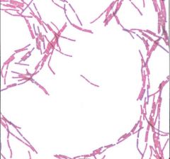
simple stain- streptobacillus |
|
|
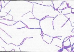
spirillum |
|
|
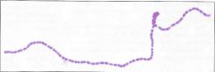
spirochete |
|
|
stain precipitate |
|
|
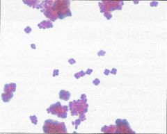
staphylococcus arrangement |
|
|
streptobacillus arrangement |
|
|
streptococcus |
|
|
tetrad arrangement |
|
|
under decolorized gram stain |
|
|
vibrio |