![]()
![]()
![]()
Use LEFT and RIGHT arrow keys to navigate between flashcards;
Use UP and DOWN arrow keys to flip the card;
H to show hint;
A reads text to speech;
265 Cards in this Set
- Front
- Back
- 3rd side (hint)
|
A microscope that allows light rays to pass directly to the eye w/o being deflected by an intervening opaque plate in the condensor |
Brightfield microscope |
|
|
|
Transport of microscope |
Both hands under or one under and one on the arm |
|
|
|
Microscope lens care |
Clean lens b4 each lab and at the end |
|
|
|
Framework of microscope |
Includes arm and a base to which all other parts are attached with older ones having a pivot point between arm and base |
|
|
|
Microscope stage |
The horizontal platform that supports the microscope slide |
|
|
|
Clamping device used for holding and moving the slide around on the stage |
Stage adjustment |
|
|
|
Knobs used to adjust and move a slide |
Stage adjustment knobs |
|
|
|
A knurled wheel on the right side of the base that regulates voltage |
Light intensity control |
|
|
|
Filter often needed to reduce the intensity of light below the lwr limit allowed by the voltage control |
Neutral density filter |
|
|
|
Different types of neutral density filters |
1) one placed over the light source on the base 2) filter built into the base |
|
|
|
All compound microscopes have |
Three lens systems: 1) the oculars (top) 2) the objectives ( magnifiers) 3) the condenser ( under stage) |
|
|
|
Ocular |
1) eyepiece on top 2) complex piece consisting of 2 or more lenses 3) usual magnification 10x 4) most modern microscopes have 2 ocular (binocular) lenses |
|
|
|
Objectives |
1) 3 or more usually present 1) low-power. 10x 2) high-dry 40x 3) oil immersion. 100x 4) rapid scanning 4x 2) attached to rotatable nosepiece for positioning over slide 3) |
|
|
|
Total magnification |
Pwr of occular lens X pwr of objective lens used |
|
|
|
Third lens system |
Condenser (under the stage) 1) collects and directs light 4m lamp to slide 2) doesnt affect magnification pwr 3) can be moved up and dwn by knob under stage ( diaphragm) |
|
|
|
Diaphragm |
1) knob, knurled ring, or lever under the stage that moves the condenser up and dwn which 2) this regulates the amt of light that reaches the slide 3) microscopes that lack a voltage control ( light intensity control) rely on diaphragm entirely for control of light intensity |
|
|
|
Diopter adjustment |
1) to bring occulars same distance 4m as eyens in and out 2) focus right eye only, w/o touching focusing knobs diopter adjustments made on left eye by turning diopter adjustment ring until sharp image seen |
|
|
|
Focusing knobs |
Fine and course adjustment knobs Moves stage up and dwn |
|
|
|
Parfocal |
A microscope that maintains focus when the objective magnification is increased |
|
|
|
Resolving power |
A lens' ability to completely seperate two objects in a microscopic field To resolve the the distance on the field |
|
|
|
Resolving power formula |
d= limit of resolution ( or the distance between the two objects d is a function of 2 properties: 1) ¥ = the wavelength of light used to observe a specimen 2) NA= numerical aperature |
|
|
|
Numerical aperature |
1) a numerical expression abt how the condenser lens concentrates and focuses light from the light source (How much light made it to objective) 2) max value when light rays are focused into a cone of light that passes through specimen into objective lens 3) light rays refracted or bent from passing 4m glass to air are lost, decreasing the NA 4) lower the NA 4m loss, the lower the resolving power |
|
|
|
Limit for most light microscopes |
1000x which is set by intrinsic property of lenses called resolving power |
|
|
|
Resolution limit of light microscope |
0.2 micrometers Any two things closer would not be seen as two distinct objects |
|
|
|
Blue filter |
Placed over light to shorten wavelength of resulting light for step 1 of 4 for max resolution |
|
|
|
Immersion oil |
1) special oil with same refractive index as glass used w/ 100x objective lens 2) oil used to max resolution step 4/4 b/c the oil forms a continuous lens system that limits loss of light b/c of refraction |
|
|
|
For magnification to increase |
Resolution must increase |
|
|
|
4 steps to maximize resolving power (resolution) |
1) blue filter over light to produce shorter wavelengths 2) condenser should b kept at highest position to maximize amt of light into objective and limit loss from refraction 3) diaphragm should not be stopped dwn too much b/c it reduces the numerical aperature 4) use immersion oil to form continuous lens system limiting loss of light |
|
|
|
Contrast adjustment |
Closing the diaphragm improves contrast |
|
|
|
Solvents for cleaning lenses |
1) green soap with warm water 2) xylene 3) acetone and alcohol sparingly Can possibly dissolve lens mounting cement |
|
|
|
Whenever ocular is removed |
A piece of lens tissue needs to be placed over open hole into microscope |
|
|
|
Main reason for starting with a low power objective |
Enables you to explore the slide to look for object to study |
|
|
|
Everytime the objective lens is changed |
Focus the condensor and adjust it for max illumination |
|
|
|
Depth of field |
The range of distance in front of and behind a focused image within which other objects will appear clear and sharply defined |
|
|
|
Parcentral |
Image will remained centered when changing objective |
|
|
|
When increasing light |
Open diaphragm first before light intensity control to extend lamp life |
|
|
|
Oil immersion lens should never |
Be used without oil |
|
|
|
Ocular micrometer |
Consists of circular disk of glass that has graduations engraved on its upper surface |
|
|
|
Before using a micrometer |
It has to be calibrated for each of the objectives using a stage micrometer |
|
|
|
Stage micrometer |
Aka an objective micrometer has lines inscribed on it that are exactly 0.01 mm (10 um) apart |
|
|
|
To calibrate the occular micrometer |
1) superimpose ocular and stage micrometers 2) determine how many how many occular graduation coincide with the stage scale graduations 3) divide how many stage grad measurement (0.01mm) by how many occular divisions fit in it (example 7) =0.00143mm 4) convert to micrometers um 1000um = 1mm = 1.43um apart in occular |
|
|
|
Umbiquitous |
Found everywhere |
|
|
|
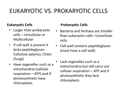
Biological domains |
1) domain bacteria 2) domain eukarya 3) domain archaea |
3 |
|
|
Domain bacteria |
1) prokaryotic cell structure -lacks organelles, organized nucleus with nuclear membrane, mitochondria, chloroplasts 2) posses 70S ribosomes that are inhibited by many broad spectrum antibiotics 3) vast majority of organisms enclosed in cell wall composed of peptidoglycan 4) bacteria and cyanobacteria are members of this domain |
|
|
|
Domain eukarya |
1) have eukaryotic cell types 2) membrane bound organelles incl. Mitochondria and chloroplasts and an organized nucleus enclosed in a nuclear membrane 3) 80S ribosomes that are not inhibited by broad-spectrum antibiotics 4) plants, animals, and microorganisms like protozoa, algae, and fungi belong 5) plants have cell walls composed of cellulose 6) fungi have cell walls composed of chitin 7) animal cells lack a cell wall structure |
7 |
|
|
what lens has the highest magnification? and what is the magnification? |
oil immersion lens = 100x |
|
|
|
magnification of the ocular |
10x |
|
|
|
objective magnification in increasing order |
4x rapid scanning ,10x low-power, 40x high-dry , 100x oil immersion |
|
|
|
resolving power |
ability of a lens to separate two objects observed under the microscope also called resolution |
|
|
|
resolving pwr based on ... |
numerical aperature & wavelength |
|
|
|
numerical aperature (NA) |
value or ability of a lens to concentrate and focus light . The more light refreaced or bent before entering the lens, the lower the NA |
|
|
|
parfocal |
image remains in focus when changing from a lwr pwr to a higher pwr |
|
|
|
effect of wavelength on resolution |
longer wavelength = decresed resolution shorter wavelength = increased resolution |
|
|
|
how does magnification effect working distance |
as mag increases, working distance decreases |
|
|
|
working distance |
distance from the objective to the slide |
|
|
|
effect of oil immersion lens wiith oil |
oil has same refractive index as glass so it eliminates loss of light due to refraction |
|
|
|
morphology |
shape and arrangement of bacteria as observed under a microscope |
|
|
|
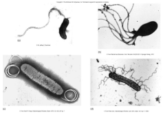
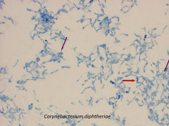
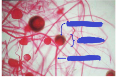
3 main shapes of bacteria |
1. Bacillus (rod) 2. coccus (sperical) 3. Spirochete (spiral, helix, or curved) |
|
|
|
coccus |
circular |
|
|
|
bacillus |
rod |
|
|
|
vibrio |
curved rod like boomerrang or comma |
|
|
|
spirilum |
spirals like lose curls |
|
|
|
spirochetes |
helical like tight curls and DNa |
|
|
|
name the 5 kingdoms of eukaryotes |
1. protozoa 2. plants, green algae 3. various algae 4. animals 5. fungi |
|
|
|
cytoskeleton of bacteria |
actin protien filaments |
|
|
|
pilus |
-sex pili - gram neg bacterium especially -way that bactreria exchange dna across -how bacteria can change dna makeup to change and become resistant etc. |
|
|
|
eukaryotic vs. prokarytic |
|
|
|
|
80S the S = |
Svedberg unit = weight unit |
|
|
|
two methods for obtaining pure cultures |
1. streak plate methods 2. pour plate method |
|
|
|
why do we put the agar pours in a water bath? |
1. media to hot to handle out of the autoclave 2. keeps media cool enough to handle, but not too cool where it will solidify |
|
|
|
water bath setting and why? |
65 degrees C to keep the media liquid b/c it solidifies at 42 degrees C |
|
|
|
what temp does media solidify? |
42 degrees C |
|
|
|
if the media were inoculated b4 it cooled... |
it would kill most bactria |
|
|
|
cooling agar also ... |
reduces condensation from forming on the lids |
|
|
|
how do you label tubes? |
write your initials, date, lab exercise, and what its innoculated with on the GLASS portion of the tube with permanent marker |
4 things to write and where to write it
|
|
|
how do you label plates? |
initials, date, lab exercise, and whats innoculated on the plate on the BOTTOM of the plate |
4 things to write and where to write it |
|
|
What does SIM media stand for? how do you innoculate it? |
S= sulfide production I= indole formation M= motility you stab itwith innoculating needle |
|
|
|
how is sulfide production shown and why in SIM media? |
it shows up as black in color b/c bacterial enzymes produce hydrogen sulfide and hy sulf reacts with the iron salts in the media and makes a black color in the media |
|
|
|
what is indole formation? |
the break dwn of amino acids |
|
|
|
how do you see motility and in what medias can you use? |
Growth of bacteria away from the stab line medias: SIM and Soft Agar plates |
|
|
|
Properties of agar |
1. polysaccharide from red algae 2. solidifies at 42 degrees C and stays solid until 100 degrees C 3. non-toxic and does not provide nutrients since its not digestible by most organisms 4. Holds nutrients and moisture 5. Lasts a week or longer if its not too thin |
5 |
|
|
what was used a lot before agar? why did they stop? |
gelatin b/c some microbes could digest it and leave a goopy mess |
|
|
|
How much agar does each media have? 1. SIM 2. Soft Agar 3. Nutrient Agar |
1. .35% 2. .7% 3. 1.5% |
|
|
|
shaking does what to soft and SIM media |
breaking b/c they are too soft |
|
|
|
Brownian movement |
not true motility organisms move b/c water current on slide or molecular bonbardment |
|
|
|
true motility |
organism has a flagella and movement can be seen in many directions |
|
|
|
name the 5 flagella arrangements |
1. polar = general term meaning that flagella are on either one or both ends of the cell 2. Monotrichous- having a single flagellum 3. lophotrichous= having bunches or tufts of flagella emerging from the same site -like a pony tail \can be on one or both ends of the cell 4. amphitrichous = flagella on both ends of the cell 5. peritrichous = flagella all over surface of the cell |
|
|
|
a. mono b. lopho c. amphi d. peri |
|
|
|
flagella staining |
really hard to do b/c you have to build up the stain on the flagella |
|
|
|
what type of disinfectant do we use on the tables? how well does it work? |
CiDecon, it is a disinfectant that doesnt sterilize but kills some bacteria and viruses but does not destroy endospores |
|
|
|
sterilization |
a process that completely destroys all viable microorganisms including bacteria, endospores, and viruses from an object or environment |
|
|
|
methods used in lab for sterilization |
1. autoclave - uses steam under pressure 121 degrees C at 25 psi for 10-40 minutes depending on the load 2. incineration - flaming the inoculating loops |
|
|
|
simple stain and how do they work |
using a simple stain o color cells to b observed under a microscope cells have an overall (-) charge so (+) charged ion are attached to the inside of the bacteria |
|
|
|
basic dyes definition and types |
the colored portion of the dye is on the (+) charged portion of teh dye 1. Methylene blue (+) chloride (-) 2. Crystal violet 3. malachite green 4. carbolfuchsin 5. safranin |
5 types with one indicating (-) compound with dye |
|
|
Acidic dyes definition and types |
Colored portion of the dye is on the (-) charged potion of the dye so it dyes the background 1. Nigrosin 2. India ink |
2 types opposite of an acid |
|
|
simple staining uses what two types of dyes |
acidic and basic |
|
|
|
crystal violet |
basic dye |
|
|
|
safranin |
basic dye, color attached to (+) portion of dye |
|
|
|
india ink |
stains background, acidic dye |
|
|
|
methylene blue |
basic dye, Methylene blue (+) chloride (-) |
|
|
|
malachite green |
basic dye |
|
|
|
carbolfuchsin |
basic dye, carbolfuchsin (+) phenol (-) |
|
|
|
paliside arrangement |
picket fence arrangement of bacillus |
|
|
|
volutins aka definition: |
metachromatic granules Inclusion bodies of bacteria that contain stored molecules |

|
|
|
pleomorphism |
cell changing shape from club, bacillus, comma, sperm, pertain to irregularity of shape |
|
|
identify |
purple 1 left = paliside arrangement red = volutins purple 2 = pleomorphism |
|
|
|
differential staining types |
1. Gram Stain -distinguishes between cells w/ thick peptidoglycan layer (G+) & thin pep. layer (G-) 2. Endospore stain - distinguishes between endospores and vegetative cells 3. Acid-fast Stain - distinguishes between cells that are acid fast and non acid fast |
3 with what it differentiates |
|
|
differential staining
|
a staining method that allows you to tell the difference between things when you are done |
|
|
|
age important for what? |
for both Gram and and endospore staining |
|
|
|
how is age important for Gram staining? |
b/c cultures older than 16-18hrs become gram variable, esp. spore formers with age comes the breakdwn of the cell wall and can no longer hold the primary stain so bacteria that have a G+ cell wall will appear G- |
|
|
|
How is age important for endosporers? |
the bacteria have to b old enough (stationary phase ) for cells to begin to form endospores. young cultures in the exponential growth phase are not yet producing endospores |
|
|
|
stage of life where bacteria can produce endospores? |
stationary phase |
|
|
|
stage of life where bacteria are growing and too young to produce endospores |
exponential growth phase |
|
|
|
name things G+ has but G- doesnt |
1. thick peptidoglycan layer 2. wall techoic acid 3. lipotechoic acid (goes inside to membrane) 4. peptidoglycan and membrane form envelope |
4 |
|
|
name things G- have but G+ dont |
1. outer membrane with top portion composed of lipopolysaccharides or LPS bottom = phospholipids 2. porin protiens through outer membrane to allow molecule in and out 3. lipoprotiens at bottom of outer membrane in between phospolipids that anchor to peptidoglycan layer 4. thin peptidoglycan layer in between cell membrane and outer membrane 5. periplasmic space between cell membrane & peptidoglycan layer |
5 with location and f(x) |
|
|
gram stain def and outcomes |
differential stain that distingushes cells with a G+ cell wall from those with a G- cell wall outcomes -G+ = retain crystal violet and stain purple - G- = lose crystal violet and stain red from safranin counterstain |
|
|
|
gram stain important basis of bacterial ....
|
classification and identification |
|
|
|
G stain practical aid in ... |
diagnosing infection and guiding drug treatment |
|
|
|
primary and secondary stain in G stain |
primary = crystal violet secondary or counterstain = safranin |
|
|
|
mordent definition and G stain mordent |
something that helps hold the stain in the cell g stain mordent= iodine dye crystals trapped in cell wall |
|
|
|
what decolorizer is used for G stain? what is its effect? |
alcohol also called grams III it weakens the outer membrane causeing cell to lose primary dye crystal violet |
|
|
|
steps of gram stain & outcomes |
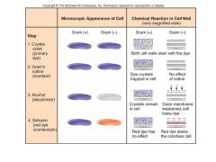
1. crystal violet primary dye - both cells purple 2. Gram's iodine (mordant) -traps color srystals in cell wall of G+ -no effect on G- -both cells purple 3. Decolorizer/ Gram's III/ alcohol -G+ crystals remain in cell -G- outer membrane weakened and loses dye -G+ cell purple , G- colorless 4. Safranin red dye counterstain -G+ - red dye has no effect and cell is purple -G- = red dye stains colorless cell making it pink or red |
|
|
|
staining method used for endospore stain and outcome |
schaeffer-fulton method -vegetative cells appear pink, endospores appear green |
|
|
|
genuses that form endospores which is aerobic or anaerobic? |
Bacillus (aerobic) and Clostridium (anaerobic) |
|
|
|
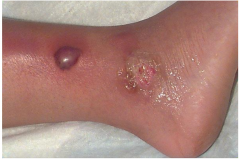
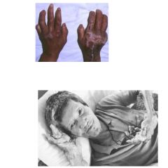
what pathogens form endospores and what do they cause? |
1. Clostridium perfringens - gas gangrene and food poisoning 2. Clostridium botulinum - botulism 3. Bacillus anthracis - anthrax |
|
|
|
causes gas gangrene |
clostridium perfringens |
|
|
|
causes anthrax |
bacillus anthracis |
|
|
|
causes botulism |
Clostridium botulinum
|
|
|
|
causes food poisoning |
clostridium perfringens
|
|
|
|
aerobic or anaerobic: clostridium |
anaerobic
|
|
|
|
aerobic or anaerobic:
Bacillus |
aerobic
|
|
|
|
similarity between bacillus and clostridium? difference? |
both form endospores bacillus aerobic clostridium anaerobic |
|
|
name it and its cause |
gas gangrene and Clostridium perfingens (per-fing-ens) bacteria |
|
|
name it and its cause |
gas gangrene, clostridium perfingens bacteria |
|
|
|
name the 3 types of botulism and their cause |
1. foodbourne 2. Infant 3. Wound cause: the bacterium Clostridium botulinum |
|
|

name it and its cause
|
foodborn botulism bacteria Clostridium botulinum |
|
|
name it and its cause
|
infant botulism bacteria Clostridium botulinum |
|
|
name it and its cause
|
wound botulism
bacteria Clostridium botulinum |
|
|
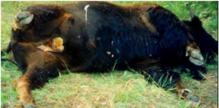
name it and its cause
|
bison infected with anthrax Bacillus anthracis |
|
|
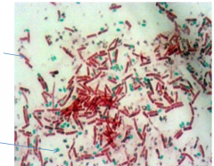
what kind of stain? name the green and pink structures |
endospore stain pink= vegetative cells green = endospores |
|
|
|
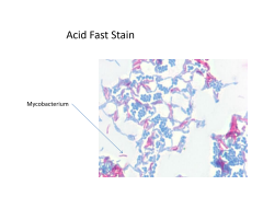
Culture for this stain is difficult to grow and has to grow on herat and brain fusion media |
acid-fast stain |
|
|
|
acid-fast stain and outcome |
differentitates acid-fast bacteria and non-acid fast bacteria acid-fast= dark pink non-acid-fast= blue |

|
|
|
what bacteria did we use i lab for acid-fast staining? |
mycobaterium lacticola |
|
|
|
name 3 pathogenic acid-fast bacteria and what they cause |
1. Nocardia - nocardia 2. mycobaterium leprae - leprosy 3. mycobaterium tuberculosis - TB |
|
|
|
primary stain we used in lab for acid-fast staining |
carbolfuchsin |
|
|
|
Acid-fast cells tend to ... |
stick together |
|
|
|
acid-fast stain procedure |
1. cover heat fixed smear with with carbolfuchsin while slide is over boiling water bath with saturated paper towel for 5 minutes (heat is mordant) 2. when cool wash with water and shake off excess water 3. decolorize with acid-alcohol for approx. 1 min. Check by tilting the slide and add more acid-alcohol until no more stain runs off. 4. stop decolorization by briefly rinsing with water 5. counterstain methylene blue for 30 secs. 6. rinse briefly with water to remove excess methyl blue 7. blot dry with bibulous paper and examine under oil lens |
|
|
|
what is the mordent we used for the acid-fast stain |
heat |
|
|
|
what decolorizer is used in acid-fast staining |
acid-alcohol |
|
|

what type of stain and name the bacterium |
|
|
|
name it and its cause |
nocardia , caused by nocardia acid-fast |
|
|
name it and its cause
|
leprosy, mycobacterium leprae, acid-fast |
|
|

name it, its cause , and 2 other facts
|
Tuberculosis, mycobacterium tuberculosis 1. people will waste away if not treated 2. a lot of antibiotics used to treat wont work b/c it has become resistant |
|
|
|
Gram Stain Procedure |

1. crystal violet -30 secs 2. wash - 2 secs 3. grams iodine - 1 min 4. wash - 2 secs 5. decolorize with alcohol or grams III for 5-15secs or till solvent flows colorlessly 6. wash -2 secs 7. counterstain with safranin- 1 min 8. wash - 2 secs 9. blot dry with bibulous paper |
|
|
|
Simple Staining Procedure |
1. Prepare smear using aseptic technique and air dry 2. when dry stain with methyl blue for 1 min 3. briefly wash off stain with water 4. water drops carefully blotted off slide with bibulous paper |
|
|
|
Endospore Staining procedure |
1. cover smear with small piece of paper towel and saturate with malachite green. Do this over a beaker of boiling water. use additional stain as needed to keep paper towel saturated. -5 mins 2. after slide cooled, remove paper towel and rinse with water - 30 secs 3. counterstain with safranin -30 secs 4. rinse briefly to remove safranin 5. blot dry with bibulous paper and examine under oil |
|
|
|
kinyoun acid-fast method |
modification to Ziehl-Neeson staining method in which concentrations of primary stain basic fuchsin (instead of carbolfuchsin) and phenol increased -increased concentrations of primary stain = mordant -safer b/c phenol fumes not made during staining |
|
|
|
Ziehl-Neeson staining method
|
primary stain carbolfuchsin containing phenol heated for 5 mins with boiling beaker -phenol and heat facilitate penetration of stain into cell (mordant= heat) - heat acting as mordant makes the mycolic acid and and cell wall lipids more permeable to the stain -problem : phenol can vaporize when heated, making toxic fumes that are toxic to the eyes and mucous membranes |
|
|
|
mordant |
facilitates the penetration of stain into a cell makes cell or cell structure more permeable to stain |
|
|
|
why are some bacteria called acid-fast ? |
because subsequent treatment of the cells with acid-alcohol does not remove the stain |
|
|
|
mycolic acid |
one of the cell wall lipids of mycobacterium and some nocardia that is a waxy material that is composed of fatty acids and fatty alcohols with hydrocarbon chains up to 80 carbons in length -sig effects staining |
|
|
|
mycology |
study of fungi |
|
|
|
fungi kingdom |
myceteae (eumycota) |
|
|
|
mycosis |
fungal infection |
|
|
|
How many species and name groups and group consstituents |
100,000 species 1. macroscopic fungi (mushroom, puffballs, gill fungi) 2. microscopic fungi (molds and yeasts) |
|
|
|
majority of fungi are... |
unicellular or colonial |
|
|
|
a few fungi have cellular... |
specialization |
|
|
|
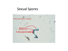
yeasts |
1. unicellular and mostly carry out asexual reproduction (blastospores (buds) while some carry out sexual (ascus/asci) |
|
|
|
pseudohyphae |
from budding yeasts that form a chain that look like a false hyphae |
|
|
|
molds |
cells are hyphae (mycelium) /microscopic filaments |
|
|
|
name the two types of hyphae |
1. septate- a division in filament with separated nuclei and pores for inbetween 2. non-septate- nuclei just float on dwn |
|
|
|
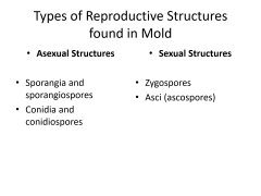
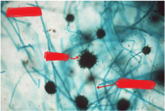
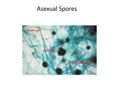
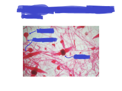
name the types of reproductive structures found in mold |
|
|
|
|
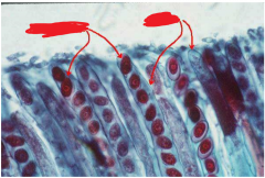
what are the asexual structures in mold? |
1. sporangia and sporangiospores 2. conidia and conidiospores |
4, the structure and the spores |
|
|
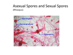
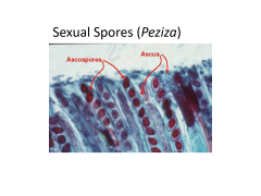
what are the sexual structures found in mold? |
1. zygosporangium &Zygospores 2. Asci and ascospores |
4, the structure and its spore |
|
Name the type of fungus, sexual or asexual, name the structures listed, what do they look like? |
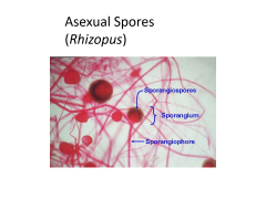
common bread mold/ looks like lollipops |
|
|
Name the type of fungus, sexual or asexual, name the structures listed |
|
|
|
Name the type of fungus, sexual or asexual, name the structures listed, what do they look like?
|
common bread mold, bats or birds |
|
|
Name the type of fungus, sexual or asexual, name the structures listed, what do they look like? like?
|
pea pods |
|
|
Name the type of fungus, sexual or asexual, name the structures listed, what do they look like?
|
|
|
|

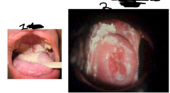
name #1 ,where its at, symptoms, and what causes it? |
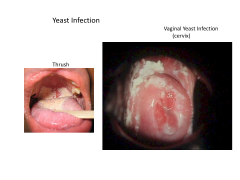
Thrush 1. caused by candida albicans 2. occurs in thick, white, adherent growth on the mucous membranes of mouth and throat |
|
|
name #2, ,where its at, symptoms, and what causes it? |

Vulvovaginal yeast infection painful, inflammatory condition of the female genital region that causes ulceration and whitish discharge -caused by candida albicans |
|
|
|
cutaneous candidiasis |
occurs in chronically moist areas of skin and in burn patients caused by candida albicans |
|
|
|
candida albicans |
-accounts for 80% of nosocomial fungal infections -accounts for 30% of deaths from nosocomial infections -can be pathogenic -normal flora of oral cavity, genitalia, large intestine, or skin of 20% of humans |
|
|
|
name 3 infections caused by candida albicans |
1. thrush 2. cutaneous candidiasis 3. vulvovaginl yeast infection |
|
|
Name it & what causes it, and where the infection is
|

|
|
|
|
where does the cap of a tube to be inoculated go ... |
in the little finger of the loop hand |
|
|
|
obligate (strict) aerobes |
-O2 is the final electron acceptor in the electron transfer chain in cellular respiration these organisms will not grow without O2 -pseudomonas and bacillus |
|
|
|
Obligate Anaerobes |
-dnt have the enzymes catalase and superoxide dismutase that break dwn the harmful forms of O2 -will not grow and will die in presence of O2 -have to be cultivated in a Gas Pak using Brewers agar b/c Gas Pak generated hydrogen and carbon dioxide -gas pak converts the O2 in it & the hydrogen it generates into water -clostridiums are these -use anaerobic respiration -occur in soil, rumen and in anaerobic sewage digesters -only found among the prokaryotes and some protozoa |
|
|
|
Gas Pak |
- used to cultivate obligate anaerobes - converts O2 in the jar using the hydrogen it generates to form water -gives off CO2 and H2 when water added to the gas pak genereator envelope -catalyst chamber has palladium pellets -has an oxygen indicator strip that turns white when O2 is present |
|
|
|
facultative anaerobe |
an anaerobe that uses oxygen for cellular respiration but can do fermentation w/o O2 -usually grow faster/better with O2 and slower w/o O2 -E. coli in colon where there's not a lot of O2 |
|
|
|
microaerophiles |
require O2 levels of 2-10% instead of the atmospheric 20% usually incubated in a candle jar helicobacter pylori- causes gastric ulcer and grows well in stomach |
|
|
|
air is mostly ... |
nitrogen |
|
|
|
aerotolerant anaerobes |
indifferent to oxygen they can grow with or without they do not require it for energy production also called obligate fermenters lack catalase enzyme, no bubbling streptococcus & enterococcus in feces and gut bubbles= no catalase |
|
|
|
catalase enzyme ... |
produces the bubbling when hydrogen peroxide applied |
|
|
|
TYGA shakes |
Tryptone glucose yeast extract -it is innoculated in liquid form, shaken to disperse organisms, then allowed to solidify |
|
|
|
FTM tubes |
fluid thioglycollate -contains resazurin- leaves a pink band at the top of the media where O2 is present |
|
|
|
Brewer's Anaerobic Agar |
-also contains thioglycollate like FTM tubes which reduces the O/R potential also has resazurin like FTM as an O2 indicator |
|
|
|
anaerobic respiration |
process in which organic compounds like nitrates and sulfate replace O2 as the terminal electron acceptor in the electron transport chain |
|
|
|
catalase degrades... |
hydrogen peroxide H2O2 into O2 and water H2O |
|
|
|
peroxidase degrades |
hydrogen peroxide as well like catalase |
|
|
|
superoxide dismutase |
will act on the superoxide anion and convert it to O2 and hydrogen peroxide after its converts it to H2O2 catalase further degrades it |
|
|
What is the full name of the media and the O2 requirements of the tubes? |
FTM - Fluid thioglycollate media 1. anaerobic 2. obligate aerobe 3. aerotolerant anaerobe 4. facultative anaerobe 5. microaerophile |
|
|
|
lysozyme |
1. enzyme found in slaiva, tears, and egg whites 2.breaks dwn Beta 1-4 bond in peptidoglycan causing cell to lysis and die |
|
|
|
resistance of bacteria to a lysozyme |
most resistant = G- generally b/c of LPS layer more resistant = G+ with teichoic acid somehow prevents break dwn least resistant aka screwed = G+ with no teichoic acid |
|
|
|
where does lysol get its name |
from lysozymes b/c it also causes cells to lyse |
|
|
|
why staph aureus causes eye infections |
b/c it contains teichoic acid |
|
|
|
what % of a G+ cell wall is peptidoglycan |
90% |
|
|
|
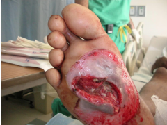
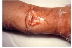
how to treat lrg wound infection with biofilm |
1. scrub wound to physically break up the biofilm 2. use xylitol, witch hazel (hammamelitannin), and lactoferrin (a protein found in milk and saliva that kills bacteria by sequestering the iron that they need to f(x) normally) on the wound 3. Use antibiotics with them at the same time. |
|
|
|
what is the max resolution of the light microscope? |
2 um |
|
|
|
two adjustments that can be made to the condenser and their effect |
1. move it up and dwn - to focus light-increases NA and resolving pwr 2. opening and closing the diaphragm with in the condenser - closing it = regulating light intensity and helps contrast opening it= increases light intensity and increases NA |
|
|
|
when using the oil immersion lens name 4 procedures that can be used to achieve max resolution |
1. opening the diaphragm 2. refocusing the condenser 3. using a blue or green filter 4. increasing voltage |
|
|
|
highest magnification objective lens |
oil 100x |
|
|
|
second highest magnification lens |
high-dry 40x |
|
|
|
second to lowest magnification |
low-power 10x |
|
|
|
lowest mag lens |
rapid scanning 4x |
|
|
|
lens with shortest working distance |
oil 100x |
|
|
|
a diaphragm is used to regulate light passing through this lens |
condensor |
|
|
|
ocular lens mag |
10x |
|
|
|
the total mag capabilty of a microscope is limited by ... |
its resolving power |
|
|
|
max mag of most light microscopes |
1000x |
|
|
|
can you focus can you rotate without striking the slide |
yes |
|
|
|
the resolving pwr of a microscope is a f(x) of its... |
magnifying pwr of its lenses and the numerical aperature of its lenses |
|
|
|
how is air contamination prevented |
by tilting an agar plate lid at a diagonal angle and the loop sterilized immediately before |
|
|
|
disinfectants are effective against what? what not? |
vegetative cells and viruses not=endospores |
|
|
|
table cleaner disinfectant we use is ... |
cidecon
|
|
|
|
the cap of a tube for innoculation should be... |
removed and held with the fingers of the loop hand |
|
|
|
define colony |
a visible mass of cells usually resulting from the division of a single cell and the # of cells in a colony can exceed one billion |
|
|
|
what is a necessary part of obtaining a pure colony |
dilution |
|
|
|
advantages of streak plate over pour plate |
quick& requires less materials |
|
|
|
before innoculating and pouring molten nutrient agar what should it be cooled to and why? |
50 degrees C so its cool enough to hold and in liquid state long enough to inoculate before it hardens if it isnt it will also cause condensation to occur on the cover of the plate and cuase microbes to spread randomly |
|
|
|
why are plates inverted during incubation? 3 |
condensation from agar lid will go onto agar surface 1) dispersing the organisms 2) disrupting the desired growth patterns and 3 preventing the formation of individual colonies |
|
|
|
two reasons for heat fixing |
1. to adhere the bacteria to the slide 2. the preserve the structural integrity of the bacteria |
|
|
|
concentrations of agar in: SIM: soft agar pours: nutrient agar plates: |
SIM: 0.35% soft agar pours: 0.7% nutrient agar plates: 1.5% |
|
|
|
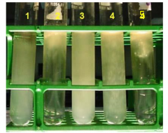
which of the two are motile P. vulgaris or M. luteus |
P. vulgaris |
|
|
|
SIM media is black means |
P. vulgaris produces hydrogen sulfide which reacts with the iron salts in the media to make a black color |
|
|
|
Which step in gram stain more prone to error? |
Decolorization b/ C if you Wecolorize too much you could wash away the dye-mordant complex and you couldn't see Any G+ even if they were there |
|
|
|
define morphology |
the shape and arrangement if bacteria as observed under a microscope |
|
|
|
staphylococcus morphology |
1. coccus 2. in clusters 3. G+ |
3 |
|
|
streptococcus morphology |
1. in chains 2. G+ 3. coccus |
|
|
|
vibrio |
curved rods like a comma, |
|
|

|
|
|
|

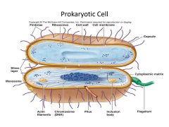
name M and its f(x) |
slime layer helps it stick to surfaces |
|
|
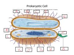
name A and its f(x)
|
Fimbrae 1. not all bacteria have these 2. helps stick to surfaces 3. have an advantage over other bacteria to cause an infection 4. hardest to get rid of 5. slime layer that accumulates in bird baths |
5 |
|

name B and its f(x)
|
Ribosomes 1. protein sythesis 2. no endoplasmic reticulum but still need to make proteins 3. 70S |
3 |
|

name H and its f(x)
|
Inclusion Bodies -not all have these but some do to store stuff -called metachromatic granules too |
|
|

name D and its f(x)
|
cell membrane 1. all cells have it w/ or w/o cell wall 2. plasma membrane 3. makes us vulnerable to osmotic pressure |
3 |
|

name I and its f(x)
|
pilus 1. sex pili 2. esp. G- bacteria 3. one way that bacteria exchange their sex DNA across in conjugation 4. how bacteria change their DNA to become resistant and etc. 5. passes plasmid through pili |
5 |
|
|
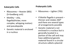
eukaryotic ribosomes |
80S ribosomes not inhibited by broad-spectrum antibiotics
|
|
|
|
animal cells lack a ... |
cell wall |
|
|
|
DomainArchaea: Exhibit characteristics of pro and eukaryotes |
Like Bacteria:
1. Simple cell structure w/o organized nucleus or organelles 2. 70S ribosomes Like Eukaryotes: 1. Protien makeup and morphology of ribosomes more so like eukaryotes 2. Ribosomes not sensitive to antibiotics 3. Have cell wall but NOT composed of peptidoglycan |
bacteria has 2 eukaryotes has 3 |
|
|
protist means |
first animal unicellular, lacking a cell wall, mostly heterotrophic |
|
|
|
colony definition |
avisible mass of cells usually resulting from the division of a single cell andthe number of cells in a single colony can exceed one billion (10⁹)
|
|
|
|
Turbidity
|
- the cloudiness or haziness of a fluid caused by large numbers of individual particles
|
|
|
|
Morphological aspects of individual bacterial colonies developed on agar media ...
|
Usually differ from one species to another
1. regularity of its edge 2. pigmentation 3. the configuration and texture of its surface 4. its elevation |
4 |
|
|
Robert KochFirst prob
|
1. was to make a way to grow bacteria in a culture so he could separate the species
|
|
|
|
Original cultures by Koch used ...
|
pieces of carrots or potatoes, or the addition of gelatin to meat broths
|
|
|
|
Who suggested the use of agar-agar as a solidifying agent. why?
|
-Wife of an early coworker Frau Hesse
-Agar better b/c unlike gelatin it could not be degraded by pathogens Koch was studying |
|
|
|
Purity of the culture further improved when R.J. Petri ...
|
who worked in Koch’s lab Introduced covered dish that protected the nutrient surface of media 4m contamination
|
|
|
|
No single media will ...
|
support the growth of all bacteria
|
|
|
|
Using the # of colonies as an indicator, which habitat sampled in class appeared to have the most bacteria? why? |
fingertips -b/c they are always brushing against and touching things that the other things tested may not have. |
|
|
|
In what ways do the macroscopic features of bacterial colonies differ from those of molds? |
1. bacteria have a range of yellow/white while mold colonies are darker 2. dif 3D formations |
2 |
|
|
why is the level of contamination measured as # of colonies rather than size of colonies? |
b/c each colony reps a single cell that was originally present , while the colony size reps the growth rate of the microbe. |
|
|
|
how can microbial levels be controlled on surfaces in the environment? |
using antimicrobial cleaners or controlling environmental conditions like pH and humidity if possible. |
|
|
|
respiration and photosynthesis with prokaryotes vs eukaryotes |
prokaryotes carry out photosynthesis & respiration as well but lack the organelles that eukaryotes have to do it like mitochondria and chloroplasts |
|
|
|
prokaryotes vs eukaryotes:
motility mechanisms
|
Eukaryotes: 1. Have cilia 2. Have a think whipping flagella 3, Have pseudopodia Prokaryotes: 1. Have a thinner flagella that rotates 360 degrees |
|