![]()
![]()
![]()
Use LEFT and RIGHT arrow keys to navigate between flashcards;
Use UP and DOWN arrow keys to flip the card;
H to show hint;
A reads text to speech;
71 Cards in this Set
- Front
- Back
|
What six parts of the pelvic girdle are used for sexing? |
|
|
|
What is the sub-pubic angle? |
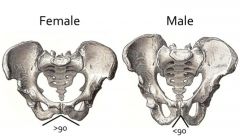
The angle made by the inferior borders of the articulated pubis bone |
|
|
What is the pubis body width? |
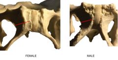
The measurement from the midpoint of the pubic symphasis to the obturator foramen |
|
|
What is the ventral arch? |
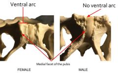
The rough project of bone visible on the anterior surface of the pubis bone |
|
|
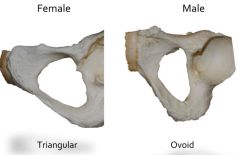
Obturator foramen |
t |
|
|
Acetabulum |

|
|
|
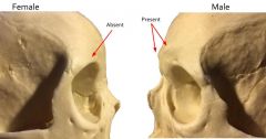
Supraorbital torus |
|
|
|
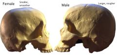
Cranial vault |
|
|
|
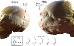
Nuchal Crest |
|
|
|
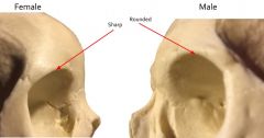
Suborbital Margin |
|
|
|
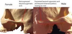
Zygomatic Arch |
|
|
|
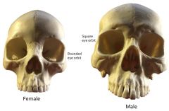
Eye Orbit Shape |
|
|
|
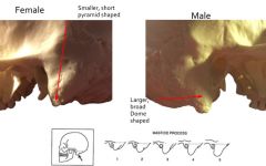
Mastoid Process |
|
|
|
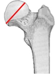
Maximum Diameter for Head of Femur (for sex comparison) |
|
|
|
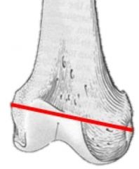
Maximum Bicondylar width (for sex comparison) |
|
|
|
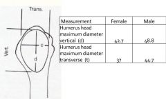
Maximum Diameter of Head of Humerus (for sex comparison) |
|
|
|
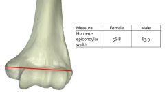
Epicondylar Width (for sex comparison) |
|
|
|
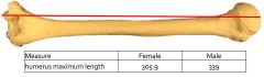
Maximum length of Humerus (for sex comparison) |
|
|
|
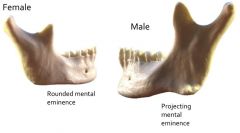
Mental eminence (for sex comparison) |
|
|
|
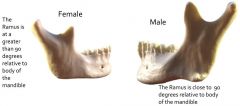
Ramus angle to mandible (for sex comparison) |
|
|
|
Cranial Index |
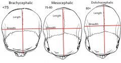
B/L x 100 |
|
|
Os Japonicum |
*bipartite (2) or tripartite (3) zygomatic bone Greater frequency in: Japanese - 7% Korean |
|
|
Humoral Septal Aperture |
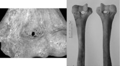
A small hole that forms at the elbow joint in the distal end of the humerus. More often in female skeleton. |
|
|
Agenesis of 3rd molar |
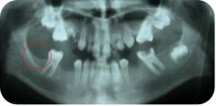
Loss of 3rd molar - failure of tooth development Commonly influenced by genetics Little or no selection for trait - good enough for non-metric identifier |
|
|
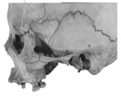
Metopic Suture |
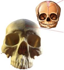
Non-closure of the frontal suture, which normally closes by 12 |
|
|
Bifurcated Rib |
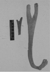
Congenital abnormality Usually unilateral Usually asymptomatic |
|
|
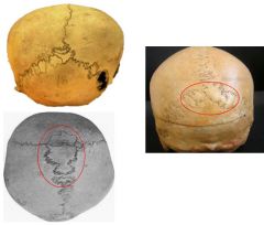
Epipteric bone (Flower bone) |
Specific type of Wormian bone Junction of parietal, frontal, greater wing of sphenoid, squamous portion of temporal bone Does not indicate ethnicity or race Some genetic heritability |
|
|
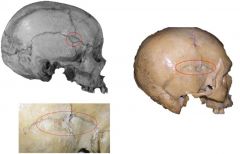
Os Inca Bone |
Large wormian bone at lambda Relatively high frequency in: Peruvian mummies Some modern day Andean Mountains Populations |
|
|
Lumbarization of S1 |
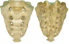
Sacrum is 5 fused sacral vertebrae Typically fused by age 23 |
|
|
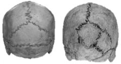
Wormian bones (extra sutural bones) |
Extra bone pieces that occur within a suture in the cranium; mostly frequently in lamboid, occasionally in sagittal & coronoal Any size Not indicator of weak skull spot. |
|
|
Sacralization of L5 |
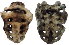
Fusion of the 5th lumbar vertebrae with the first segment of the sacrum |
|
|
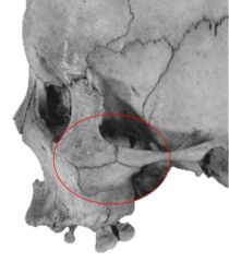
H-Form Pterion |
|
|
|
Rocker Jaw |
Rounded inferior and dorsal aspect of the mandible Appears in most Polynesian groups |
|
|
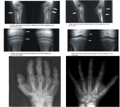
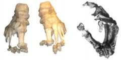
Epiphyseal union - drawing & photo |
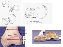
A lot of bones in children fuse to become a single bone Epiphysial closure indicates end of bone growth, allowing for age of death to be determined by what has and has not fused. Females typically fuse earlier. |
|
|
When are developmental traits used for aging? What do they include? |
Used on sub-adults Tooth eruption & Ephiphyseal union |
|
|
Epiphyseal Union X-ray samples |
|
|
|
Bone resorption |
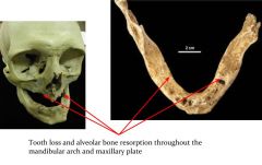
Accompanies loss of permanent teeth Associated with old age BUT cannot provide accurate age range. Loss of permanent teeth and stimulus of chewing causes bone loss. |
|
|
Decidious Dentition Tooth Eruption |
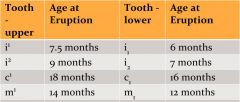
2nd molars erupt at 24 months |
|
|
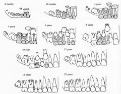
Permanent Dentition |
|
|
|
Degenerative Traits |
|
|
|
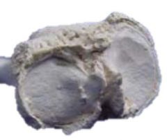
Pubic Symphyseal Face Stages |
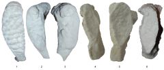
Undergoes regular changes from 18 on |
|
|
Dental Attrition |
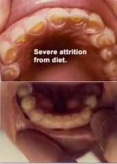
Usually associated with adults Loss of permanent teeth associated with adults Cannot accurate age range Leads to loss of outer white tooth enamel and exposure of yellowish dentine of pulp cavity; more exposed with age Does not take diet into consideration |
|
|
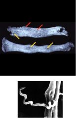
Stages of Bone Healing |
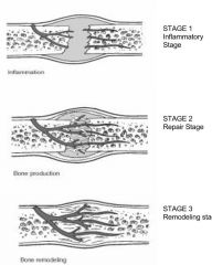
Stage 1: 2-4 weeks, overlaps with stage 2 Stage 2: 1-2 months Stage 3: months to years |
|
|
Leprosy |
Disease caused by bacteria Primarily damages the peripheral nerves and mucosa of the upper respiratory tract skin lesions form and eventually causing permanent damage to the skin, nerves, bones and eyes.Once in the bone nodules form. |
|
|
Rheumatoid arthritis |
An inherited disease/side effect of Lyme disease. May be triggered by microorganisms Affects more joints, more symmetric Skeletal changes: epiphyseal lipping, ankylosing spondylitis, vertebral column fusion |
|
|
Periostitis |
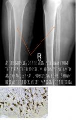
Outer surface of bone is reabsorbed in a wormy/dendritic pattern Periosteum separates from bone Immature bone fills gap Bone begins to bulge and thicken |
|
|
Anemia |
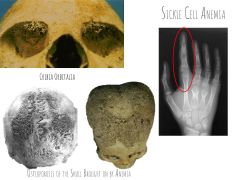
Possible explanation of osteoporotic lesion on superior aspect of orbit |
|
|
Vertebral osteophytosis |
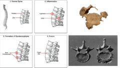
Degeneration of spinal column Leaking disc fluid leads to osteophyte formation on vertebral bodies May result in vertebral fusion |
|
|
Treponemal diseases |
Diseases that change bone tissue Changes to bone that are connected to where lesions form. Include: syphilis, TB, leprosy |
|
|
Syphilis |
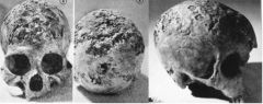
Skulls with characteristic lesions |
|
|
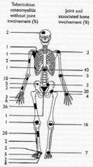
Tuberculous - where it can affect bone |
|
|
|
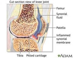
Osteoarthritis |
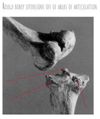
Caused by trauma or wear-and-tear on synovial joints like knees, wrists, & fingers Epiphyseal lipping or osteophyte growth can be caused by it. |
|
|
Gout |
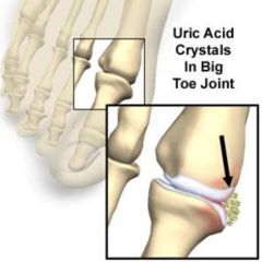
Medical condition caused by elevated levels of uric acid in blood Uric acid crystallized and deposits in joints, tendons and surrounding tissues Creates arthritic conditions, ultimately alteration of the shape of the bones |
|
|
Pott's disease |
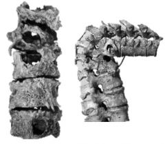
Extrapulmonary TB that invades the spine Weakens vertebrae to collapse point Creates severe bend in spine |
|
|
Avascular necrosis |
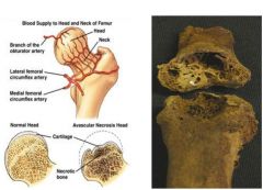
Version of osteoarthritis that is randomly distributed throughout the body |
|
|
Teeth.... |
|
|
|
Uses for teeth |
|
|
|
Anatomy of Teeth |
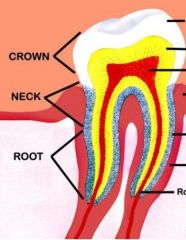
Crown made of enamel which is resistant to most decomposition processes DNA is in root |
|
|
Permanent Teeth |
|
|
|
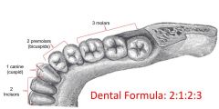
Adult Dental Formula |
|
|
|
Deciduous Dental Formula |
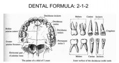
incisors and canines mini versions of adult ones 2nd molar is replicated and becomes 1st permanent molar |
|
|
Tooth Types |
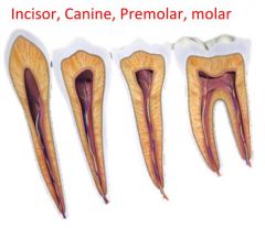
Usually 32 in adult mouth Differ in: Size, shape, root type |
|
|
Order of Deciduous Eruption |
After 2 years, interval of 4 before permanent start to erupt |
|
|
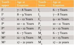
Eruption of Permanent Teeth |
|
|
|
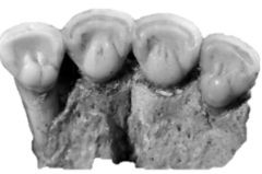
Molar cusp patterns |
Anthropological use: distinguishing modern populations, assessing primate ancestry Limited in prehistoric population due to grit-related attrition of cusps Maxillary molars have different patterns from mandibular patterns |
|
|
Shovel-shaped teeth |
Especially in populations of Asian decent: Asian groups, Native American groups, indigenous groups of C. and S. America Tongue side extension of incisor lateral borders Occasionally cheek side extension of lateral borders: double shovel-shaped incisor (usually on top) |
|
|
Morphological Teeth Variation |
|
|
|
Cultural Deformation |
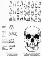
Filed, chipped, drilled, incised (often decorative) Multiple populations Mostly incisors (easier to work on, most visible) Interproximal grooves (cavities, absesses, pain relief efforts) |
|
|
Facial Reconstruction |
Methods - sculpting clay to skull/reproduction; drawing soft tissues on picture of skull First introduced in late 1800s, into medico-legal in early 1900s 50% favorable results Various number of tissue markers (9-32), 17 avg. Lots of formulas and guides |
|
|
Handedness |
Historically, it was believed that the side of the skeleton that had the most bone mass would indicate handedness BUT Not supported by studies, No measurements of value in determining handedness from the skeleton |
|
|
Body Weight Estimation |
Four Factors Considered - Sex, Muscle Markings, Height, Skeletal Robusticity Determine sex and stature, consult H/W charts Clothing - Belts, dresses that pinch at waist ONLY ABLE TO ESTIMATE WITHIN A BROAD RANGE WITH MEASUREMENTS AND WEIGHTS OF SPECIFIC BONES |