![]()
![]()
![]()
Use LEFT and RIGHT arrow keys to navigate between flashcards;
Use UP and DOWN arrow keys to flip the card;
H to show hint;
A reads text to speech;
27 Cards in this Set
- Front
- Back
|
regrowth rate of eyelashes |
2 weeks if cut , 2 months with epilation |
|
|
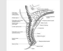
anatomy of eyelid |
skin, SUBCUT, O.O , TARSUS, conj |
|
|
lid crease |
1-skin 2-O.O 3- Levator app 4-mullers muscles 5-conj |
|
|
modified glands |
for eyelashes: 1- molls> sweat glands 2-zeis> sebaceous glands for tarsus meibomien gland for conj: wolfring and kruase > sebaceous glands |
|
|
eye lid anatomy |
|
|
|
layers of eyelid 1- skin |
1-epidermis 2- dermis 3- adnexa |
|
|
epidermis layers BASIC PAINT GETS KRAPPY |
1- B asal > col 1 layer cells attached to BM by hemidesmosomes P rickle > 3-5 cell polygonal layer, connected by desmosomes , acanthosis = increase thickness G ranular > 1-2 layers , contain hyalokeratin granules Keratin> ( keratin fibers - cell organelles ) hyperkeratosis = inc thickness parakeratosis = retained organelles dyskeratosis : keratinization of any other layers |
|
|
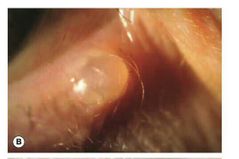
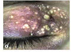
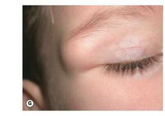
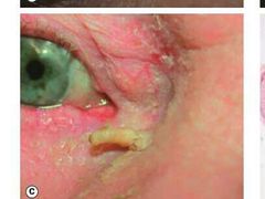
1- chalazion > terminology |
1- chalazion : sterile gramulomatous inflammation of meibomien gland = meibomien cyst 2- marginal chalazion : infl of glands of zeis 3- hordeoulum internum ( infected chalazion ) |
|
|
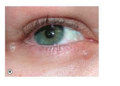
treatment : |
away from lid margin : 1- not infected : 1- conservative : >warm compresses >trial of expersion with sterile cotton tips >antiinflmmatory ointments >incision and curretage 2- infected : >with associated orbital cellulitis : oral antibiotics >no cellulitis : topical antibiotic drops and ointment 3- prophylaxis : treatment of blepharitis 4- margical chalazion : >conservative > intralesional steroids : >shave curettage or I&c using a horizontal conj incision or vertical incision at lid margin |
|
|
intralesional steroid treatment |
0.2-2 ml of TA diluted with lidocaine to conc of 5 mg/ml injected with 27/30 guage needle SE > localized depigmentation , fat atrophy , retinal artery occlusion |
|
|
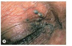
cyst of moll : dilated sweat gland at lid margin |
|
|
eccrinr hydrocytoma : dilates sweat gland away from the lid margin |
|
|
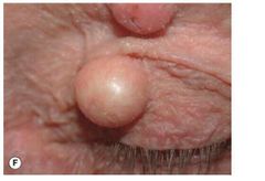
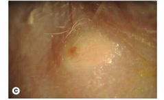
cyst of zeis : non trnslucent cyst at lid margin |
|
|
cysts at lid margin |
translucent > cyst of moll non translucent> cyst of zeis |
|
|
milia : obstructed pilosebaceous units by keratin |
|
|
epidermal inclusion cyst : downgrowth of epidermis into dermis after trauma or surgery |
|
|
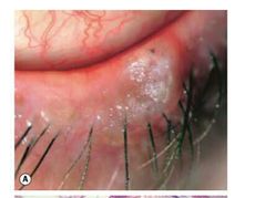
comedone: dilated ducts of hair follicles with retained secretions open : black heads closed: white heads |
|
|
dermoid cyst |
|
|
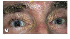
xanthelasma : >hyperlipidemia > arcus sinilis >treatment: excision for cosmesis microdissection with flap if large lesion chemical peeling |
|
|
epidermoid vs dermoid |
epidermoid in infants , only contains keratin dermoid , in adults , contains skin + hair |
|
|
sebaceous cyst key feature : dilated gland orifice |
|
|
lesions caused by blocked duct of pilosebaceous unit |
1- comedone : blocked by sebum and keratin , open duct with retianed oxidized materials >black , obstructed > white 2- milia: retained keratin 3- sebaceous cyst : blocked pilosebaceous unit , visible gland orifice |
|
|
xanthelasma vs chalazion |
xanthelasma > lipid is intracellular , in foam cells (lipid laden histiocytes ) chalazion > lipid seeps out extracellularly , surrounds by epitheloid histiocytes |
|
|
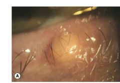
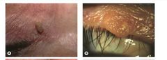
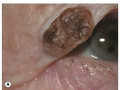
squamous papilloma : vascular CT core covered by epithelium types : sessile , peduculated , horn-like treatment : simple excision , chemical , electrocautry cause : HPV |
|
|
saeborrheic keratosis : stuck on appearance hyperpigmented , oily lesion benign treatment : shave biopsy , electrocautery , cryo , chemical peeling |
|
|
actinic keratosis : > in elderly >premalignant >hyperkeratotic plaque |
|
|
benign vs malignant |
>squamous cell papilloma ( cutaneous horn )> squamous cell carcinoma >basal cell papiloma ( saeborrheic keratosis ) > BCC > |