Reading...
![]()
Play button
![]()
Play button
![]()
Use LEFT and RIGHT arrow keys to navigate between flashcards;
Use UP and DOWN arrow keys to flip the card;
H to show hint;
A reads text to speech;
88 Cards in this Set
- Front
- Back
|
skeletal muscle
|
- strong, quick discontinuous voluntary contraction
- bundle of fibers - muscle cells - myofibril |
|
|
cardiac muscle
|
- strong, quick continuous involuntary contraction
|
|
|
smooth muscle
|
- weak, slow involuntary contraction
|
|
|
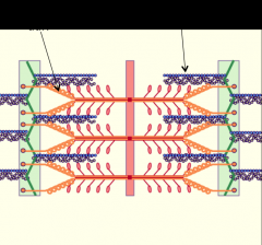
myofibril
|
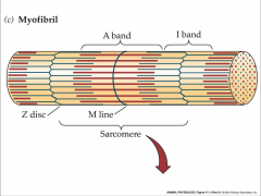
- rods of proteins inside muscle fibers
- myofilaments = thick and thin filaments - arranged in specific way = thick filament surrounded by six thin filaments |
|
|
sacromere
|
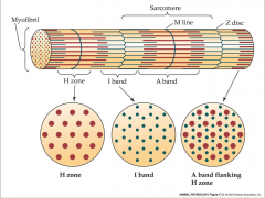
- functional unit of muscle
- z line and I band to the next z line - I band split between 2 sacromeres - sacroplasmic reticulum - terminal sisterny = stores calcium - I tubule, terminal sisterny, sacroplasmic reticulum = triad |
|
|
thick filament
|
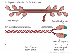
- made of myosin
- myosin composed of 6 polypeptides - myosin lines up tail to tail = heads pointed in opposite directions - 4 light chains and 2 heavy chains |
|
|
thin filament
|
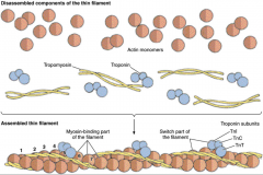
- made of many proteins
- actin composed of globular actin - actin has myosin binding site - tropomyosin wraps around actin and covers binding site - troponin has 3 subunits - tropomyosin exposes binding sites when calcium present |
|
|
troponin 3 subunits
|
- TnT = binds to tropomyosin
- TnC = binds to calcium - TnI = inhibitory |
|
|
titin
|

- huge protein that helps center thick filament
- elastic element - links thick filaments to Z lines - largest protein in human genome |
|
|
nebulin
|
- acts as molecular ruler
- determines how long the thin filament will be - actin binding protein |
|
|
sliding filaments
|
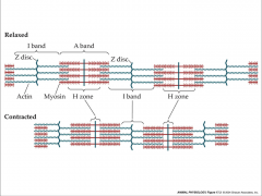
- produce muscle contraction
- H zone and I band disappear - sacromere shortens = thin filament pulled over thick filament |
|
|
cap-Z
|
- binds to thin filaments and stabilizes it on the Z line
- caps the plus ends of actin filaments at Z-disk |
|
|
muscle contraction
|

- dystroglycan complex = proteins that interact with extracellular matrix and bound to sacroglycan complex
- sacroglycan = enormous gene that is capable of having mutations - muscle dystrophy = dystrophin is absent or mutated |
|
|
sequence of muscle contraction
|
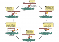
- rigor is a transient state
- ATP binding dissociates myosin from actin - myosin ATPase hydrolyzes ATP to ADP and P - hydrolization causes myosin to have a cocked position - myosin binding site on actin binds to actin binding site on myosin making a cross bridge - conformation change called a power stroke and ADP is released - binds to ATP and myosin releases from actin |
|
|
power stroke
|
- thin filament is pulled over the thick filament toward the M line
|
|
|
regulation of contraction
|
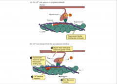
- myosin binding site on actin is controlled
- tropomyosin covers the myosin binding site and won't let myosin connect to actin - Ca binds to troponin causing a conformational change that pulls down the tropomyosin exposing myosin binding sites |
|
|
vertebrate plan
|
- based on muscles organized into motor units
- motor units = motor neuron and all muscle fibers it innervates |
|
|
motor end-plate
|
- region of the muscle cell membrane covered by the terminal bud
- has clefts and ridges = junctional folds |
|
|
T tubule
|
- contains dihydropyridine receptor (DHPR)
- blocks channel for ryanodine |
|
|
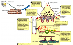
excitation-contraction coupling sequence
|
- action potential in motor neuron triggers exocytosis of ACh
- ligand-gated channels bind ACh and open generating an action potential - action potential propagates over cell membrane and depolarizes the t-tubules - depolarization reaches the DHPR and causes a conformation change that opens a RyR calcium channel of the SR and Ca diffuses out of SR into cytoplasm - Ca ions bind to troponin and tropomyosin moves to expose myosin-binding sites on actin - acetylcholinesterase in the extracellular matrix of synaptic cleft hydrolyzes ACh to terminate the action potential - cross bridges go through several cycles as long as Ca remains bound to troponin - once wave of depolarization ceases, DHPRs return to their original conformation and RyR Ca channels close - as ATP-depenedent Ca pumps decrease the Ca concentration in cytoplasm, Ca leaves TN, TM blocks myosin binding sites on actin, and contraction ends |
|
|
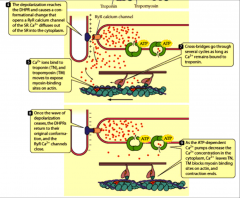
excitation-contraction coupling
|
- Ca is stored in the SR both free and bound to the protein calsequestrin
- ATP dependent Ca pumps are continuously active, before, during, and after contraction - each ATP hydrolyzed, 2 Ca are moved from cytoplasm into the SR |
|
excitation-contraction coupling diagram
|
excitation-contraction coupling diagram
|
|
|
whole skeletal muscles
|
- prime mover
- synergist - antagonist - fixator |
|
|
prime mover
|
- agonist
- produces most of force |
|
|
synergist
|
- aids prime mover
- stabilizes the nearby joint - modifies direction of movement |
|
|
antagonist
|
- opposes prime mover
- prevents excessive movement and injury |
|
|
fixator
|
- prevents movement of bone
|
|
|
force
|
- generated only by contracting
- lengthen passively |
|
|
contraction
|
- tension generated by a muscle during cross bridge activity
- may or may not involve shorteining |
|
|
twitch
|
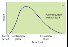
- mechanical response of a muscle to a single action potential
- latent period - contraction phase - relaxation phase |
|
|
latent period
|
- period of time that elapses between the generation of an action potential and the start of the contraction
- Ca release cross bridge formation |
|
|
contraction phase
|
- starts at the end of latent period and ends at tension peak
|
|
|
relaxation phase
|
- period of time from end of the tension peak until the end of the contraction
|
|
|
types of muscle contraction
|
- isotonic
- isometric |
|
|
isotonic contraction
|
- changes in length
- muscle attempts to move a load that is equal to or less than the tension generated by muscle - tension in the muscle remains constant despite a change in muscle length - shortening can occur only when a muscle's maximal force of contraction exceeds the total load on the muscle |
|
|
types of muscle contractions
|
- concentric = shortening
- eccentric = lengthening |
|
|
concentric
|
- tension generated is sufficient to overcome the resistance, and the muscle shortens as it contracts
- occurs throughout the length of the muscle, generating tension at musculo-tendinous junction, causing the muscle to shorten and changing the angle of the joint - cross-bridge cycling |
|
|
eccentric
|
- tension generated is insufficient to overcome the external load on the muscle and the muscle fibers lengthen as they contract
- an opposing force is greater than the force generated by the muscle - used as a means of decelerating a body part or object, or lowering a load gently rather than letting it drop, or hiking uphill - unknown mechanism - leads to minor muscle damage that causes soreness following exercise |
|
|
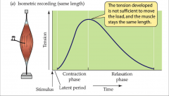
recording isometric and isotonic contraction
|
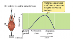
- tension developed is sufficient to move the load, and the muscle shortens
- latent period is followed by rise in tension - not enough tension to move a load - plateau = force produced by the muscle remains constant - isotonic/same tension allowing the muscle to shorten |
|
|
work of contraction
|
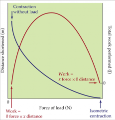
- isotonic contractions show that muscle shortens the greatest distance with no load
- shortens progressively shorter distances with increasing loads - multiply the force developed by distance shortened for each load gives a curve that represents work performed by muscle |
|
|
muscle force
|
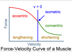
- decreases with increased velocity of contraction during concentric contraction
- increases with increased velocity of contraction during eccentric contraction |
|
|
isometric contraction
|
- tension without changing length
- muscle attempts to move a load that is greater than the tension generated by the muscle - muscles of hand and forearm grip an object, joints of hand don't move but muscles generate sufficient tension to prevent the object from being dropped |
|
|
power
|
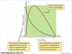
- equal to force times velocity
- muscle generates no power at either isometric force (due to zero velocity) or maximal velocity (due to zero force) - maximal force produced by a muscle is proportional to cross-sectional area of its contractile elements - velocity of shortening is influenced by the myosin isoforms expressed by the motor units of muscle |
|
|
tension/muscle force
|
- amount a muscle can do depends on its volume
- force/cross sectional area - tension generated by a muscle fiber that is directly proportional to number of attached cross bridges |
|
|
threshold
|
- minimal stimulus needed to depolarize the sarcolemma
- point at which sodium ions start to move into the cells = depolarization - ability to reach threshold is determined by the magnitude of stimulation and duration of stimulation |
|
|
length-tension relationship
|
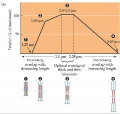
- tension that a muscle generates varies with its length
- found when a muscle is under isometric contraction and maximum activation of the muscle - in a singe muscle fiber, peak force is noted at a normal resting length - bell-shaped curve - too much overlap of thick and thin filaments results in less tension - overlap of thick and thin filaments is ideal to generate maximal force - sacromere set longer than ideal length doesn't have enough overlap so fewer sites of cross sectional formation |
|
|
length-tension for isometric contraction
|
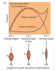
- shows tension produced by a muscle when it is set a different lengths prior to simulation
- shorter = tension drops - maximal tension was achieved when muscle was set a lengths near normal relaxed lengths |
|
|
motor units
|
- skeletal muscles of vertebrates
- independent - single alpha motor neuron and all the corresponding muscle fibers it innervates - when activated all of its fibers contract |
|
|
motor unit recruitment
|
- progressive activation of a muscle by successive recruitment of contractile units/motor units to accomplish increasing gradations of contractile strength
- each vertebrate muscle twitch fiber is innervated by a single axon that branches to make many synaptic contacts at the middle of multiple fibers |
|
|
tension varies
|
- varying the frequency of impulse in a single motor unit
- varying number of active motor units |
|
|
multiple fiber summation
|
- weak signal is sent by the CNS to muscle
- smaller motor units are stimulated first - smaller motor units are more excitable than the larger ones - as strength of signal increases = more motor units are excited in addition to larger ones - largest motor units having as much as 50 times the contractile strength as the smaller ones - as more and larger motor units are activated = force of muscle contraction becomes progressively stronger |
|
|
size principle
|
- allows for a gradation of muscle force during weak contraction
- occur in small steps which then become progressively larger when greater amounts of force are required |
|
|
contractile and elastic components
|
- to achieve maximal tension all structures in series must be stretched taut
- sustained high calcium concentration must be present from multiple individual twitches |
|
|
summation
|
- muscle is stimulated repeatedly
- stimuli arrive one after another within a short period of time - twitches can overlap and result in a stronger muscle contraction |
|
|
tetanus
|
- stimuli continue to be applied frequently to a muscle over a prolonged period of time
- muscle will eventually reach a plateau - twitches fuse |
|
|
tension force
|
- minimum produced by single twitch in smallest motor unit
- maximum produce by simultaneous fused tetanic contraction in all motor units |
|
|
fiber firing
|
- 1/3 of fibers in muscle firing at once under conscious muscle exertion
- actual number firing affected by various physiological and psychological factors = Golgi tendon organs and Renshaw cells - low level of contraction is protective mechanism to prevent avulsion of tendon - force generated by 95% contraction of all fibers is sufficient to damage the body |
|
|
mechanical model of muscle
|
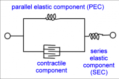
- contractile component = muscle fiber
- series elastic component = tendon - parallel elastic component = muscle membrane |
|
|
elastic elements
|
- series elastic elements
- non-contractile component of muscles - lies in series - store energy when stretched - tendons - cross bridges between actin and myosin also contribute |
|
|
parallel elastic elements
|
- non-contractile component of a muscle
- provides resistive tension when muscle is passively stretched - muscle membranes which lie in parallel to muscle - Hook's law |
|
|
Hook's law
|
- F=KL
- F is force exerted on spring - K is constant - L is displacement |
|
|
viscous resistance
|
- muscle cells contents become compressed
- increase with maximal force - contributes to passive resistance - produce by parallel elastic components |
|
|
parallel fibers
|
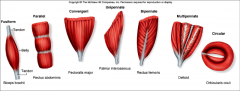
- parallel to longitudinal axis of the muscle
- sartorius, masseter, biceps branchi - each fiber attached to its own tendon with tendons converging on a common point - gets shorter and increases diameter when contracts - fibers shorten in direction parallel to direction shortening of muscle - located in positions requiring longer movements with less power of faster movements - greater length and less cross sectional area = greater velocity |
|
|
convergent fibers
|

- fibers spread over a broad area
- all the fibers converge at one common attachment site - fibers typically spread out, like a fan or a broad triangle, with tendon at apex - pectoralis major muscle - versatility because stimulation of only one portion of muscle can change direction of pull - pull in different directions - allow maximum force production |
|
|
pennate fibers
|

- at angle to longitudinal axis of muscle
- rectus femoris, deltoid - attach to a common tendon - direction of shortening of individual fibers is different from direction of shortening of whole muscle - fewer sarcomeres in series - cannot shorten as much as parallel - located in positions requiring small but powerful movements - fatigue quickly - force produced is greater than force produced by parallel - greater cross sectional area and less length = greater force |
|
|
angle of pennation
|
- greater it is the smaller the amount of effective force transmitted to tendon
- increases as tension progressively increases in the muscle fibers - contains more muscle fibers - produces more tension |
|
|
muscle architecture
|
- muscle force is proportional to physiologic cross-sectional area
- muscle velocity is proportional to muscle fiber length |
|
|
contraction and temperature
|
- most efficient at 38.5 C
- elevated muscle temperature causes a shift in force-velocity curve - increased maximum isometric tension - increased maximum velocity of muscle shortening - requiring less motor unit to sustain a given load - body temperature too high = heat exhaustion or heat stroke |
|
|
2 types of skeletal muscles
|
- red muscles
- white muscles |
|
|
red muscles
|
- contain myoglobin
- highly vascularized - receiving and using more oxygen than white muscles - require lower minimum rate of stimulation for tetanic fusion - contain many mitochondria - gets most of its ATP form oxidative phosphorylation = rapid - sustain contraction longer without fatiguing - produce large tension |
|
|
white muscles
|
- contain little myoglobin
- more rapidly contracting fast muscles - poorly vascularized - contain few mitochondria - ATP from glycolysis - produce small tension |
|
|
muscle fibers specialized
|
- different muscles contain different types of fibers
- specialized for slow or quick response |
|
|
3 different forms of myosin
|
- type 1
- type 2a - type 2x |
|
|
type 1
|
- slow oxidative
- hydrolyze ATP slowly - slow Ca uptake by SR - slow contraction - long duration of twitches - high resistance to fatigue - many mitochondria, myoglobin, and capillaries - red muscle - small fiber diameter - posture - slow myosin ATPase activity - specialized for endurance - require constant oxygen = no lactate dehydrogenase - energy from aerobic metabolism - slow cross-bridge formation |
|
|
type 2a
|
- fast oxidative glycolytic
- hydrolyze ATP rapidly - fast Ca uptake by SR - fast contraction - short twitch duration - intermediate resistance to fatigue - many mitochondria, myoglobin, and capillaries - red muscle - intermediate fiber diameter - standing, walking, rapid repetitive movements - generate great deal of power - myosin ATPase activity is high - energy from glycolysis and oxidative phosphorylation |
|
|
type 2x
|
- fast glycolytic
- hydrolyze ATP rapidly - fast Ca uptake by SR - fast contraction - short duration of twitch - low resistance to fatigue - few mitochondria, myoglobin, and capillaries - white muscle - large fiber diameter - jumping, bursts of high speed locomotion - generate great deal of power and force - myosin ATPase activity high - energy from glycolysis |
|
|
motor unit 1
|
- small alpha motor neurons
- innervate relatively few muscle fibers - form motor units that generate small forces - innervate small red muscle fibers = slow oxidative - small motor units - especially important for activities that require sustained muscular contraction - more excitable |
|
|
motor unit 2
|
- intermediate alpha motor neurons
- properties that lie between those for the other two - use type 2a - fast fatigue resistance motor units - not quiet as fast as FF units - generate about twice the force of a slow motor unit |
|
|
motor unit 3
|
- large motor neurons
- innervate large, more powerful motor units - type 2x - fast fatigable motor units - especially important for brief exertions that require large forces - fire last - small axon diameter - least excitable |
|
|
plasticity
|
- muscle capable of changing in both mass and cellular characteristics
- change mass by hypertrophy - atrophy occurs when muscle fibers lose actin and myosin or from loss of cells |
|
|
endurance training
|
- elicits changes in fiber type, capillary density, mitochondrial density
- proportion of type1 fibers remain unchanged - proportion of type 2a fibers increase - proportion of type 2x fibers decrease - changes in gene activity - increase density of capillaries - exercised muscle produce and release cytokine vascular endothelial growth factor - increases aerobic capacity of muscle fibers by increasing mitochondria and lipid droplets - increased Mhc 2a isoform and decreased amount of Mhc 2x isoform |
|
|
vascular endothelial growth factor
|
- increases with exercise
- increases less in trained muscles than in untrained muscles |
|
|
hypertrophy
|
- adding structural proteins
- occurs in cardiac muscle - doesn't add cell numbers by mitosis |
|
|
regulating muscle mass
|
- myostatin
- PI3-K-Akt1 pathway |
|
|
myostatin
|
- prevents from getting to much muscle
- decreases PI3 - Akt1 pathway - negative growth regulator - binds to a R on muscle PM - initiates an intercellular signaling pathway - controls cell growth - decreases amount of fat deposited between muscle fibers - limits protein production and satellite cell activation |
|
|
PI3-K-Atk1 pathway
|
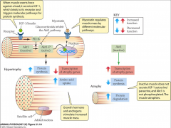
- provides molecular signals that regulate balance between synthesis and degradation
- insulin like growth factor (IGF-1) secreted when struiated (skeletal and cardiac) muscle exerts force against a load - insulin also activates - IGF-1 binds to R - activates phophoinositol 3-kinase (PI3-K) that phophorylates Akt-1 making it active - increased protein synthesis by entering nucleus and binding to genes that make products that control protein degradation |
|
|
muscle energetics
|
- ATP is immediate source of energy for powering muscle contraction
- ATP binding required for detachment of myosin and actin - ATP hydrolysis activates acting binding site on myosin - ATP drives the ATPase-Ca pump that transports Ca into the SR |
|
|
3 biochemical mechanisms produce ATP in muscle
|
- use of phosphagen creatine phosphate
- aerobic glycolysis - aerobic catabolism |