![]()
![]()
![]()
Use LEFT and RIGHT arrow keys to navigate between flashcards;
Use UP and DOWN arrow keys to flip the card;
H to show hint;
A reads text to speech;
48 Cards in this Set
- Front
- Back
|
Eyespots |
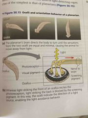
Pair of ocelli near head of planarians Simplest form of light detecting organs Allow them to move away from light and seek shade |
|
|
Compound eyes |
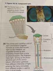
Insects, crustaceans, and some worms Consist of several thousand light detectors called ommatidia Effective at detecting movement Insects have good color vision and some can see into UV range |
|
|
Single lens eyes |
Found in jellies, spiders, molluscs, and all mammals Work like camera: iris changes diameter of the pupil to control how much light enters |
|
|
Pupil |
Opening through which light enters |
|
|
Iris |
Expands/contracts to let more/less light in Changes diameter of pupil Formed by the choroid |
|
|
Conjunctiva |
Mucous layer covers front of eye and lines eyelids |
|
|
Sclera |
Collagen and elastic fibers, connective tissue At the front, forms the transparent cornea |
|
|
Choroid |
Thin pigmented layer Forms the iris in the front |
|
|
Retina |
Just inside the choroid Made of neurons and photoreceptors |
|
|
Lens |
Transparent disk of protein Divides eye into 2 cavities: Aqueous humor (watery substance in front of eye) and vitreous humor (jellylike behind the lens) |
|
|
Retina contains... |
Bipolar cell: receives info from several cones or rods Ganglion cell: gather info from several bipolar cells Horizontal and amacrine cells: integrate info across retina |
|
|
Pathway of vision |
Light hits retina 2 types of photoreceptors (rods and cones) Info is relayed to the optic nerve and then brain |
|
|
Optic disk |
Blindspot In retina Space where there are no photoreceptors so light is not detected |
|
|
2 photoreceptors |
Rods: sensitive to light, do not distinguish colors (125 million) Cones: provide color (6million) |
|
|
Rhodopsin |
Visual Pigment that consist of retinal (retinal is a light absorbing pigment bound to the membrane protein opsin and a derivative of vitamin A) Absorption of light causes a shape change in retinal In rods |
|
|
Retinal |
Light absorbing pigment bonded to membrane protein opsin Exist as 2 isomers Light shifts from cis (bent arrangement) to trans (straight arrangement) This change activated opsin protein |
|
|
Rods |
Important for night vision(shades of gray) Sensitive to dim light, but no colors Contain rhodopsin Provide peripheral vision and the perception of motion |
|
|
Cones |
Located primarily in fovea centralis (macula, acute vision) Activated by bright light, sensitive to different wavelengths of light Allow for fine detail and color |
|
|
3 kinds of cones |
Blue, green, red Composed of 11 cis-retinal bound to an opsin (various opsin structures to absorb different pigments) Various combination of stim produce different colors |
|
|
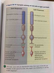
Transduction in the dark |
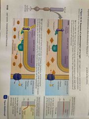
GMP binds to Na channels and keeps them open Na flows into cell depolarization it |
|
|
Transduction in the dark |

GMP binds to Na channels and keeps them open Na flows into cell depolarization it |
|
|
Transduction in light |
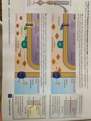
When light hits photoreceptors, rhodopsin, transducin , phisphodiesterase is activated These convert cGMP to GMP When cGMP levels fall, cGMP no longer occupies the binding sites on the channels, causing channels to close and hyperpolarize |
|
|
Glutamate |
In the dark, rods and cones continue to release glutamate with bipolar cells In light, they shut off glutamate release |
|
|
Different pathways of rods and cones |
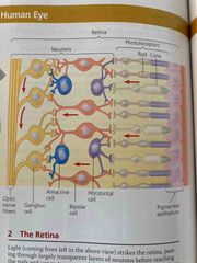
Info directly from photoreceptors to bipolar cells to ganglion cells Or Horizontal cells carry signals from one rod or cone to other photoreceptors and to several bipolar cells |
|
|
Gang lions axons are |
Optic nerve |
|
|
Lateral inhibition |
When a rod or cone stims horizontal cell and that cell then inhibits more distant photoreceptors and bipolar cells This is how Horizontal cells enhance contrast of image (light appears lighter and dark appears darker) Repeated by interaction amacrine cells and ganglion cells (amacrine distribute info from bipolar cells to several ganglion cells |
|
|
Receptive field |
Ganglion cells define part of visual field Sensitivity of cones vs rods is due to how directly they connect to ganglion cells As many as 150 rods May synapse on same ganglion cell (blurred vision) Some cone cells in fovea centralis activate only one Ganglion cell (sharper vision, smaller receptive field) |
|
|
Optic chiasm |
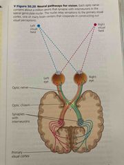
Where optic nerve meets at base of cerebral cortex Left to right side and right to left side |
|
|
Fovea |
Center of visual field, contains no rods but has a high density of cones |
|
|
Function of lens |
Focusing of image on retina occurs by changing shape of lens and pupil Focuses light rays onto retina (assisted by cornea and aqueous/vitreous humors) Image is inverted on retina and then changed by brain |
|
|
Near vs distant vision |
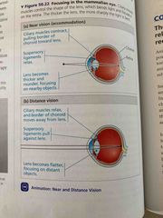
Near: ciliary muscles contract pulling choroid toward lens, suspension ligament relax, lens becomes thicker and rounder Distant: ciliary muscles relax, choroid moves away from lens, suspense ligaments pull against lens, lens becomes flatter |
|
|
Nearsighted myopia |
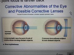
See close objects better than distance Eyeball is elongated |
|
|
Farsighted hyperopia |
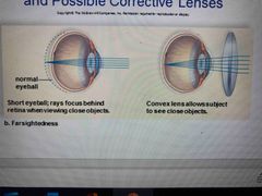
Cannot see close up Eyeball is shortened |
|
|
Color vision |
Photopsins: pigments formed when retinal binds to 3 distinct opsin proteins |
|
|
Color blindness |
Abnormal color vision due to mutations in genes for one or more photopsin proteins Usual deficiency in one type of cone Red-green most common type (x-linked recessive so usually in males) |
|
|
Retinal d/o |
Diabetic retinopathy: capillaries to retina damaged Macular degeneration: cones are destroyed Detached retina: following trauma |
|
|
Glaucoma |
Fluid builds up in eye destroying nerve fibers w peripheral boss ion (aqueous humor) |
|
|
Cataracts |
Cloudy spots on lens UV light, diabetes, alcohol use, and smoking |
|
|
Gustation (taste) |
Dependent on detection of chemicals (tastants) |
|
|
Olfaction (smell) |
Dependent on detection of odorant molecules |
|
|
Taste in mammals |
5 perceptions: salty, sweet, sour, bitter, and unami(elicited by glutamate also called savory) receptors for all five tastes Individual taste cell expresses one receptor type and detects only one of the five types Sweet or bitter solely on what neurons are activated |
|
|
Taste buds |
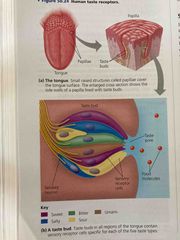
Organized Taste receptors cells are Modified epithelial cells with microvilli Most have projections called papillae Located in several areas of tongue Any region of taste bud can detect five taste |
|
|
3 types of receptors |
1. G protein coupled receptors: sweet, umami, and bitter 2. Transient receptor protein (TRP): sour, similar to capsaicin and other thermoreceptors 3. Sodium channel: salt |
|
|
Olfactory receptors |
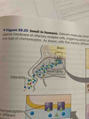
Neurons that line nasal cavity (each one has about 5 olfactory cilia) Binding to cilia triggers transduction pathway sending action potentials to brain Smell and taste are independent but interact |
|
|
Odorant receptor |
Odorant diffuses biding to GPCR protein on plasma membrane of olfactory cilia Triggers transduction leading to cyclic AMp CAMP opens channels in plasma membrane permeable to Ca and Na |
|
|
Humans can distinguish thousands of different of odors |
Directly connected to limbic system connecting to memory and emotions |
|
|
Anosmia |
Born without sense of smell Sense of smell Devine’s at age 60 |
|
|
Covid 19 and smell |
Olfactory do not express gene that encodes ACE2 (which is needed for covid to enter cell) Gene is expressed in cells that provide metabolic and structural support to olfactory neurons Covid does not permanent damage neurons (anosmia) |