Reading...
![]()
Play button
![]()
Play button
![]()
Use LEFT and RIGHT arrow keys to navigate between flashcards;
Use UP and DOWN arrow keys to flip the card;
H to show hint;
A reads text to speech;
86 Cards in this Set
- Front
- Back
|
If the heart were to stop, what three organs would suffer the most damage from loss of oxygen?
|
Brain, kidneys and heart muscle.
|
|
|
Where is the heart located?
|
It sits on the diaphragm in the mediastinum, between the two lungs in front of the esophagus.
|
|
|
What is the pericardial sack?
|
A bag that surrounds the heart made up of 2 linings. The outer lining is a fibrous dense irregular connective tissue. The inner lining is a simple squamous epithelium that is serous, makes fluid to reduce friction.
|
|
|
Heart Walls: Describe the Outer Wall
|
A simple squamous, serous (makes fluid) epithelium. Also called the "Visceral Pericardium"
|
|
|
Heart Walls: Describe the Middle Layer
|
The middle layer is a thick cardiac muscle called "Myocardium"
|
|
|
Heart Walls: Describe the Inner Layer
|
It is a simple squamous epithelium, also called "Endocardium". It CAN also be called "Endothelium" which is a term for the special anti-clotting epithelium that lines the heart and all arteries & veins.
|
|
|
What are the tissues of the Heart Valves?
|
Dense irregular connective tissue covered with endothelia.
|
|
|
Describe the left side of the heart
|
The left side of the heart receives blood from the lungs and must pump it to the body, so it has very thick, muscular walls. It pumps the blood via the Left Atrioventricular Valve, or bicuspid valve or Mitral Valve.
|
|
|
Describe the right side of the heart
|
The right side of the heart receives blood from the body and sends it to the lungs. It receives the blood from the Inferior & Superior Vena Cavas and out the Pulmonary trunk to the right and left pulmonary Arteries
|
|
|
What is Chordae Tendonae?
|
It is the "anchor" that holds the valve closed and attaches it to the papillary muscle. It is also known as the "heart strings".
|
|
|
Why is it important to take good care of your teeth?
|
A dental infection can travel straight to your heart muscle and infect it. An infected heart muscle can weaken it.
|
|
|
What is ligamentosum arteriosum?
|
A ligament running from the aorta to the pulmonary trunk. It was an open artery in the fetal stage called the ductus arteriosus and closes at birth.
|
|
|
What is a heart attack?
|
If any of the coronary arteries are blocked.
|
|
|
What is V-fib?
|
If the Left Anterior descending artery is blocked.
|
|
|
Describe the cardiac muscle.
|
Cardiac muscle cells have an automatic ion release and contraction. When two cardiac muscle cells are connected, they will both beat at the rate of the faster cell. When placed in a petri dish with the right conditions, it will beat on it's own.
|
|
|
What is the natural pacemaker.
|
The sinoatrial node. Calcium is released sending the signal down the heart to the atrioventricular node where the AV bundle (called Purkinjee fibers) splits and sends the signal down each side and back up to the ventricles.
|
|
|
What is the normal heart rate range. Give an example of each.
|
Normal heart rate range is 100 to 65. Infants are normally around 100 and a young, in season, athlete could be around 60 or 65.
|
|
|
What is below normal heart rate?
|
Below 65 is called Bradycardia.
|
|
|
What is above normal heart rate?
|
Above 100 is called Tachycardia. Should look into medications, are they in pain or stressed?
|
|
|
In an electrocardiogram, an EKG (ECG), what is "P"?
|
Right after the SA node fires, sodium (Na+) enters the cells in the atria and is represented by the P in an EKG.
|
|
|
In an EKG, what is Q?
|
When potassium (Ka+) leaves the atria.
|
|
|
IN an EKG, what is R?
|
When sodium (Na+) enters the ventricles. It should be big, sharp and organized.
|
|
|
In an EKG, what is S?
|
Q and S represent the same thing. When potassium (Ka+) leaves the atria.
|
|
|
In an EKG, what is T?
|
When potassium leaves the ventricles.
|
|
|
In an EKG, what is the distance between the P and R?
|
It represents the time it takes for the signal to get from the SA node to the ventricles.
|
|
|
In an EKG, what would indicate a big P wave?
|
The atria is too large, possibly indicating the valve isn't closing all the way.
|
|
|
In an EKG, what would indicate a wide R wave?
|
The AV bundles are sending the signals at different times, indicating a heart attack happened. One of the bundles has to bypass scar tissue, arriving later than the other.
|
|
|
In an EKG, what is fibrillation?
|
Unorganized, not pumping.
|
|
|
In an EKG, what is asystole?
|
Dead. No contraction.
|
|
|
In an EKG, what is "ST elevation"?
|
It indicates a heart attack. In slang, it is referred to as a "tombstone" by how it appears on the EKG.
|
|
|
What is a circumflex artery?
|
An artery that runs around the side of the heart to the posterior side.
|
|
|
Define Systole
|
Contracting
|
|
|
Define Diastole
|
Relaxed heart between contractions
|
|
|
What are the steps of the Cardiac Cycle?
|
1. Ventricular Filling (SA node fires) (AV valves open)
2. Atria Contracts (last 30% of blood into ventricles) 3. Ventricular Systole (pressure in ventricle rises) A. AV valves close B. Eject blood into arteries 4. Ventricles now empty (ish) - AV valves open again |
|
|
What are the body's concerns for the brain stem in order?
|
1. Blood O2 delivery to the brain
2. Blood pressure - too low, could faint - too high, could cause artery damage 3. Heart Rate is last |
|
|
What could be the results of artery damage?
|
Kidney disease
blindness hardening of the arteries which could cause heart attack or stroke |
|
|
How do we calculate Cardiac Output?
|
Heart Rate X Stroke volume = CO
|
|
|
What is stroke volume?
|
Blood from the left ventricle in 1 beat
|
|
|
How many liters of blood does the average person have?
|
The average person pushes a volume of 5 to 6 liters of blood throughout their body in 1 minute.
|
|
|
How do we calculate Stroke volume?
|
End diastolic volume minus end systolic volume.
(How full it can get minus what is left in the ventricle after it is emptied) This tells us how effective it is at pushing out the blood and keeping it out. |
|
|
What affects the filling of the ventricles?
|
Vein pressure (wounds or lack of water)
Time available to fill the heart (if it's beating at a faster rate, there's less time to fill) |
|
|
What affects emptying of the ventricles?
|
Contraction strength
- calcium, epinephrine and stress make it stronger - the fuller it is, the stronger it is - artery pressure (up) emptying (down) |
|
|
What are the 2 centers in the Medulla Oblongata that control Heart Rate?
|
Cardiac Acceleratory Center and the Cardiac Inhibitory Center
|
|
|
What are the 3 locations that send input to the Medulla?
|
Carotid Sinus (baroreceptors - BP, O2, CO2
Aortic Arch - BP & O2 detectors Right Atrium - vein pressure |
|
|
What happens if BP or O2 go down?
|
The result goes to the Cardiac Acceleratory center which triggers the sympathetic NS and epinephrine is released on the heart to increase the heart rate & cardiac output which increases blood pressure
|
|
|
What happens if BP or O2 go up?
|
The result goes to the Cardiac Inhibitory Center which triggers the parasympathetic NS and the vegas nerve releases acetylcholine on the heart which slows the heart rate.
|
|
|
What other factors affect the heart rate?
|
Temperature - if it's high, the HR is high
if it's low, the HR is low Calcium causes it go up Emotions - excited or scared, HR goes up, depressed causes HR to go down. |
|
|
What is Circulatory Shock?
|
The body's tissues aren't getting enough blood, so not enough oxygen. There is a significant drop in blood pressure.
|
|
|
What causes circulatory shock?
|
Heart stopping or failing
Serious blood loss Serious Dehydration (heat stroke) Serious Allergy - fluid leaves the vessels and goes to tissue instead due to inflammation and edema |
|
|
What is the biggest risk with circulatory shock and why?
|
Kidney damage due to vasoconstriction. The heart rate rises and the arteries get really tight. All the arteries but the brain, heart and lungs vasoconstrict, leaving the kidneys without blood.
|
|
|
What are the layers of the arteries and veins?
|
Tunica Intima (inner) - single layer of endothelia (anti-clotting)
Tunica Media - (middle layer) smooth muscle & elastin Tunica Adventitia - dense, irr CT & collagen (very strong) |
|
|
What are the layers of the capillaries?
|
Only one layer - the Tunica Intima only - simple squamous endothelia.
|
|
|
Describe arteries.
|
Carry blood away from the heart. Has a thick wall to resist B.P. Has a large tunica media. Is capable to dilating and constricting.
|
|
|
What are the largest arteries in the body?
|
Carotid and Aorta. They have a large amount of elastic CT in the Tunica Media to stretch with the pulse. Has a strong recoil. Has a vasovasorum.
|
|
|
What is a vasovasorum?
|
The blood supply needed for the largest arteries in the body because the walls are so thick.
|
|
|
What is anastomosis?
|
Separation and rejoining of arteries, often when there is a blockage.
|
|
|
Why are capillaries so thin walled?
|
To allow for diffusion.
|
|
|
Describe the vein.
|
Very little muscle. Has very little pressure. Relies on valves and outside pressure to return blood to the heart against gravity. Skeletal muscles & the diaphragm help to do that. To decrease friction the size of the vein is increased.
|
|
|
What are the biggest veins in the body?
|
Vena Cavae.
|
|
|
What is blood pressure?
|
The force on the artery walls from blood flow. The further away from the heart, the weaker it is.
|
|
|
What is the systolic reading in blood pressure?
|
It is the top number (the higher number) and it is the force during a surge of blood.
|
|
|
What is the diastolic reading in blood pressure?
|
It is the lower number and it is the force between surges, normal pulse.
|
|
|
What causes blood pressure to go higher.
|
Being tired or stressed.
|
|
Identify this
|
A venule or small vein
|
|
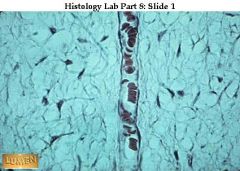
Identify this
|
Capillary
|
|
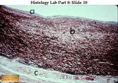
Identify this - what are a, b & c?
|
Aorta - the largest elastic artery
a. is the tunica intima, b. is the tunica media and c. is the tunica adventitia |
|
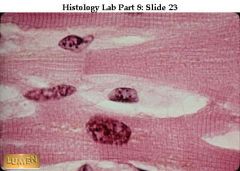
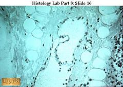
Identify this
|
Cardiac muscle
|
|
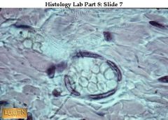
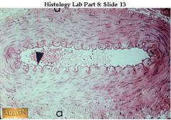
Identify this
|
lymphatic vessel (looks empty - is filled with fluid to bring back to the heart)
|
|
Identify this
|
muscular artery
|
|
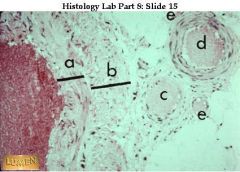
What are a, b and d?
|
a. is the vein's media, b. is the adventitia and d is an artery
|
|
|
What is hypertension?
|
High blood pressure
|
|
|
What is hypotension?
|
Low blood pressure
|
|
|
What is orthostatic hypotension?
|
Low blood pressure from standing up
|
|
|
What affects Blood Pressure?
|
Blood volume and friction (Peripheral Resistance) and viscosity
|
|
|
Why does blood volume affect blood pressure?
|
If there is less blood, then the BP goes down. There is less blood volume if there is a lot of sweating or blood loss.
If there is more blood, then BP goes up. IV fluids can increase it or retaining fluids in the kidney |
|
|
Why does friction or Peripheral Resistance affect BP?
|
If the artery or vein is smaller, less room for the cells to move meaning more friction and BP goes up. If it's larger, then BP goes down.
|
|
|
What is viscosity and how does it affect BP?
|
Viscosity is the thickness of the blood. If there are more cells than normal caused by leukemia or blood doping, then BP goes up. If there are more proteins in the blood caused by inflammation, then viscosity and BP go up.
|
|
|
What are the controls for blood pressure?
|
Cardiac output, control for blood volume and vasomotor control.
|
|
|
What is the control for cardiac output to control blood pressure?
|
If the heart rate increases, then the cardiac output increases and BP increases. The reflexes in the carotid sinuses and the aortic arch tell the heart rate to go up or down to control the cardiac output.
|
|
|
What's the control for blood volume?
|
If BP goes down, the medulla tells the hypothalamus to make you thirsty. Then the hypothalamus signals the pituitary gland to release an anti-diuretic hormone on the kidneys which then retain H2O which causes the arteries to vasoconstrict so there's less room, more friction and the BP goes up.
|
|
|
What is the stress related control for blood volume?
|
If the pituitary gland releases aldosterone on the kidneys via the adrenal gland, then the kidneys retain sodium & H2O and lose potassium and BP goes up.
|
|
|
What is the vasomotor control for blood pressure?
|
If BP is down, the medulla signals the sympathetic NS which then releases epinephrine on the arteries (except those in the coronary system, lungs and brain). They vasoconstrict creating less room and more friction and BP goes up.
|
|
|
Name three more things that affect vasomotor control (and therefore BP)
|
1.Chemoreceptors, which detect O2 levels and CO2 levels. If O2 levels are down or acid levels are up, they will send a signal to vasoconstrict.
2.Temperature - hot, dilate, cold, constrict 3. Emotions - scared, constrict, angry, dilate |
|
|
Is there a local control to dilate an artery?
|
Yes, the endothelial cells can detect O2 levels and temperatures. If they detect warmth, they will release a gas called Nitric Oxide on the smooth muscle cells to relax them and it dilates the artery.
|
|
|
What happens in the capillaries?
|
Fluids filter out of the capillaries due to the pressure difference. The tissue outside the capillary has a pressure of 0 and the pressure inside is anywhere from 25mm to 10mm Hg. O2, nutrition and cells stay behind because they are too big.
|
|
|
How does fluid return to the heart?
|
60% of fluids return to the venule by osmosis. Clotting cells, antibodies and albumin attract the fluid to return to the capillaries. The other 40% returns via the lymphatic vessels.
|