![]()
![]()
![]()
Use LEFT and RIGHT arrow keys to navigate between flashcards;
Use UP and DOWN arrow keys to flip the card;
H to show hint;
A reads text to speech;
16 Cards in this Set
- Front
- Back
|
Describe the functions of the nervous system
|
Communication
1. Between periphery of body and brain - provides information (SENSORY) - e.g. pain, pressure, temperature, touch, conscious proprioception 2. Between brain and periphery of body provides: (MOTOR) - Voluntary control of effectors (skeletal muscles) in the periphery = somatic - Involuntary control of effectors (smooth muscle, cardiac muscle, glands) in periphery = autonomic |
|
|
What makes up the somatic nervous system?
|
- 31 pairs of spinal nerves (spinal segments)
• 8 cervical • 12 thoracic • 5 lumber • ~ 5 sacral • 1+ coccygeal - 12 pairs of cranial nerves |
|
|
Describe a basic scheme of the nervous system
|
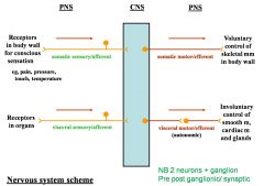
- Somatic and visceral systems are largely the same
- However, visceral systems are fed by autonomic nerves and therefore the visceral motor is made up of 2 nerves fibres rather than one, and an extra synapse |
|
|
Describe the formation of spinal nerves
|
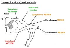
- The dorsal root contains only sensory fibres, which synapse at the dorsal root ganglion
- The ventral root contains only motor fibres - The two roots come together to form a mixed spinal nerve - Spinal nerve gives off a mixed dorsal ramus and a mixed ventral ramus |
|
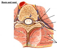
Label this diagram:
|

|
|
|
What is the lateral horn?
|
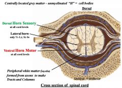
- Found between the ventral and dorsal horns of grey matter
- Found only at levels T1-L2 and S1-S4 = autonomic |
|
|
Describe the basic pathophysiology of polio
|
- Infection attacks only the cells of the anterior horn
- This means that there are no motor signals to smooth muscle, causing paralysis and muscle wastage |
|
|
Describe the pathophysiology of saryngomyelia
|
- Cleaved dorsal horn → no sensory input
- Means that keep burning themselves etc as no feeling |
|
|
Describe the morphology of grey matter
|
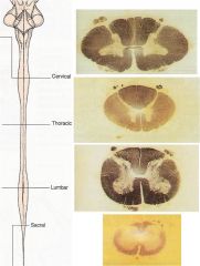
- Different shapes at each anatomical segmental level
- Note the large anterior horns for the cervical and lumbar plexi i.e. the arms and legs |
|
|
Describe the topographical anatomy of the spinal cord
|
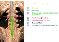
- True spinal cord ends at L1/L2 in the adult (and L3/L4 in the child)
- The dura and arachnoid mater i.e. the foetal sac end at S2 |
|
|
Describe the spinal meninges
|
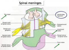
- Dura mater is a thick fibrous sheath surrounding the spinal cord and the spinal nerves and their rami
- Arachnoid mater is a thin, web-like layer that also covers the spinal nerves and rami - Pia mater closely adheres to the tissue of the spinal cord and forms denticulate ligaments, which anchors the cord to the arachnoid and dura maters |
|
|
Describe the arterial supply to the vertebral column
|
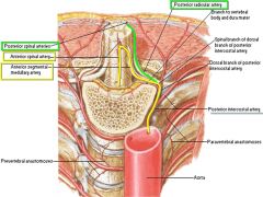
- Anterior spinal artery formed by the union of vertebral arteries run from the medulla of the brainstem to the medullary cone of the spinal cord
- 2 posterior spinal arteries are branches of either the vertebral artery of the posteroinferior cerebellar artery - Also anterior and posterior segmental branches which are derived from spinal branches of: • ascending cervical • deep cervical • vertebral • posterior intercostal • lumbar arteries - Most of these are branches from the aorta |
|
|
Describe the venous drainage of the spinal column
|
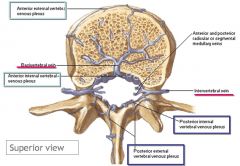
- 3 Anterior and 3 posterior spinal veins form internal (anterior and posterior) venous plexi. Note that these veins (in the epidural space) are valveless
- Also external (anterior and posterior) venous plexi - Basivertebral and intervertebral veins are involved in marrow drainage |
|
|
What is a nerve plexus?
|
- Peripheral nerves that divide and join other nerves to weave a network
- Plexus permits individual nerve fibres to pass from one peripheral nerve to another, allowing redistribution of nerve roots - The only parts of the body that are not covered by plexi are T1 to L2 segmental innervation of skin and muscles |
|
|
Describe the pathophysiology of Herpes zoster
|
- If Herpes zoster is caught it is never fully removed from the body but can remain latent in nerve cell bodies
- They can remain there for many years without causing symptoms - Years or decades after a chickenpox infection, the virus may break out of nerve cell bodies and travel down nerve axons to cause viral infection of the skin in the region of the nerve. - The virus may spread from one or more ganglia along nerves of an affected segment and infect the corresponding dermatome causing a painful rash. |
|
|
Describe the 3 classifications of nerve injury
|
1. Neuropraxia = 1st degree
- No axon degeneration - Rapid recovery 2. Axonotmesis - Wallerian degeneration - Endoneurium survives = weakness and tingling due to damage and degeneration - Nerve fibre will eventually grow back down Schwann cell tube unless the tube is cut and feeling will be restored 3. Neurotmesis = 5th degree - Division of a trunk = nerve ends detatched |

