Reading...
![]()
Play button
![]()
Play button
![]()
Use LEFT and RIGHT arrow keys to navigate between flashcards;
Use UP and DOWN arrow keys to flip the card;
H to show hint;
A reads text to speech;
44 Cards in this Set
- Front
- Back
|
size of normal kidney
|
length 9 -12cm
width 4 - 6cm depth <3.5cm (differences >2cm imply a pathology) |
|
|
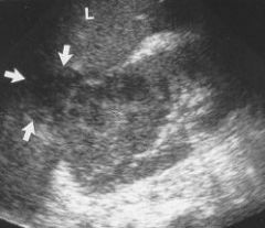
most common renal mass is _____.
|
Simple Cysts (typical features of a cyst
seen) |
|
|
Simple Cysts may become ____ or
____ and get internal echoes and septae |
infected; haemorrhagic
|
|
|
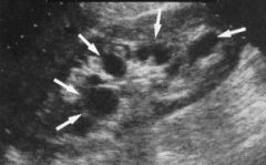
about 50% of people
over the age of 50 have renal________. |
cysts
|
|
|
this cyst has become
infected as indicated by the ___ level within it |
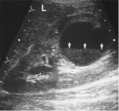
debris
|
|
|
although most cysts are cortical, sometimes they are associated with the renal pelvis (parapelvic cysts).
|
these may be single or multiple and may look like a hydronephrosis
|
|
|
APCKD?
|
Adult polycystic kidney disease
|
|
|
what is APCKD?
|
inherited, autosomal
dominant disorder, so scan relatives usually bilateral enlargement with multiple cysts may see cysts in other organs may be hypertensive slow progression to renal failure kidney may be large |
|
|
Renal Calculi
|
Seen if large enough, but small ones easily missed, so an
IVU/CT KUB/Urography may be indicated. |
|
|
Ureteric Calculus
|
a calculus has become lodged in the lower end of the
ureter leading to a dilation of the ureter |
|
|
Renal Tumour
|
Very difficult to differentiate malignant renal tumours from benign.
|
|
|
Renal cell carcinoma
|
the most common
solid renal neoplasm generally a disease of the middle aged and elderly more common in men than women |
|
|
Renal cell carcinoma
|
in this example the
carcinoma can be seen invading the liver usually a heterogeneous solid mass RCC may develop cystic areas and calcifications within it |
|
|
Wilms tumour (nephroblastoma) commonly found in ___.
|
children
|
|
|
paediatric patients who has Wilm's tumour often
|
has haematuria and palpable abdo mass
tumour appears solid, large well-defined and heterogeneous (areas of haemorrhage and necrosis are common) kidney often obliterated |
|
|
what is hydronephrosis?
|
an abnormal dilatation of the renal collecting system. can be graded as mild (2mm seperation), moderate
(entire sinus seperated), severe (calyceal involvement) usually due to an obstruction e.g. calculus, pelvic malignancy, but other causes are known |
|
|
when there is a mickey mouse sign, there is ___.
|
Hydronephrosis
|
|
|
normal measurements of thyroid
|
12-20mm thick (2cm
is max. allowed), 20-25mm wide, 30-70mm long |
|
|
Thyroid Disease
|
usually nodular in form rather than diffuse
• very difficult to differentiate malignant from benign • however, the vast majority are benign |
|
|
Malignant thyroid disease:
|
microcalcification
• poorly marginated • hypo- or isoechoic • more commonly single nodule seen (can get mixed malignant and benign) • irregular or incomplete halo • enlarged lymph nodes |
|
|
Benign thyroid disease:
|
calcification tends to be
coarse or peripheral • usually well defined • hypo-, iso and hyperechoic • often multiple nodules • complete halo • normal nodes |
|
|
most common thyroid pathology is
|
Hyperplasia
(non-neoplastic thyroid enlargement e.g. hyperplasia) |
|
|
May get cystic areas in hyperplasia, so called
_______-, with projections into the cyst |
colloid nodules
|
|
|
_______ may also be seen in hyperplasia.
|
calcification
|
|
|
When a benign colloid nodule or an adenomatous noduleundergoes degeneration it may be mistaken for a cyst, butthese will have ______ and _______.
|
thick walls; internal debris
|
|
|
Solitary nodules
|
are either benign adenomas or carcinomas
|
|
|
adenomas can be _______ or ________
(depends on the tissue they are derived from) and look exactly the same as benign nodular hyperplasia |
follicular; non-follicular
|
|
|
most solitary nodules will be ________.
|
adenomas
|
|
|
Adenomas are prone to undergo _____ and haemorrhage
|
cystic degeneration
|
|
|
how is Papillary carcinoma spread?
|
spreads via the lymphatics, so the
cervical nodes will probably be involved |
|
|
Papillary carcinoma most commonly affect ______ although it can also affect all pt groups.
|
young females
|
|
|
Follicular carcinoma is the ______ most common thyroid carcinoma.
|
second
|
|
|
Follicular carcinoma look similar to
|
follicular adenomas
|
|
|
Follicular carcinoma are characterised by
|
vascular invasion and tend to
spread via the blood stream so mets. are more distant need FNA to really differentiate |
|
|
medullary carcinoma
|
fairly rare, very slow
growing, usually involves lymphatics |
|
|
anaplastic thyroid carcinoma
|
usually a
disease of the elderly, a very agressive tumour and presents as a rapidly growing |
|
|
fine needle aspiration is probably the best
way to make a _______ • However, ultrasound still has a role to play in |
differential diagnosis
|
|
|
detection and localisation of neck masses under ______
• differentiation of malignant from benign (but not easy). |
• FNA guidance
|
|
|
Thyroiditis
|
diffuse thyroid diseases using leading to
general enlargement of the gland |
|
|
`thyroiditis ,may be (3) types:
|
acute, subacute or chronic
|
|
|
Thyroiditis are diagnosed
|
clinically or by FNA
on US gland will appear thicker, and may get reduced reflectivity |
|
|
an eg of thyroiditis is
|
Hashimotos disease
|
|
|
Hashimotos disease aka
|
chronic lymphocytic thyroiditis
|
|
|
Hashimotos disease
|
affects young to middle aged women
painless, diffuse gland enlargement with coarse parenchymal pattern, may see a few discrete nodules, may have lymphadenopathy patient will have hypothyroidism difficult to differentiate from other diffuse disease |