Reading...
![]()
Play button
![]()
Play button
![]()
Use LEFT and RIGHT arrow keys to navigate between flashcards;
Use UP and DOWN arrow keys to flip the card;
H to show hint;
A reads text to speech;
68 Cards in this Set
- Front
- Back

Label
|
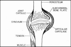
-
|
|
|
What type of Collagen is present in Cartilage?
|
Type II Collagen
|
|
|
Discuss the Synovium of joints
|
-Arranged in Villi projection lined by Synovial epithelial cells
-Synovial fluid is very low in protein but has alot of mucin -Cartilage does not have its own blood supply & must rely on the Synovium for nutrition |
|
|
Define Gout
|
Hyperuricemia & deposition of Monosodium Urate Crystals in joints, resulting in recurrent bouts of Acute Arthritis
|
|
|
Describe the etiology of Primary Gout
|
Multifactorial hereditary disease due to over-synthesis of uric acid from purine nucleotides
Most common in MALES |
|
|
What are 3 possible causes of Secondary Gout?
|
1. Leukemia = due to increased nuclear cell turnover -> increased Purine metabolism
2. Decreased renal urate excretion in renal disease 3. Lesch-Nyhan Syndrome = deficiency of HGPRT causing hyperuricemia -Mental retardation -Spasticity -X-linked |
|
|
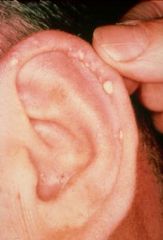
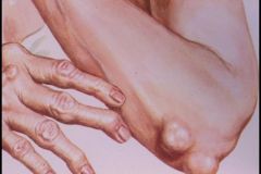
Gout
-Podagra = inflammed MTP joint of big toe |
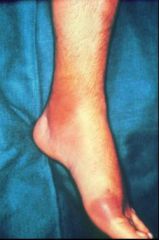
What type of arthritis?
|
|
|
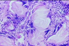
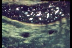
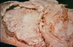
Gout
-joint surface is covered with white chalky material = Urate crystals = Tophi -Tophi = clusters of Urate crystals surrounded by fibroblasts, lymphocytes, & Giant cells located in cartilage or soft tissues |
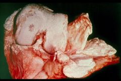
What type of arthritis?
|
|
|
Gout
|
What type of Arthritis are Tophi associated with?
|
|
|
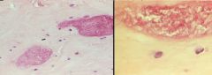
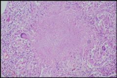
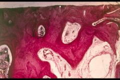
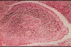
Tophus in Gout
-center is accumulation of Urate crystals -chronic lymphocytic infiltration -MNGC's are present |
What is this?
|
|
|
Gout
-Podagra = Metatarsophalangeal (MTP) joint in the big toe |
What type Arthritis?
|
|
|
What are 3 complications of Hyperuricemia?
|
1. Chronic renal failure requiring dialysis
2. Atherosclerosis 3. Deformity due to destruction of articular surfaces |
|
|
What is Lesch-Nyhan Syndrome?
|
X-linked absence of Hypoxanthine-guanine Phosphoribosyl Transferase (HGPRT)
1. Hyperuricemia -> Gout 2. Mental retardation 3. Spasticity 4. Self-mutilating behaviors |
|
|
Pseudogout = Chondrocalcinosis
|

What type of arthritis is this?
|
|
|
What is the pathogenesis of Pseudogout?
|
Calcium Pyrophosphate crystal deposition into Articular Cartilage
|
|
|
Basophilic, rhomboid crystals that are weakly positively birefringent
|
Pseudogout
|
|
|
Pseudogout:
1. commonly what joint? 2. what age? |
1. knee
2. > 50; Both sexes affected equally |
|
|
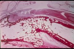
Pseudogout = Calcium Pyrophosphate crystals
-no inflammation -cartilage looks normal -large deposits of acellular material |
What is seen here?
|
|
|
Pseudogout = Chondrocalcinosis
-Calcium Pyrophosphate Crystals |
Weakly Positively Birefringent
|
|
|
Pseudogout
|
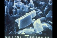
Basophilic Rhomboid Crystals under scanning EM
|
|
|
Calcium Hydroxyapatite deposition arthritis
|
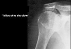
What is "Milwaukee Shoulder"
|
|
|
Calcium Hydroxyapatite deposition arthritis = Milwaukee Shoulder
|
This was taken from a shoulder, what type of Arthritis?
|
|
|
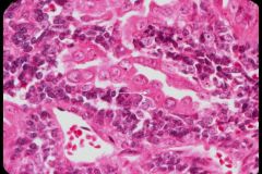
Septic Arthritis
Monoarticular MCC -Gonococci = MCC in urban population -S. aureus -Strep -Mycobacterium |
What is this?
How do you know? Most common causes? |
|
|
Septic Arthritis
-subarticular cartilage surface is lined by PMN's & bacteria |
What is this showing?
|
|
|
Tuberculous Arthritis of the Spine = Pott's disease
|
What is this?
|
|
|
Tuberculous Arthritis
-Granuloma with Caseous Necrosis in the center surrounded by MNGC's |
What is this? How do you know?
|
|
|
A young man has Arthritis & Achilles tendon periostitis, Urethritis, & Conjunctivitis. What is the disease? What usually precedes the reaction? What HLA is it associated with?
|
Reiter's Syndrome
Venereal disease (C. trachomatis) HLA-B27 = autoimmune component |
|
|
Osteoarthritis:
1. cause? 2. Gender? 3. Age? |
1. "wear & tear"
2. Females 3. usually > 50 |
|
|
Most common form of Arthritis
|
Osteoarthritis
|
|
|
What is the pathogenesis of Osteoarthritis?
|
Proteolytic degradation of cartilage matrix -> loss of resilience of Articular cartilage -> surface irregularity of Subchondral Bone
|
|
|
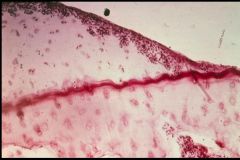
Osteoarthritis
-loss of Proteoglycans -shrinkage of the cartilage & the chondrocytes tend to cluster |
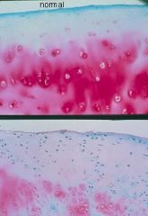
What is the bottom pic showing?
|
|
|
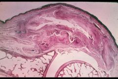
Osteoarthritis
*Fibrillation = clefts develop in the surface of Cartilage |
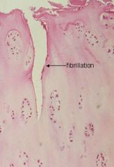
What arthritis is Fibrillation common in?
|
|
|
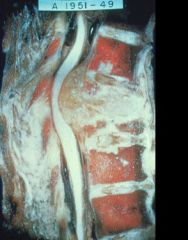
Osteoarthritis
-Green = Collagen = worn away -Osteophyte = bone spur = mushroom-like mass on the lateral edges of the joint |
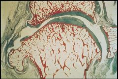
What is this from? How do you know?
|
|
|
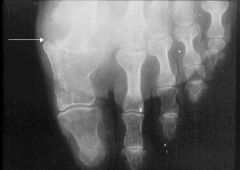
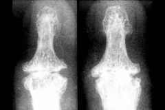
Hebreden's node = DIP joint Osteophyte
Osteoarthritis |
What is this called? What arthritis?
|
|
|
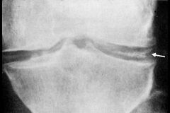
What is a Bouchard node? What arthritis is it seen in?
|
Osteophyte at the PIP joint
Osteoarthritis |
|
|
Osteoarthritis
Osteophytes are present |
What type of Arthritis? How do you know?
|
|
|
Eburnation = polished, ivory-like appearance of bone -> due to rubbing of bone on bone
Osteoarthritis -Subchondral bone sclerosis = red |
What is seen at the arrow? What type of Arthritis?
|
|
|
Type of arthritis in which the joint pain becomes worse later in the day after use
|
Osteoarthritis
|
|
|
What are the risk factors for Rheumatoid Arthritis?
|
1. HLA-DR4
2. EBV infection |
|
|
Rheumatoid Arthritis:
1. Gender preference 2. Age of onset |
1. Females (4:1)
2. 20-50 years of age |
|
|
Describe the pathogenesis of Rheumatoid Arthritis
|
1. B cells in the joint produce RF complexes
-RF complexes are IgM auto-Ab's against the Fc receptor of IgG -RF complexes are also present in the serum of 70-90% of cases -Type III HS 2. RF complexes activate complement = attracts Neutrophils -Neutrophils produce acute inflammation of Synovial tissue -Neutrophil phagocytosis of RF complexes produces Ragocytes 3. Chronically inflammed Synovial tissue proliferates (forms Pannus) -Pannus releases cytokines that destroy Articular Cartilage -End result is reactive fibrosis & joint fusion (Ankylosis) |
|
|
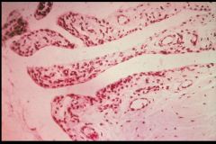
Rheumatoid Arthritis
Villi that extend into the Synovial Cavity are enlarged due to Chronic Inflammation |
Type of Arthritis? How do you know?
|
|
|
Rheumatoid Arthritis
|
This is an inflammed Synovial Villus...what type of Arthritis?
|
|
|
Pannus = proliferation of the Synovium & granulation tissue over the Articular Cartilage of the joint
Rheumatoid Arthritis |
What is this called? What is it? What type of arthritis?
|
|
|
Subcutaneous Rheumatoid Nodule
|
What is seen here?
|
|
|
Rheumatoid Nodule
-central FIBRINOID NECROSIS surrounded by epitheloid macrophages, lymphocytes, & granulation tissue |
What is this?
|
|
|
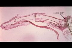
Subluxation
Rheumatoid Arthritis |
What is happening here? What type of arthritis is it common in?
|
|
|
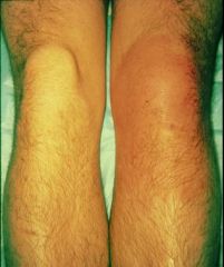
Rheumatoid Arthritis
|
What type of arthritis is this common in?
|
|
|
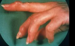
Ulnar deviation of the fingers + Subluxation of the knuckles
Rheumatoid Arthritis |
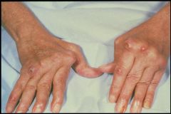
What is this pathology called? What type of arthritis?
|
|
|
Ankylosis
Rheumatoid arthritis |
This is fibrous fusion of the joints, what is the proper term? What type of arthritis?
|
|
|
Ankylosis = bony fusion across the joint space
Rheumatoid arthritis |
What has happened here? What type of arthritis?
|
|
|
Rheumatoid Arthritis
|
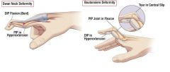
What are these 2 deformities common in?
|
|
|
What is Still's Disease?
|
Rheumatoid Arthritis in Children with RF usually absent
-often preceded by generalized lymphadenopathy & hepatosplenomegaly & acute onset marked by fever |
|
|
HLA-B27 associated disease in which the spine & sacroiliac joints are affected & can lead to rigidity & fixation of the spine as a result of bone fusion
-may be associated with IBD |
Ankylosing Spondylitis
|
|
|
Rheumatoid Arthritis + Splenomegaly + Neutropenia = ?
|
Felty's Syndrome
|
|
|
RA with marked chronic inflammation & enlargement of Lacrymal & Salivary glands. Associated with dryness of eyes & mouth
|
Sjogren's syndrome
|
|
|
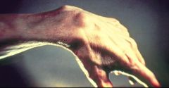
Erythema Chronicum Migrans
Lyme Disease = Borrelia Burgdorferi |
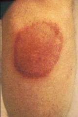
What is this called? What is it due to?
|
|
|
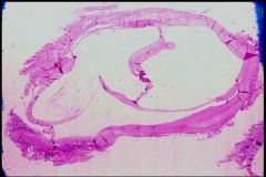
Borrelia Burgdorferi = Spirochete
Lyme Disease |
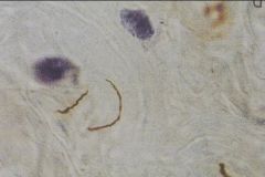
What is this? What does it cause?
|
|
|
How is Lyme Disease contracted?
|
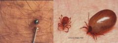
Ixodes dammini tick
|
|
|
What are the late sequelae of Lyme Disease?
|
1. Polyarticular arthritis
2. Neuropathy 3. Cardiac Arrhythmias |
|
|
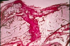
Ganglion Cyst of the wrist = Small, non-neoplastic cystic tumor arising in the joint capsule or adjacent tendon sheath, usually in the wrist
|
What is seen here?
|
|
|
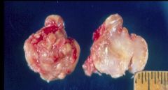
Surgically removed Ganglion Cyst
|
What is this?
|
|
|
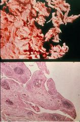
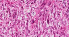
Giant Cell tumor of tendon sheath (Pigmented Villonodular Tenosynovitis)
-Benign tumor of giant cells in the vicinity of a joint. This lesion is probably non-neoplastic, and is closely related to pigmented villonodular synovitis. The latter condition is characterized by an exuberant, heavily pigmented villous synovial overgrowth containing pigmented cells, lipid-laden histiocytes, and multinucleated giant cells |
What are these?
|
|
|
Giant Cell Tumor of Tendon Sheath
-Benign tumor of giant cells in the vicinity of a joint. This lesion is probably non-neoplastic, and is closely related to pigmented villonodular synovitis. The latter condition is characterized by an exuberant, heavily pigmented villous synovial overgrowth containing pigmented cells, lipid-laden histiocytes, and multinucleated giant cells |
What is this?
|
|
|
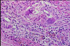
Biphasic soft tissue tumor
-Epithelial cells forming glands + intervening Spindle cells |
Synovial Sarcoma
|
|
|
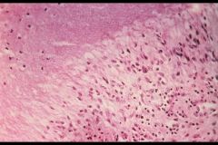
Synovial Sarcoma
-A highly malignant soft tissue tumor arising in the vicinity of a joint. It is characterized by a biphasic microscopic pattern with intermixed atypical spindle cells and nests of epithelial cells with cleft-like spaces |
What is this?
|
|
|
Synovial Sarcoma
-A highly malignant soft tissue tumor arising in the vicinity of a joint. It is characterized by a biphasic microscopic pattern with intermixed atypical spindle cells and nests of epithelial cells with cleft-like spaces |
What soft tissue tumor is this?
|
|
|
Arthritis in which the joint pain is more severe in the morning but resolves within a couple hours
|
Rheumatoid Arthritis
|