![]()
![]()
![]()
Use LEFT and RIGHT arrow keys to navigate between flashcards;
Use UP and DOWN arrow keys to flip the card;
H to show hint;
A reads text to speech;
30 Cards in this Set
- Front
- Back
|
Tympanometry components |
Air pump, loudspeaker, microphone (with probe tip measuring the sound pressure level SPL) |
|
|
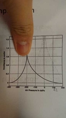
Tympanogram (x axis) |
Middle ear pressure in daPa |
|
|
Tympanogram (y axis) |
Immitance (compliance/flexibility) |
|
|
Static acoustic compliance |
Maximum compliance value |
|
|
Static acoustic compliance normal range and pathologies |
0.3-1.6
Less than 0.3 (otitis media, otosclerosis, thick eardrum)
More than 1.6 (ossicular chain discontinuity, thinned eardrum) |
|
|
Eer canal volume (ECV) normal range and pathologies |
0.2-2.0ml Less than 0.2ml: cerumen/ foreign body More than 2.0ml: eardrum perforation/ PE tube |
|
|
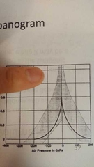
Type A tym - Normal -Peak compliance 0.30-1.50 ml @ 0 daPa -Peak pressure:+100 to -100 daPa (up to -150 daPa for child) |
|
|
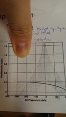
Type Ad tym - Highly compliant - Path: ossicular chain discontinuity/ flaccid eardrum |
|
|
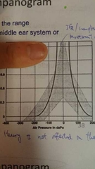
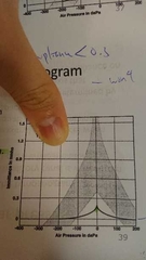
As tym - Shortened v-shape - Indicate stiffness of the middle ear - Path: otosclerosis/ scarred eardrum |
|
|
Type B tym - No sharp peak/little variation in impedance -Indicate severely restricted mobility of eardrum - Path: middle ear effusion/ perforated eardrum/ blocked probe/ blocked ear canal |
|
|
Type C tym - Peak beyond -100/-150 daPa - Path: eustachian tube dysfunction/ ceetain stages of middle ear infection |
|
|
Malleus muscle, innervation |
Tensor tympani, nerve V (trigeminal nerve) |
|
|
Stapes muscle, innervation |
Stapedius, nerve VII (facial nerve) |
|
|
Acoustic reflex (normal value, mechanism) |
- Above 70 dbHL - Stapedius muscle contracts and pulls the stapes away from the oval window. - This stiffens the ossicular chain. - Decreasing the vibration to cochlea. - Happens to both ears even though the stimulus is presented in one ear only. |
|
|
Acoustic reflex arc |
Acoustic Reflex Measures: Pathways • Afferent structures consist of the cochlea and the auditory portion of the eighth cranial nerve.• Afferent pathways carry information from the ear to the brain. • The cochlear nucleus (nuclei) in the brainstem contribute (s) to the acoustic reflex arc in both ipsilateral and contralateral measurement conditions. • Additional neurons in the trapezoid body and medial superior olivary complex contribute to the acoustic reflex pathways in the contralateral measurement condition. Acoustic Reflex Measures: Pathways• A descending efferent pathway passes from the brainstem back to the ear. • The descending (efferent) pathway includes motor fibers within the 7th (facial) cranial nerve, particularly a small branch that innervates the stapedius muscle. • Ipsilateral acoustic reflex pathways remain on one side of the body and are sometimes described as an uncrossed acoustic reflex. • Contralateral pathways are often described as crossed acoustic reflexes. |
|
|
ART normal value |
Acoustic reflex threshold • 80-105 dB ART for 0-50 dB HTL• SL = 55-85 dB |
|
|
How to administer acoustic reflex decay test? |
Acoustic reflex threshold is determined for one ear with a stimulus, like a 1000 Hz tone. Next, the same stimulus is presented continually for 10 seconds at an intensity level 10 dB higher than the threshold.
|
|
|
Administering OAE and normal value |
Otoacoustic emissions
- OAE measurements begin with insertion of probe into ear canal. - Probe presents stimulus + detects very faint sounds from cochlea. - 有hair cell motility (lengthening and shortening of hair cells) 先有OAE - OAE normal level: 0-15 dB SPL - >6dB=OAE present |
|
|
Acoustic reflex decay test usage |
To indicate auditory nerve or auditory brainstem problem rather than inner ear |
|
|
Tympanogram usage |
To imdicate middle ear involvement |
|
|
Types of OAEs |
Spontaneous OAEs (SOAEs) Transient evoked OAEs (TEOAEs) Distortion product OAEs (DPOAEs) |
|
|
TEOAE value |
- Presented at 80 dBSPL - 0-4000 Hz of TEOAE energy recorded |
|
|
DPOAEs value |
- High fequency to low frequency=1.2 - Intensity level at 55 to 65 dBSPL - DPOAEs recorded at 500-8000 Hz |
|
|
OAE usage |
- To distinguish sensory (inner ear hair cell problem) from neural problem - Indirectly indicate normal middle ear function - Provide evidence that outer hair cells for that portion of the cochlea is intact |
|
|
ABR mechanism and administration |
Auditory brainstem response - Reflects auditory nerve fibre and pathways withij auditory brainstem - Summed electrical activity from nerve fibres firing is picked up with electrodes on the forehead and near the ears - These electrical reaponses must pass through brain tissue to electrodes on skin - ABR repeatedly recorded at progressively lower stimulus intensity for click and tone burst stimuli at different frequencies |
|
|
ABR wave types and meaning |
I: auditory nerve near cochlea III: pons of auditory brainstem V: midbrain of auditory brainstem |
|
|
Interpreting ABR |
Latency analysis Amplitude analysis |
|
|
ABR usage |
Estimate auditory thresholds in infants and young children |
|
|
Hearing Ax for children below 6 mos |
Acoustic immitance (equivalent volume of ear canal, static acoustic compliance of middle ear tympanometry, acoustic reflex) Otoacoustic emissions ABR |
|
|
Acoustic reflex presence test usage |
Absence=abnormal middle ear functioning |