![]()
![]()
![]()
Use LEFT and RIGHT arrow keys to navigate between flashcards;
Use UP and DOWN arrow keys to flip the card;
H to show hint;
A reads text to speech;
28 Cards in this Set
- Front
- Back
|
Functions of autonomic nervous system |
Maintains internal body system in state of homeostasis Controls response to external environment Involuntary |
|
|
Divisions of ANS |
Sympathetic (thoracolumbar) Parasympathetic (craniosacral) Enteric (independent, controls secretion and motility in gut) |
|
|
Basic organization of autonomic fibers/ganglion |
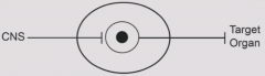
Two step signaling process. CNS neuron has preganglionic neuron which projects to ganglion. Ganglion has postganglionic neuron which projects to target organ. Target organs are smooth/cardiac muscles and secretory glands. |
|
|
Location of sympathetic and parasympathetic ganglia |
Sympathetic ganglia are right outside or very close to spinal cord. Postganglionic fibers are long. Parasympathetic ganglion are located near or within target organs. Postganglionic fibers are short. All autonomic ganglia are located outside the blood brain barrier although preganglionic neuron is within blood brain barrier. |
|
|
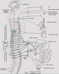
Possible destinations of presynaptic sympathetic fibers |

1. Synapse on sympathetic chain ganglion 2. Go up or down some ganglia before synapsing on sympathetic chain ganglion - generally to sweat glands, piloerector muscles, as well as vasculature for blood pressure. 3. Pass through chain and synapse in celiac, superior mesenteric, and inferior mesenteric ganglion. |
|
|
Somatic and sympathetic simple spinal cord circuits |
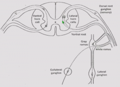
Sensory cell bodies synapse in dorsal horn. Interneuron goes to upper motor neuron cell body in ventral horn which projects out. Autonomic sensory neurons are in dorsal root ganglion also. Make single synapse on lateral horn cells. Postganglionic cells project through ventral root. Peel off through white ramus to enter sympathetic chain ganglion. Postganglionic neuron either synapses in dorsal root and passes through gray ramus to target, travel up and down to different ganglions, or go to collateral (celiac, sup mes, inf mes) ganglion. |
|
|
Basic layout of parasympathetic division |
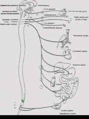
Preganglionic parasympathetic neurons are located in accessory nuclei in brainstem/cervical and sacral regions. Long preganglionic fibers synapse in discrete ganglion or in target tissue. Postganglionic fibers are shorter. |
|
|
Basic layout of enteric division |
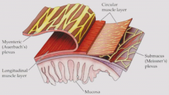
From inside to out: Mucosal layer - produces secretions and is responsible for absorption. Submucosal (Meissner's plexus) Circular muscle Myenteric plexus Longitudinal muscle Plexi have parasympathetic and sympathetic activity. |
|
|
Function of Enteric Nervous System |
Sensory neurons detect presence of food. System performs peristalsis independent of consciousness and CNS. |
|
|
Autonomic control of enteric nervous system |
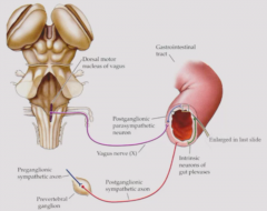
Although functions autonomously, can be modified/turned off by vagal input or sympathetic from prevertebral ganglion. |
|
|
Generalization of function of PS vs S |
Fight/flight (sympathetic) - Induced by rapid change in environment - pupil dilation, increased flow to skeletal muscle and brain. inhibit defecation, urination, digestion. Rest/digest (parasympathetic) - Defecation, urination, digestion. |
|
|
Second property of PS vs S function |
Generalized response - sympathetic system. All functions is carried out simultaneously and by same system. Discrete/localized response - parasympathetic system. Functions are not elicited in generalized way. |
|
|
Does activity of sympathetic or parasympathetic ever completely turn off? |
No - there is tonic activity in these systems.
Rest/digest response increases tonic activity of parasympathetic neurons and decreases tonic activity of sympathetic neurons. Fight/flight response decreases tonic activity of parasympathetic neurons and increases tonic activity of sympathetic neurons. |
|
|
Parasympathetic and sympathetic preganglionic neurotransmitter |

Presympathetic neurotransmitter is ACh for both parasympathetic and sympathetic system. |
|
|
Parasympathetic and sympathetic postganglionic neurotransmitter |
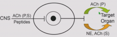
Parasympathetic postganglionic neurotransmitter is ACh. Sympathetic postganglionic neurotransmitter is usually NE, except for cholinergic synapses for sweat glands and peripheral microvasculature. |
|
|
What does target response of autonomic nerve fiber depend on? |
Depends on the receptor |
|
|
Adrenal chromaffin cell |
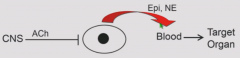
Sympathetic neuron-like cell which is innervated by preganglionic cholinergic neurons. When stimulated, releases Epi and NE directly into blood which reaches target organ via bloodstream. Reinforces generalized sympathetic discharge. Also has different kinetics - takes time to clear from body. Slower, longer-lasting sympathetic response. Embryologic origin is similar to peripheral neurons. |
|
|
Receptors on autonomic ganglion |
Nicotinic AChR - depolarize postganglionic neuron Muscarinic AChR Neuropeptides are co-released at ganglion and have slow modulatory roles. |
|
|
Autonomic receptors on target organ |
Parasympathetic - mAChR Sympathetic - NE acts on alpha and beta adrinergic receptors, ACh activates mAChR. |
|
|
Convergence of inputs at Autonomic Ganglia |
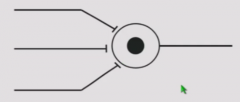
Multiple preganglionic fibers innervate single neuron (S - 50:1, PS 2:1) Because multiple subthreshold signals needed to activate postganglionic fiber |
|
|
Divergence of inputs at autonomic ganglion |
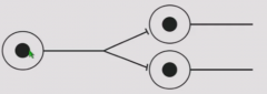
Single preganglionic fibers innervate multiple postganglionic neurons (S 1:200, PS 1:3) Amplification mechanism - small signal can affect multiple postganglionic neurons. Makes sympathetic response generalized. |
|
|
Electrophysiology of autonomic ganglion - experimental set up |
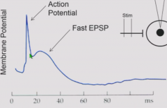
Put electrode into postganglionic neuron and record membrane potential and action potentials. Stimulate white ramus and look at response. Stimulate at time 0. Delay for propogation ofaction potential down preganglionic neuron, release and action of neurotransmitter. Causes potential change in postganglionic cell that causes AP if exceeds threshold. |
|
|
Autonomic ganglion electrophysiological event following action potential |
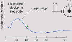
Following AP, a second depolarizatoin (fast EPSP) that occurs which takes 40 ms to return to baseline. Occurs due to ACh released from preganglionic axon. If place Na channel blocker, action potential is abolished but fast EPSP still results. |
|
|
Autonomic ganglion - final electrophysiological event |
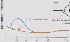
If then add curare or hexamethonium to block nicotinic receptors, fast EPSP is abolished but slow hyperpolarization (slow IPSP) is still visible. This is potential induced by ACh acting at muscarinic receptors. |
|
|
Summary of elements of electrpohysiological activity at autonomic ganglion |
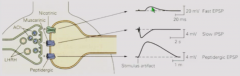
1. Fast EPSP - induced by ACh binding to nicotinic receptors. Induces action potential if reaches threshold. 2. Slow IPSP - induced by ACh binding to metabotropic receptors. Hyperpolarizes cell for a couple of seconds afterwards. 3. Peptidergic EPSP - Moderately large, slow (1 minute) depolarization due to binding of peptide. |
|
|
What kind of post synaptic potentials do muscarinic receptors induce? |
Can be slow EPSP or slow IPSP, depending on which metabotropic receptor there is. |
|
|
Effect of slow potential on electrophysiology of postganglionic neuron |
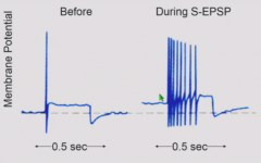
Slow EPSPs make postganglionic neuron hyperexcitable - more action potentials generated from same stimulus. Opposite is true for slow IPSPs. |
|
|
Ionic basis of F-EPSP, S-IPSP, S-EPSP, and P-EPSP (potential, concductance changes, channel, and blocker) |
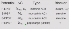
F-EPSP - Nicotinic ACh, increase in K and Na conductance. Blocked by curare and hexamethonium (C6) S-IPSP - Muscarinic ACh, increase in K conductance. Blocked by atropine. S-EPSP - Muscarinic ACh, decrease in K conductance. Blocked by atropine. P-EPSP - Peptidergic (LHRH) receptor, decrease in K conductance. |