Reading...
![]()
Play button
![]()
Play button
![]()
Use LEFT and RIGHT arrow keys to navigate between flashcards;
Use UP and DOWN arrow keys to flip the card;
H to show hint;
A reads text to speech;
25 Cards in this Set
- Front
- Back
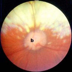
This is a view of the fundus (posterior aspect of the retina) of the eye from a dog.
a. What causes the bright (yellow/white) region to be seen in the top part of the image, above the structure labeled b? b. The structure labeled b is the optic disc. The axons of what specific cells of the retina exit the eye at the optic disc? |
a. tapetum (lucidum)
b. ganglion cells |
|
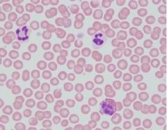
Identify the three white blood cells seen in this blood smear.
|
-2 Neutrophils
-1 Eosinophil |
|
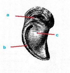
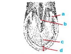
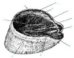
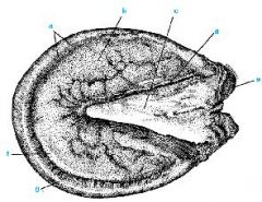
This picture shows you the ground surface (bottom surface) of one half of a cloven hoof from an ox. What are the name for the regions of the hoof indicated by the red arrows?
|
a. bulb
b. white region c. sole |
|
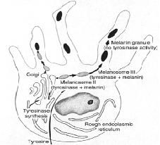
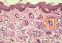
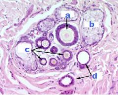
What are these extensions?
What layers are they located between? Where is theMelanin "injected" into? What is the embryonic origin of melanin? |
-Cytoplasmic extensions of Melanocytes btw stratum basale&stratum spinosum
-Stratum basale&stratum spinosum -Neural crest cells |
|
|
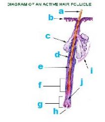
a. hair
b. surface of epidermis confluent with outernsheath c. sebaceous gland units d. outer root sheath e. inner root sheath f. Region of hair elongation and inner sheath production g. hair bulb h. dermal papilla i. arrector pili m. j. germinal matrix |
|
|
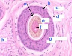
Hair & Hair Follicle - cross-section
|
|
|
a. Cuticle
b. Hair Follicle c. Dermis d. Dermal Sheath e. Cortex f. Medulla g. Outer Epithelial Sheath h. Sebaceous gland |
|
|
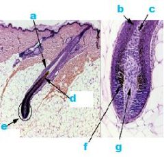
a. Root Sheaths
b. Forming Cortex&Cuticle c. Forming Medulla d. Maturing Hair Shaft e. Germinal Matrix (Epithelia portion of hair bulb) f. Melanocytes (in Germinal M) g. Dermal Papilla (CT) |
|
Compound Hair Follicle
|
a. Guard Hair
b. Sebaceous Gland c. Wool Hair d. Sweat Gland |
|
|
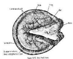
a. Bar
b. Frog c. Sole d. Wall |
|
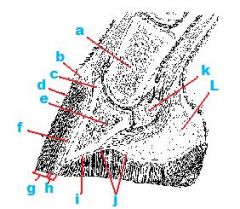
Corium=DERMIS
|
a. Middle Phalanx
b. Perioplic Corium c. Coronary Corium d. Stratum Externa e. Distal Phalanx f. Laminae g.Stratum Medium h. Stratum Internum i. Corium of Sole j. Sole Papillae k. Distal Sesamoid Bone L. Digital Cushion |
|
|
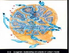
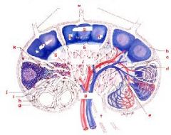
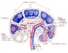
a. Germinal Center
b. Subcapsular Sinus c. Medullary Sinus d. Medullary Cord e. Trabecula f. Capsule g. Afferent Lymphatic Vessle h. Efferent Lymphatic Vessles |
|
|
|
|
|
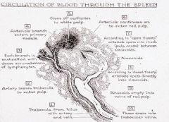
Circulation
|
|
|
|
|
|
Here we see only what part of the epidermis?
|
Keratinized
|
|
|
|
|

|
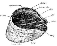
a. Coronary Region
b. Periople Region c. Laminar Region |
|
|
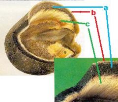
a. Stratum Medium
b. Stratum Externum c. Stratum Internum (Insensitive Laminae) |
|
|
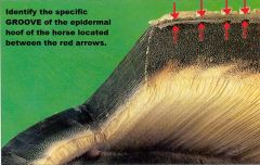
Perioplic Groove
|
|
|
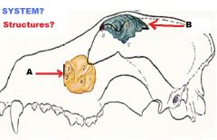
Respiratory System
Paranasal Sinuses A. Maxillary Sinus B. Frontal Sinus |
|

|
|
|
|
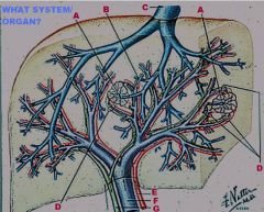
Digestive System
Liver A. Central vv. B. Interlobular v. C. Hepatic V. D. Portal Triad E. Hepatic A. F. Portal V. G. Bile Duct |
|
|
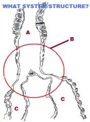
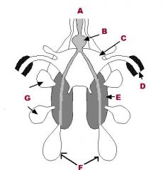
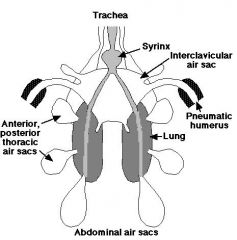
Avian Respiratory System
A. Trachea B. Syrinx C. Principle Bronchii |
|
|
|