![]()
![]()
![]()
Use LEFT and RIGHT arrow keys to navigate between flashcards;
Use UP and DOWN arrow keys to flip the card;
H to show hint;
A reads text to speech;
22 Cards in this Set
- Front
- Back
|
For Tissue preparation: |
Formaldehyde, Glutaraldehyde and Paraformaldehyde = Cross Link Protein Amine Group. |
|
|
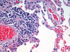
Hematoxylin is what type of dye (Acidic or Basic) and what structres does it stains, and what color? What are some examples of the structures it dyes? |
Hematoxylin is a Basic Dye with +ve charge. It stains Negative charged structures Blue. Examples: (heterochromatin,nucleoli, rough ER,ribosomes) |
|
|
Eosin is what type of dye (Acidic or Basic) and what structres does it stains, and what color? What are some examples of the structures it dyes? |
Eosin is an Acidic Dye with -ve charge. It stains Positive charged structures Red. Examples: (heterochromatin, nucleoli, rough ER, ribosomes)cytoplasmicfilaments,membranouscomponents {esp.mitochondria}, extracellular filaments. |
|
|
T/F Dueto chemical interactions, different stains have affinities to certain cellularcomponents and therefore specificstains help us identify specific components of cells. |
True |
|
|
For Staining, the cellular structures that are basophillic in nature are what (acidic or basic)? |
•Basophilic cellular components–“likesbasic stain” therefore acidic |
|
|
Hematoxylin and Eosin are most common stains. What color do they stain the cellular structures and what structures usually do they stain? |
•Hematoxylinstains nuclei blue |
|
|
You are in the laboratoy and want to stain glycoprotein at the following cellular regions: basal lamina, cell surface and mucous. What stain would you use? |
Periodic Acid Schiff (PAS) |
|
|
* Staining Technique usedto distinguish collagen (blue) in the extracellular matrix is? |
Trichrome. |
|
|
Name the cellular components where these Special Stains would stain: |
Special HISTOCHEMICAL STAINS: |
|
|
What microscopic technique is used to study living cells? Would you stain living cells? |
Phase Contrast: •Nostaining •Usedfor living cells •Cellularcomponents diffract light beam True. |
|
|
You are in a lab and want to study living tissue. You utilize a radioactive substance to incorporate into living cells to study method used to tagging antibodies or to study mitotically active cells. What technique is it called and what radioactive substance is it? |
Autoradiography |
|
|
Mitochondria is stained ___________________? |
Mitochondria is stained Eosinophilic. Eosinophilic is acidic dye and therefore the structure being stained is Basic and as a collorary Acidophilic. |
|
|
TheRNA in the ribosomes of rough ER are basophilic / acidophilic |
•TheRNA in the ribosomes of rough ER are basophilic •Thereforecytoplasm with a lot of rough ER appears blue |
|
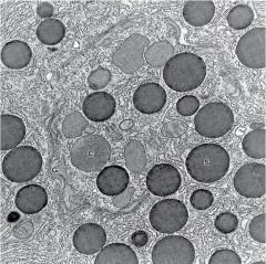
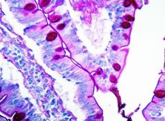
What structured do you see in this images? |
Lysosomes |
|
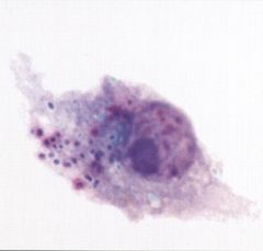
What structure looks dense and basophilic under the light microscopy? |
Lysosomes |
|
|
•Gaucher Disease•Niemann-Pick Disease Mucolipidosis II•Metachromatic Leukodystrophy•Mucopolysaccharidoses •Hurler Syndrome •Fabry disease. Are all example of what type of disease? |
Lysosomal Storage Disease. |
|
|
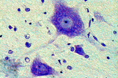
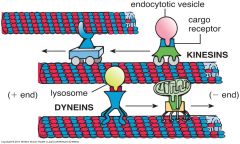
What component is it?
|
Proteosomes |
|
|
What are some examples of Ubiquitine Proteosome disorder? Microtubules are made from? |
•Astrocytomas •Parkinson’sdisease (Lewy bodies) •Alzheimer’sdisease (amyloid) Breakdown of very long chain Fatty Acids is through Beta Oxidation and occurs in Proxisomes. Intermediate Filaments Keratin |
|
|
Elongation of Microtubules occurs from -ve direction because +ve end is capped? T/F |
* F it is opposite. * Cilia purpose is Motile Cell Surface Adaptations. |
|
|
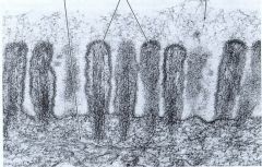
__________ assist also in the reproductive systemto move along the egg. In respiratory tract the ________ assists with moving alongthe mucus. In brain __________ helps with movement of CSF. _____ are found on the surface of the cell. |
Cilia. Cilia is made of Microtubules. Cilia is made of 9+2 configuration of Microtubules. |
|
|
________________ help aid in theabsorption of the nutrients. |
Micrivilli. |
|
|
Give the areas the following intermediate filaments would be found? |
Glial Filaments - Glial Cells Tonofilaments - Epithelium |