Reading...
![]()
Play button
![]()
Play button
![]()
Use LEFT and RIGHT arrow keys to navigate between flashcards;
Use UP and DOWN arrow keys to flip the card;
H to show hint;
A reads text to speech;
57 Cards in this Set
- Front
- Back
|
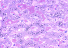
What is the primary cell type in the liver?
|
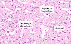
Hepatocytes (may be binucleate or mononucleate)
|
|
|
What kind of cells comprise the parenchyma of the liver?
|

Hepatocytes
|
|
|
How are hepatocytes arranged?
|

Anastomosing cords of cells with intervening sinusoids
|
|
|
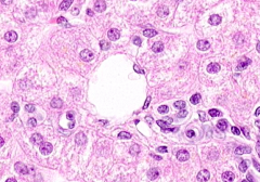
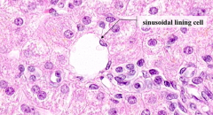
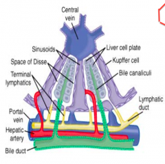
What are sinusoids?
|

Vascular spaces lined by endothelium of sinusoidal lining cells
|
|
What lines a sinusoid?
|

Sinusoidal Lining Cells (basophilic nuclei)
|
|

What should we know about how endothelial cells lining the sinusoids are held together?
|
They are NOT held together by extensive tight junctions
|
|
What do the nuclei in the lumen of the sinusoids belong to?
|

WBCs and sinusoidal macrophages (Kupffer cells)
|
|

What do Kupffer cells originate from?
|

Monocytes
|
|
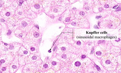
What is one function of the Kupffer cells?
|

Endocytose particles and cell debris
|
|
|
Where is the perisinusoidal space (of Disse)?
|
Between the sinusoidal lining cells and the hepatocytes
|
|
|
What is the space between the sinusoidal lining cells?
|
Perisinusoidal Space (of Disse)
- Intermediate compartment facilitating the exchange of materials between hepatocytes and bloodstream |
|
|
What is the function of the Perisinusoidal Space (of Disse)?
|
Serves as an intermediate compartment facilitating the exchange of materials between hepatocytes and the bloodstream
|
|
|
What kinds of cells reside in the Perisnusoidal Space of Disse?
|
Ito cells (hepatic stellate cells, retinoid storing cells)
|
|
|
What three structures lie within the hepatoduodenal ligament?
|
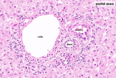
- Common Bile Duct
- Hepatic Portal Vein - Proper Hepatic Artery |
|
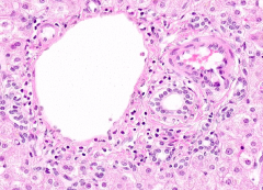
What are the structures in this slide?
|

Branches / tributaries of these structures:
- Common Bile Duct - Hepatic Portal Vein - Proper Hepatic Artery (Lymphatic vessels and nerves are present in the portal area, but are more difficult to identify) |
|
|
What type of epithelium lines the lumen of a bile ductule?
|

Simple Cuboidal
|
|
|
In the classic liver lobule concept of liver histology, blood flows from the periphery of a hexagonal area toward what structure?
|
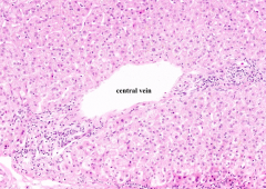
Central vein
|
|
|
Central veins join together, ultimately to form what large blood vessel?
|
Hepatic veins which ultimately lead into the IVC
|
|

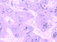
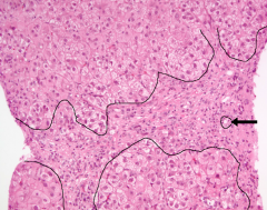
What do you notice about the tissue of the liver?
|
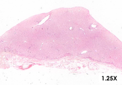
The tissue is rather homogenous in appearance
|
|

What do you notice about the 1 µm tissue section of the liver?
|
- Staining intensity is not uniform
- Demonstrates that hepatocytes in different zones of the liver can differ with respect to metabolic activity, glycogen storage, and organelle composition |
|
What does this darker zoomed in area of the 1 µm tissue section of the liver show?
|
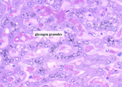
Hepatocytes containing black material - these are glycogen granules
|
|
What does this lighter zoomed in area of the 1 µm tissue section of the liver show?
|
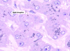
- Fewer granules are present in these cells
- Pink-staining lipid droplets make the hepatocyte cytoplasm appear foamy |
|
|
How can you see the bile canaliculi between adjacent hepatocytes?
|

- Using the light microscope and an oil immersion objective
- It is easier to see them on EM (as pictured) |
|
|
What types of intercellular junctions are found at the margins of the canalicular area of adjacent hepatocytes?
|
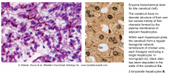
- Tight junctions
- Gap junctions |
|
|
What are the exocrine functions of the liver?
|
Bile synthesis and secretion
|
|
|
In the portal lobule concept of liver histology, bile flows from the periphery of a triangular area bounded by three central veins toward what structure?
|
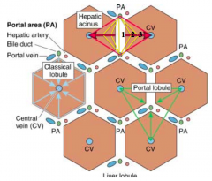
Bile ductule
|
|

What is shown in this slide of the liver stroma?
|
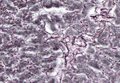
Silver-stained reticular fiber, composed of type III fibrillar collagen
|
|
|
What is cirrhosis?
|

- A progressive liver disease characterized by diffuse damage to parenchymal cells and fibrosis
- Alcohol by-products or viral-induced inflammation may damage hepatocytes and activate Kupffer cells and Ito cells |
|
|
What do Ito cells do when activated? Results?
|
- Transform from fat- and retinoid-storing cells to myofibroblasts that secrete ECM components into the peri-sinusoidal space
- Broad fibrous bands form in the tissue, sinusoids narrow, and there is increased resistance to blood flow |
|
|
What physical signs might result if the perisinusoidal space of Disse is blocked by ECM components (cirrhosis)?
|
- Jaundice - yellowing of skin, sclera and other tissues
- Ascites - fluid accumulation in the body cavity - Liver failure |
|
|
Why does jaundice occur from cirrhosis?
|
Hepatocyte damage results in bilirubin accumulation in the tissues, resulting in yellowing of the skin, sclera, and other tissues
|
|
|
Why does Ascites occur from cirrhosis?
|
Blood flow from the gut to the liver is diverted due to increased pressure
|
|
|
Why does liver failure occur from cirrhosis?
|
Damage to hepatocytes
|
|
|
What is the structure of the gallbladder? Location?
|
Small, blind-ended sac attached to the underside of the liver
|
|
|
What is the surface of the gallbladder covered in?
|
- Surface attached to the liver is covered with Adventitia
- The free surface is covered in Serosa |
|
|
What is adventitia? Which part of the gallbladder is covered in it?
|
- Outermost CT that surrounds an organ or vessel which is not covered by serosa
- Covers the surface of the gallbladder attached to the liver |
|
|
What is serosa? Which part of the gallbladder is covered in it?
|
- Outermost covering (visceral peritoneum) of an organ that lies within the body cavity
- Consists of a simple squamous epithelium (mesothelium) that secretes a watery serous fluid - Covers the free surface of the gallbladder |
|
|
What is the function of a serosa?
|
- Simple squamous epithelium (mesothelium) that secretes a watery serous fluid
- Allows the organs of the body cavity to move past each other with minimal friction |
|
|
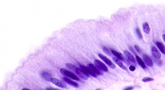
What type of epithelium lines the lumen of the gallbladder?
|
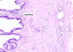
Simple columnar epithelium
|
|

The specimen was obtained from the wall of a gallbladder facing which surface?
|
Attached - gallbladder wall blends with the CT
|
|
|
What is the function of the gallbladder?
|
Store and concentrate bile
|
|
|
How does the gallbladder store and function bile? What structural features facilitate this function?
|
- Concentrates bile by absorbing water
- Apical microvilli on its epithelial cell increases the surface area for this function - Water is transferred to capillaries in the lamina propria |
|
|
When lipid enters the small intestine, the enteroendocrine cells of the duodenum secrete what hormone? Function?
|
Cholecystokinin causes contraction of smooth muscle in the gallbladder facilitating release of bile.
|
|

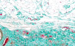
What does this slide of the gallbladder show?
|

- Olive green: smooth muscle of gallbladder wall
- Bright green: collagen fibers - Red: nuclei and erythrocytes |
|
The specimen was obtained from the wall of a gallbladder facing which surface?
|

Free - adipose tissue is present beyond the serosa
|
|
|
How is the pancreas subdivided?
|
Parenchyma is subdivided into lobules by CT trabeculae (septae)
|
|
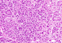
What can you see in this slide of the pancreas?
|

Pancreatic acinar cells (grape-like clusters of secretory cells)
|
|
|
What are the definitive, secretory cells of the body?
|
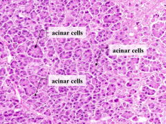
Pancreatic acinar cells
|
|

The basal cytoplasm of acinar cells is basophilic due to the presence of which organelle?
|

RER
|
|

The apical cytoplasm of acinar cells is eosinophilic due to the presence of which organelle?
|

Zymogen granules
|
|
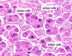
What is the principle product of the pancreatic acinar cells?
|

Digestive enzymes
|
|
|
What are the diagnostic feature of the pancreas? What are they a component of?
|
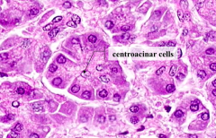
Centroacinar cells - component of the intercalated ducts
|
|
|
What forms the beginning of the duct system?
|

Centroacinar cells
|
|
|
What do cells of the intercalated ducts contribute to the exocrine secretion of the pancreas?
|
Bicarbonate and water
|
|
|
Intercalated ducts join to form what? What does that structure combine to form? Which drains into?
|
- Intercalated ducts join to form Intralobular ducts
- Inbralobar ducts join to form Interlobular ducts - Interlobular ducts drain into the Main Pancreatic Duct |
|
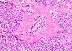
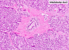
What is this a picture of?
|

Interlobular Duct
|
|
|
What type of duct is present in the parotid gland but absent from the pancreas?
|
Striated Ducts
|