Reading...
![]()
Play button
![]()
Play button
![]()
Use LEFT and RIGHT arrow keys to navigate between flashcards;
Use UP and DOWN arrow keys to flip the card;
H to show hint;
A reads text to speech;
56 Cards in this Set
- Front
- Back
|
know how ischemia leads to cell death
|
|
|
|
define
necrosis ischemia hypoxia infarct |
Necrosis
- Death of cells prior to death of animal Ischemia - Lack of blood flow - Common cause of necrosis - Worse than hypoxia Hypoxia - Absence of oxygen - Anytime you have ischemia, you have hypoxia but you can have hypoxia w/o ischemia Infarct - Focal area of ischemic necrosis |
|
|
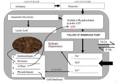
what is the pathogenesis of ischemic cell injury & death
|

|
|
|
what is reperfusion injury and how does it cause damage
|
restored blood flow to ischemic tissue increases damage
mechanisms - reperfusion provides oxygen -> abundant free radicals - neutrophil infiltration damages viable tissue |
|
|
what are the gross changes associated with cell death
|
bad smells, ewwy-gooeyness...
softening - must be differentiated from autolysis color change - lighter or darker than normal +/- peripheral rxn ulceration - only on epithelial or mucosal surfaces (full thickness necrosis) |
|
|
what are the microscopic changes associated with cell death
|
nuclear
- pyknosis (shrunken nucleus) - karyorrhexis (fragmented nucleus) - karyolysis (faded or absent nucleus) cytoplasmic - increased eosinophilia (loss of ribosomes and alteration of proteins) loss of internal structures |
|
|
cells do not show signs of death unless _____
|
the animal continues to life for several hours
|
|
|
define pyknosis, karyorrhexis, and karyolysis
|
pyknosis
- shrunken, densely basophilic nucleus karyorrhexis - fragmented nucleus karyolysis - faded or absent nucleus |
|
|
what does normal tissue stain like for H&E
|
hematoxylin
- stains DNA, RNA, and mineral blue Eosin - stains proteins pink to red |
|
|
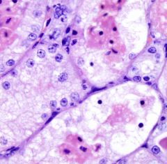
cell death
karyolysis (faded or absent nuclei) bright red area is dead - loss of ribosomes/ alteration of proteins - loss of internal structures |
|

|
cell death
bright red area is dead - loss of ribosomes/ alteration of proteins - loss of internal structures |
|

|
karyorrhexis
- fragmented nuclei |
|
|
pyknosis
- shrunken, densely basophilic nucleus |
|
|
describe the host reaction to necrosis
|
local inflammation
- stimulated by release of cell contents => hyperemia (red line surrounding necrotic tissue) => leukocyte inflitrate (white line surrounding necrotic tissue) systemic reaction - only happens if significant amt of cell contents enter circulation - systemic inflammatory response syndrome |
|
|
morphologic classification of necrosis
|
coagulative necrosis
- cells and tissues are dead but maintain their architecture - commonly caused by ischemia (infarct - focal) or toxins (diffuse) liquefactive necrosis - complete loss of architecture - necrotic neutrophils in fluid - commonly caused by bact infection and/or neutrophilic infiltrate - abscess (tissue liquefies b/c neut release juices that digest tissue) - malacia of CNS (not due to bact or neutrophils) caseous necrosis - crumbly, cheesy texture - caused by specific bacterial infections (esp Mycobacterium spp) - loss of architecture, but tissue is solid and friable - frequently present at center of granuloma - ex TB/ caseous lymphadenitis fat necrosis - very poor prognosis - enzymatic: pancreatitis - firm chalky white spots in abdominal fat, normally clear adipocytes become red (dead) then blue (mineralized), +/- inflammatory rxn - nutritional/toxic *fish eating carnivores -> yellow orange fat due to free radical damage - nutritional/toxic (ruminants) * firm opaque abdominal fat that may obstruct intestines, less inflammatory than pansteatitis fibrinoid necrosis - only in blood vessels - vessel wall is necrotic and hypereosinophilic (resembels fibrin) - due to circulating infectious agents, toxins, or hypertension gangrene (ischemic necrosis) - dry (coagulative) - wet (liquefactive) - gas (necrosis with gas bubbles) |
|
|
what morphoologic classifications of necrosis can occur in any tissue?
which can only occur in fat which can occur only in blood vessels |
any tissue
- coagulative - liquefactive - caseous fat only - fat necrosis vessels - fibrinoid necrosis |
|
|
coagulative necrosis
|
coagulative necrosis
- cells and tissues are dead but maintain their architecture - commonly caused by ischemia (infarct - focal) or toxins (diffuse) |
|
|
liquefactive necrosis
|
complete loss of architecture
- necrotic neutrophils in fluid commonly caused by bact infection and/or neutrophilic infiltrate EX: - abscess (tissue liquefies b/c neut release juices that digest tissue) - malacia of CNS (not due to bact or neutrophils) |
|
|
caseous necrosis
|
crumbly, cheesy texture
caused by specific bacterial infections (esp Mycobacterium spp) loss of architecture, but tissue is solid and friable frequently present at center of granuloma ex - TB - caseous lymphadenitis |
|
|
fat necrosis
|
very poor prognosis
enzymatic: pancreatitis - firm chalky white spots in abdominal fat - normally clear adipocytes become red (dead) then blue (mineralized), +/- inflammatory rxn nutritional/toxic - fish eating carnivores - yellow orange fat due to free radical damage - nutritional/toxic (ruminants) * firm opaque abdominal fat that may obstruct intestines, less inflammatory than pansteatitis |
|
|
pathogenesis of pancreatitis related fat necrosis
|
pancreatitis
=> release of enzymes (lipases) => digestion of abdominal fat into free fatty acids => FFA precipitate calcium to form soap (saponification) see chalky white saponification in abdominal fat |
|
|
pathogenesis of fat necrosis in carnivores that eat fish
|
diet high in rancid fish
=> excess oxidized fats => antioxidant deficiency => free radical injury of adipose tissue responsive to Vit E/ Selenium looks like pancreatitis but with yellow pigment (ceroid) |
|
|
pathogenesis of fat necrosis in ruminants
|
unknown pathogenesis
predisposing factors are grazing fescue, channel island breeds, increasing age see very firm, opaque abd fat that may obstruct intestines looks similar to pancreatic fat necrosis but less inflammatory |
|
|
fibrinoid necrosis
|
similar to Beagle-oid or Sadie-oid
only occurs in blood vessels vessel wall is necrotic and hypereosinophilic (resembles fibrin) caused by circulating infectious agents, toxins, or hypertension |
|
|
gangrene
|
ischemic necrosis
dry gangrene - coagulative necrosis - typically ischemic - ex: ergotism/ frostbite wet gangrene - liquefactive necrosis - typically involves bacteria - ex: dog bite gas gangrene - necrosis with gas bubbles (formed by certain bacteria) - ex: blackleg due to Clostridium chauvoei |
|

|
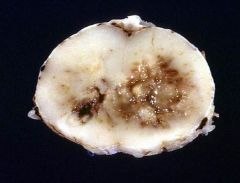
coagulative necrosis (lower left)
tissue is dead but still see tubules and glomeruli notice line of inflammation around area of necrosis |
|

|
coagulative necrosis
notice the line of inflammation around area of necrosis |
|

|
necrosis
|
|

|
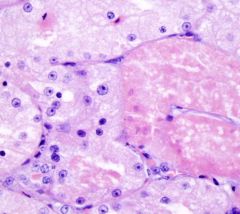
coagulative necrosis
live hepatocytes on either side and dead hepatocytes down the middle |
|

|
coagulative necrosis
|
|
|
liquefactive necrosis (malasia)
In CNS, not always an abscess when see tissue liquefaction - Call malasia - See liquefaction with even ischemic damage in CNS - Not always caused by bacteria |
|
|
what type of cell death is affecting my brain cells right now
|
liquefactive necrosis (aka malasia)
|
|

|
liquefactive necrosis
|
|

|
liquefactive necrosis
notice loss of architecture |
|

|
liquefactive necrosis
notice loss of architecture |
|

|
liquefactive necrosis
notice loss of architecture |
|

|
liquefactive necrosis
notice loss of architecture |
|

|
caseous lymphadenitis
Corynebacterium pseudotuberculosis |
|

|
caseous lymphadenitis
Corynebacterium pseudotuberculosis |
|

|
caseous lymphadenitis
Corynebacterium pseudotuberculosis |
|

|
caseous necrosis
Mycobacterium tuberculosis |
|

|
caseous necrosis
|
|

|
caseous necrosis
|
|

|
enzymatic fat necrosis
pancreatitis => release of enzymes (lipases) => digestion of abdominal fat into FFA => FFA precipitate Ca2+ to form soap |
|

|
enzymatic fat necrosis
pancreatitis => release of enzymes (lipases) => digestion of abdominal fat into FFA => FFA precipitate Ca2+ to form soap |
|

|
enzymatic fat necrosis
pancreatitis => release of enzymes (lipases) => digestion of abdominal fat into FFA => FFA precipitate Ca2+ to form soap |
|

|
nutritional fat necrosis
diet high in rancit fish => excess oxidized fats => antioxidant deficiency => free radical injury of adipose tissue (ceroid pigment in necrotic fat) usually responsive to Vit E/ Selenium |
|

|
nutritional fat necrosis
diet high in rancit fish => excess oxidized fats => antioxidant deficiency => free radical injury of adipose tissue (ceroid pigment in necrotic fat) usually responsive to Vit E/ Selenium |
|

|
nutritional fat necrosis ruminant
unknown pathogenesis but assoc with grazing fescue, genetics, increasing age |
|

|
nutritional fat necrosis ruminant
unknown pathogenesis but assoc with grazing fescue, genetics, increasing age |
|

|
dry gangrene - ergotism
coagulative necrosis (usually ischemic) |
|

|
dry gangrene - ergotism
coagulative necrosis (usually ischemic) |
|

|
dry gangrene
coagulative necrosis (usually ischemic) |
|

|
wet gangrene - liquefactive necrosis
necrosis with inflammation |
|

|
gas gangrene
necrosis with gas bubbles blackleg (clostridium chauvoei) happens so fast animal dies before cell changes |
|
|
morphologic classifications of necrosis & their causes (quick and dirty)
|
Coagulative Necrosis – ischemia/ toxin
Liquefactive Necrosis - bact Caseous Necrosis – some bact Fat Necrosis - pancreatitis Fibrinoid Necrosis – toxin infection Gangrene - professor |