![]()
![]()
![]()
Use LEFT and RIGHT arrow keys to navigate between flashcards;
Use UP and DOWN arrow keys to flip the card;
H to show hint;
A reads text to speech;
360 Cards in this Set
- Front
- Back
|
Anatomy |
(Greek "to cut apart")- the study of the form, or structure of body parts and of how these parts relate to one another. Static image. |
|
|
Physiology |
the study of the functioning of the body's structural machinery - how the parts of the body work and carry out their life-sustaining activities. Dynamic processes. |
|
|
Gross anatomy |
study of large body structures visible to the naked eye. |
|
|
Regional anatomy |
all structures in one part of the body are studied at the same time. |
|
|
Systemic anatomy |
various systems of the body are studied. |
|
|
Microscopic anatomy |
examination of body tissues using a microscope |
|
|
Cytology |
study of the cells of the body |
|
|
Histology |
study of the tissues of the body |
|
|
Embryology |
developmental changes occurring before birth |
|
|
Pathology |
disease related changes |
|
|
Molecular biology |
subcellular level |
|
|
Complimentarity of structure and function |
function always reflects structure. what a structure can do depends on its specific form. |
|
|
Hierarchy of Structural Organization |
the human body incorporates many levels of structural complexity |
|
|
Atoms |
building blocks of matter |
|
|
Molecules |
water, sugar, proteins -- groups of atoms |
|
|
Organelles |
basic components of microscopic cells |
|
|
Cells |
living structural and functional units of an organism |
|
|
Tissues |
groups of similar cells having common structure and function. Four basic types. |
|
|
Organ |
complex physiological processes become possible. Discrete structure composed of at least two tissue types; four tissue types more common. |
|
|
Organ System |
organs that cooperate and work closely together to accomplish a common purpose. |
|
|
Organism |
sum total of all levels of complexity working continuously and in unison |
|
|
Homeostasis |
ability to maintain relatively stable internal conditions despite a changing external environment. Dynamic state of equilibrium, or balance. Body is balanced when its cellular needs are adequately met and functional activities are occurring smoothly. Virtually every organ system plays a role in maintaining the internal environment. |
|
|
Maintenance of boundaries |
keeps the internal environment separate and distinct from the external environment. Cell members contain its contents while admitting needed substances and restricting the entry of unnecessary or harmful substances. |
|
|
Movement |
all activities promoted by the muscular system such as walking, running, movement of blood, food, urine, through the internal organs, etc. |
|
|
Responsiveness |
ability to sense change and respond to it |
|
|
Digestion |
breakdown of ingested food into usable molecules |
|
|
Metabolism |
all chemical reactions occurring w/in the cells. Depends on other systems and is regulated by the endocrine system. |
|
|
Excretion |
removal of unusable waste products |
|
|
Reproduction |
formation of offspring |
|
|
Growth |
increase in size |
|
|
Nutrients |
chemical susbstances used for energy and cell building and maintenance carbohydrates (CHO)-major energy fuel; proteins/fats - building cell structures; minerals/vitamins - assist in chemical reactions |
|
|
Oxygen |
absolutely essential. Chemical reactions that release energy from food are oxidative reactions and req. oxygen. 20% of the air we breathe. |
|
|
Water |
60-80% of body weight. Provides liquid environment for chemical reactions and fluid base for body secretions/excretions. |
|
|
Body Temperature |
must be maintained around 37 C (98.6 F). Decreased temp: physiological reactions slowed. Increased temp: chemical reactions proceed too rapidly, body proteins denature. |
|
|
Atmospheric Pressure |
required for exchange of gases in the lungs |
|
|
Anatomy subdivisions |
1. Gross anatomy a. Regional anatomy b. Systemic anatomy 2. Microscopic anatomy a. Cytology b. Histology 3. Other subdivisions a. Embryology b. Pathology c. Molecular biology |
|
|
Levels of Hierarchy |
1. Atoms 2. Molecules 3. Organelles 4. Cells 5. Tissues 6. Organ 7. Organ System 8. Organism |
|
|
Maintenance of Life |
1. Maintenance of boundaries 2. Movement 3. Responsiveness 4. Digestion 5. Metabolism 6. Excretion 7. Reproduction 8. Growth |
|
|
Survival Needs |
1. Nutrients 2. Oxygen 3. Water 4. Body Temperature 5. Atmospheric Pressure |
|
|
Nervous and endocrine |
Communications w/in the body are essential for homesostasis. Accomplished chiefly by the ____and ____ systems. |
|
|
What are the three components that all homestatic control mechanisms have? |
1. Control Center 2. Receptor 3. Effector |
|
|
Control Center |
determines the set point, analyzes input and determines the appropriate response. |
|
|
Receptor |
monitors the environment, sends information or input to the control center (afferent pathway) |
|
|
Effector |
provides the means by which the control center can cause a response to a stimulus (efferent pathway). The result of the response then "feeds back" to influence the stimulus, either depressing it (negative feedback) or enhancing it (positive feedback). Most homeostatic control mechanisms are negative feedback mechanisms. The output of the system feeds back and decreases the input into the system. The net effect is to decrease the original stimulus or reduce it effects. |
|
|
All negative feedback mechanisms have the same goal -- |
prevention of sudden severe changes w/in the body |
|
|
positive feedback |
In ____ ____ mechanisms the response enhances the original stimulus and the output is accelerated. The change that occurs proceeds in the same direction as the initial disturbance, causing the further deviation from the original set point. |
|
|
episodic/infrequent |
Positive feedback mechanisms usually control ____/____ events that do not require continuous adjustments. Rarely used to promote moment to moment well being.examples: coagulation labor contractions (oxytocin) |
|
|
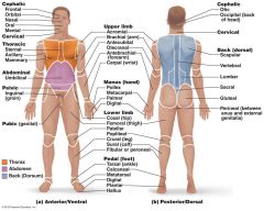
Superior Inferior |
Placement of a body structure along the long axis of the body.
|
|
|
Anterior Posterior |
Structures or surfaces are those that are most forward (face, chest, abdomen). Structures or surfaces are those toward the backside of the body. |
|
|
Medial Lateral |
Toward the midline Away from the midline |
|
|
Cephalad Caudal |
Toward the head Toward the tail Used interchangeably w/ superior & inferior
|
|
|
Dorsal Ventral |
Backside Belly side Used interchangeably w/ anterior & posterior |
|
|
Proximal Distal |
Nearer the truck or attachment end Farther from the trunk or point of attachment Used primarily to locate various areas of the body limbs |
|
|
Superficial Deep |
Toward or at the body surface Away from the body surface or more internal Used to locate body organs in terms of their relative closeness to the body surface |
|
|
anatomical position |
-body is erect w/ feet together -palms facing forward w/ the thumbs pointing away from the body |
|
|
direction |
"right & left" refer to those sides of the person or cadaver being viewed |
|
|
axial |
-makes up the main axis of the body. -consists of the head, neck, & trunk |
|
|
appendicular |
consists of the appendages or limbs |
|
|
sagittal |
runs longitudinally divides the body or organ into right & left portions |
|
|
midsagittal |
(median plane) - exactly midline & the parts are symmetrical or equal |
|
|
parasagittal |
all other sagittal planes, not equal
|
|
|
transverse
|
runs horizontally across & at a right angle to the long axis of the body or organ & divides it into superior & inferior parts |
|
|
frontal |
runs longitudinally, but the body or organs are divided into anterior & posterior |
|
|
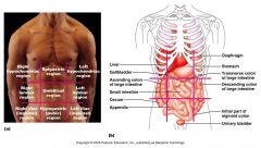
Dorsal Body Cavity |
-Located nearer to the posterior surface of the body -subdivided into cranial & vertebral or spinal -continuous w/ one another -house vital & very fragile organs - brain & spinal cord |
|
|
Ventral Body Cavity |
-More anterior & larger -major subdivisions: thoracic & abdominopelvic |
|
|
lateral pleural medial mediastinum |
The thoracic is surrounded by the ribs and muscles of the chest and is subdivided: ____ ____ cavities each containing a lung and ____ ____ which contains the heart and remaining thoracic organs (esophagus, trachea, etc) |
|
|
Abdominopelvic cavity |
Divided into the abdominal cavity which contains the stomach, intestines, spleen, liver, and other organs and the pelvic cavity which contains the bladder, some reproductive organs, and the rectum. |
|
|
Serosa or serous membranes |
the walls of the ventral body cavity and the outer surfaces of the organs are covered w/ a thin, double layered membrane |
|
|
Parietal serosa |
part of the membrane lining the cavity walls |
|
|
visceral serosa |
folds on itself to form the ____ ____ which covers the organs in the cavity. |
|
|
Parietal |
"parie" means wall |
|
|
Visceral |
"viscus" means an organ in a body cavity |
|
|
serous fluid |
The serous layers are separated by a thin lubricating fluid ____ ____. It allows the organs to slide easily across the cavity walls and one another w/o friction. |
|
|
parietal pericardium |
lines pericardial (heart) cavity |
|
|
visceral pericardium |
covers the heart |
|
|
parietal pleura |
lines thoracic wall in the pleural cavity |
|
|
visceral pleura |
covers the lungs |
|
|
pleurisy |
Inflammation of serous membranes, accompanied by a deficit of lubricating fluid results in excruciating pain as organs stick together. |
|
|
Regional Terms |
|
|
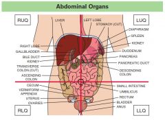
Abdominopelvic Regions |
|
|
Quadrants |
|
|
matter |
anything that occupies space and has mass |
|
|
states of matter |
1. solid 2. liquid 3. volume |
|
|
solid |
has definite shape and volume |
|
|
liquid |
has no definite shape and definite volume |
|
|
gas |
has no definite shape and no definite volume |
|
|
energy |
the capacity to do work or put matter into motion |
|
|
kinetic energy |
energy in action |
|
|
potential energy |
stored energy; has the capability to do work but is not now doing so |
|
|
forms of energy |
1. chemical 2. electrical 3. mechanical 4. radiant |
|
|
chemical |
stored in the bonds of chemical substances (food molecules, ATP) |
|
|
electrical |
movement of charged particles |
|
|
mechanical |
energy directly involved in moving matter |
|
|
radiant |
energy that travels in waves (electromagnetic spectrum) |
|
|
elements |
All matter is composed of ____, unique substances that cannot be broken down by ordinary chemical methods. The elements are listed in the periodic table. Each element is known by an atomic symbol. |
|
|
atoms |
Each element is composed of particles called ____ that display the characteristic properties of that element. |
|
|
Physical properties |
____ are things that can be detected w/ our senses or measured (color, texture, boiling point). |
|
|
Chemical properties |
____ describe how atoms react w/ other atoms. |
|
|
Protons, neutrons, and electrons |
An atom has three subatomic particles: |
|
|
Protons and neutrons |
The central nucleus of an atom is composed of____ and ____. |
|
|
Protons |
___are positively charged. |
|
|
Neutrons |
____ are neutral (no charge). |
|
|
negatively |
The nucleus is surrounded by ____ charged electrons orbiting in an electron cloud. |
|
|
Protons and electrons |
An atom has an overall neutral charge because there are equal numbers of ____ and ____. |
|
|
Atomic numbers |
Elements can be identified by their ____ ____. The atomic number is the number of protons in an element. It is written as a subscript to the left of the atomic symbol. |
|
|
carbon (C), nitrogen (N), hydrogen (H), and oxygen (O) |
The elements ____, ____, ____, and ____ make up around 96% of body weight. |
|
|
calcium (Ca), phosphorus (P), potassium (K), sulfur (S), sodium (Na), chlorine (Cl), magnesium (Mg), iodine (I), iron (Fe) |
Some elements necessary for the body are referred to as minerals. The most important minerals are ____, ____, ____, ____, ____, ____, ____, ____, and ____. |
|
|
chromium (Cr), cobalt (Co), copper (Cu), fluorine (F), manganese (Mn), molybdenum (Mo), selenium (Se), silicon (Si), tin (Sn), vanadium (V), and zinc (Zn) |
Several other elements are needed in small amounts, and are known as trace minerals: ____, ____, ____, ____, ____, ____, ____, ____, ____, _____, ____, and ____. |
|
|
molecule molecule of that element |
combination of two or more atoms held together by chemical bonds. when the two atoms are identical the resulting substance is called a _____ (H^2 is a molecule of hydrogen gas). |
|
|
compound |
when two or more different kinds of atoms bind they form molecules of a compound; chemically pure; all molecules identical (H^2O is a compund - it's a molecule of water) |
|
|
mixtures |
substances composed of two or more substances physically intermixed |
|
|
solutions |
homogeneous mixtures of components that may be gases, liquids, or solids. [air, seawater] |
|
|
solvent |
substance in a solution present in the greatest amount (dissolving medium) [water] |
|
|
solute |
substance in a solution present in smaller amounts (thing that is dissolved) [salt] |
|
|
energy levels |
The electrons in an atom are arranged in _____ ____ (or electron shells) that increase in capacity the farther they get from the nucleus. |
|
|
valence |
The outer most energy level called the ____ is the most important in determining how atoms react w/ other atoms. |
|
|
8 |
Atoms are most stable when the outermost shell has ____ electrons in it and will react w/ other atoms to try to reach this level of stability. **The exception to this is hydrogen and helium, which only have one energy level that is filled to capacity and is stable w/ only 2 electrons. |
|
|
ion |
Atoms are neutral, but they can gain or lose electrons. When this happens an atom becomes an ____, which will have a charge - either positive or negative depending upon whether electrons were lost or gained. |
|
|
ionic |
An ____ bond is a chemical bond between atoms formed by the transfer of one or more electrons from one atom to another. An example is sodium chloride (NaCl), table salt. |
|
|
True |
(T/F) Atoms can also achieve stability by sharing electrons. This means that electrons are not transferred from one atom to the other, but spend time orbiting both nuclei. |
|
|
covalent |
A ____ bond is a chemical bond formed between two atoms that are sharing a pair of electrons, each atom contributing one electron to the shared pair. If the atoms are shared equally then the resulting molecule is nonpolar (hydrogen gas). If one atom has a greater attraction for electrons (a property known as electronegativity), the electrons may be shared unequally, resulting in a polar molecule (water). |
|
|
hydrogen |
____ bonds form when a hydrogen atom, already covalently linked w/ one electronegative atom (like oxygen or nitrogen), is attracted by another electronegative atom and forms a bridge between them. These bonds are too weak to bind atoms together to form molecules, but they are important in attracting water molecules to each other and as intramolecular bonds in large biological molecules (proteins, DNA) where the hydrogen bonds stabilize the overall structure. |
|
|
anabolic |
synthesis reactions are constructive |
|
|
endergonic |
absorb/use energy |
|
|
catabolic |
decomposition reactions break things down |
|
|
exergonic |
release energy |
|
|
high heat capacity |
absorbs and releases large amounts of heat before changing appreciably in temperature itself. This prevents sudden, severe changes in body temperature from vigorous activity. |
|
|
high heat of vaporization |
large amounts of heat are needed to break water's hydrogen bonds and allow it to change from a liquid to a gas (evaporation). This makes perspiration and effective cooling mechanism. |
|
|
polar solvent properties |
the universal solvent. The chemical reactions of the body depend on the reactants being dissolved in water. Since water molecules are polar, ionic compounds and other small reactive molecules dissociate in water, causing the substance to dissolve. Water also forms hydration layers around large charged molecules, such as proteins. |
|
|
reactivity |
decomposition reactions by hydrolysis; large molecule assembly by dehydration syntesis |
|
|
acids |
____ are substances that release hydrogen ions(H+) in detectable amounts. |
|
|
hydrochloric acid (HCI) |
_____ _____ is found in the stomach. |
|
|
bases |
____ take up hydrogen ions in detectable amounts and are characterized by the [presence of hydroxyl ions (OH-) |
|
|
bicarbonate ion |
____ ____ is an important base in the body. |
|
|
hydrogen; hydroxyl |
The more ____ ions present, the more acidic a solution is; the more ____ ions present, the more basic a solution is. |
|
|
pH |
The relative concentration of hydrogen ions in body fluid is measured in concentration units called ____. |
|
|
0-14 |
The pH scale runs from ____ and is logarithmic, i/e. each unit represents a ten-fold change. |
|
|
neutral |
At a pH of 7, a solution is ____ w/ equal amounts of hydrogen and hydroxyl ions. |
|
|
acidic; basic |
Below 7 pH is ____; above 7 pH is ____. |
|
|
dissociates |
The farther away from 7 a substance is in pH, the more it ____ in solution and the stronger a substance it is. |
|
|
7.35 - 7.45 |
Blood must maintain a pH in the range of ____. |
|
|
buffers |
____ are present in the blood to prevent big shifts in pH. A buffer does this by releasing hydrogen ions when the pH rises and binding hydrogen ions when the pH drops. |
|
|
carbohydrates |
____ include sugars and starches. They are composed of carbon, hydrogen, and oxygen. |
|
|
monosaccharides |
____ are simple sugars, example glucose (the major fuel source for the body). |
|
|
disaccharides |
____ are composed of two sugar units, for example table sugar or sucrose (glucose + fructose). |
|
|
polysaccharides |
____ are composed of many sugar units, for example starch (the storage carbohydrate of plant tissues) and glycogen (the storage carbohydrate of animal tissue). The primary function of carbohydrates in the body is to serve as a source of cellular fuel. |
|
|
lipids |
____ are insoluble in water, but dissolve readily in other lipids and organic solvents like alcohol. Includes neutral fats, phospholipids, and steroids. Protect and insulate body organs. |
|
|
tryglycerides |
____ are neutral fats, composed of a backbone of glycerol w/ three attached fatty acid chains. |
|
|
Neutral fats |
____ ____ provide the body's most efficient and compact form for storing usable energy fuel. |
|
|
phospholipids |
____are similar to triglycerides except one fatty acid chain is replaced by a phosphate group. Compose a large part of cellular membranes. |
|
|
steroids |
flat molecules made of interlocking hydrocarbon rings. An example of cholesterol which is a building block of Vitamin D, steroid hormones, and bile salts. |
|
|
proteins |
____ are composed of building blocks called amino acids joined by peptide bonds. Serve as the major structural material in the body and also as enzymes. |
|
|
20 |
There are ____ different amino acids in biological proteins. |
|
|
peptides |
Small chains of less than 50 amino acids are usually referred to as ____. Proteins are chains of more than 50 amino acids and often will have 100-10,000 amino acids. |
|
|
intramolecular hydrogen |
Proteins may fold up into distinct shapes via ____ ____ bonding. The protein usually must maintain its proper three- dimensional shape in order to function. |
|
|
Fibrous |
____ proteins such as collagen make up a large part of connective tissue. |
|
|
globular |
____ proteins include hormones and enzymes. |
|
|
enzymes |
____ are biological catalysts - substances that regulate and accelerate the rate of biochemical reactions but are not used up or changed in those reactions. help the biochemical reactions of our body to proceed at a rate that supports life. will have an active site, a region that fits and interacts chemically with other molecules of complementary shape and charge. |
|
|
nucleic acids |
____ ____ such as DNA and RNA are composed of chains of nucleotides made up of a phosphate, sugar, and nitrogen-containing base. |
|
|
DNA (deoxyribonucleic acid) |
____ is the genetic material. A chromosome consists of a molecule of DNA. Certain segments of DNA are known as genes. |
|
|
gene |
A ____ provides the instructions for how to make a single protein chain. |
|
|
RNA (ribonucleic acid) |
____ assists in gene expression by carrying the code for a protein to the place in the cell that proteins are manufactured. |
|
|
ATP (adenosine triphsophate) |
___ is a nucleotide which serves as the energy carrier molecule in the cell. |
|
|
RobertHooke
|
---- was the first to observe plant cells with a crude microscope in the late 1600's.
|
|
|
Schleiden and Schwann
|
----proposed that all living things were composed of cells. |
|
|
Virchow
|
---- suggested that cells arise only from other cells.
|
|
|
cell theory
|
1. A cell is the basic structural and functional unit of living organisms.
2. The activity of an organism is dependent on both the individual and collective activities of its cells. 3. Principle of Complementarity - biochemical activities of cells are determined and made possible by thespecific subcellular structures of cells. 4. Continuity of life has a cellular basis. |
|
|
plasma membrane, cytoplasm, and nucleus
|
All cells have three major parts:
|
|
|
plasma membrane |
The ---- ---- defines the boundary of the cell and acts as a fragile barrier. The membrane is thin, elastic, and semi-permeable. It is composed chiefly of a double layer, or bilayer of phospholipid molecules with protein molecules dispersed in it. The chemical nature of the phospholipid molecules causes the membrane to self-assemble and to re-seal quickly when torn. |
|
|
fluid mosaic model
|
The ---- ---- ---- of membrane structure depicts theplasma membrane as a phospholipid bilayer (2 layers of phospholipid molecules), with the polar head exposedto water inside and outside the cell and the nonpolar tails facing each other and buried in the internal portion ofthe membrane with proteins embedded among the phospholipid molecules. The membrane is a dynamic fluidstructure of about the consistency of olive oil. The lipid molecules are free to move laterally. Some of proteinsfloat freely, while others are more restricted.
|
|
|
to serve as a boundary for the cell
|
The function of the phospholipid part is ----. It is a selective barrier, permittingsome things to go through and preventing others. Substances that can pass through the membrane are soluble inlipids, very small, or transported by a protein.
|
|
|
Globular
|
---- proteins are embedded in the bilayer. The amount of protein varies with membrane function andaverages 50% of the mass of the membrane. The proteins in a cell membrane vary depending on the type of cell.The specific collection of proteins forms a picture of what the cell can do (like a mosaic).
|
|
|
protein functions |
transport (ion channel or carrier), receptors (respond to chemical signals in the environment), attachment to cytoskeleton (maintain shape, movement), enzymes, intercellular joining, and cell-cell recognition and interaction. |
|
|
glycolipids and glycoproteins
|
Some of the lipids and proteins of the membrane have sugar molecules attached to the side facing outside of thecell, making them ---- and ---- .
|
|
|
Microvilli |
minute, finger-like projections which increase the surface area of the membrane. Found on the surface of absorptive cells such as kidney tubules and intestinal cells.
|
|
|
Membrane Junctions
|
---- two types of junctions are formed between cells that make up tissues.
(1.) junctions thatfasten cells together (2.) junctions that permit transfer of ions and other molecules between cells. |
|
|
tight junctions
|
In ---- ---- protein molecules in adjacent membranes fuse together preventing free passage of moleculesthrough intercellular space between cells. In the digestive tract they form a barrier to enzymes andmicroorganisms preventing them from entering the blood. (impermeable junction)
|
|
|
Desmosomes
|
---- are spotlike patches that act as mechanical couplings between adjoining cells. The membranes donot actually touch, but are held together by fine glycoprotein filaments stretched between button-like thickeningof the inner membranes. They are abundant in tissues subject to mechanical stress such as skin, heart, andmuscles. (anchoring junction)
|
|
|
Gap Junctions
|
---- ----function to allow direct passage of chemical substances between adjacent cells. The cells areconnected by hollow cylinders composed of transmembrane proteins. This allows ions, sugars, and other smallmolecules to pass through. In adults they are found in electrically excitable tissues such as in the heart andmuscles. (communicating junction)
|
|
|
interstitial fluid
|
The plasma membrane is a selectively, or differentially, permeable barrier that allows some substances to passwhile excluding others. Our cells are bathed in an extracellular fluid called ---- ----, derived from bloodand containing thousands of ingredients such as amino acids, sugars, fatty acids, hormones, etc. Cells must beable to get what they need by transporting substances across the cell membrane.
|
|
|
Passive Processes
|
---- ---- substances penetrate the membrane without any energy input from the cell because it justuses the kinetic energy of the particles themselves. It depends on the physical process of diffusion. Moleculesdiffuse down the concentration gradient from an area of high concentration to an area of low concentration untilequilibrium is reached. At this point, while the molecules are still moving, there is no net change inconcentration from one area to another. The greater the differences in original concentrations (steeperconcentration gradient), the faster the rate of diffusion.
|
|
|
simple diffusion
|
oxygen and carbon dioxide can pass through the membrane by ---- ---- because they are fat-soluble.
|
|
|
Facilitated diffusion
|
some molecules and ions can still move by diffusion but need helperprotein(s) to traverse the phospholipid barrier. ---- ---- can happen via a carrier protein or an ionchannel.
|
|
|
Osmosis
|
diffusion of water through a specific channel protein (aquaporin) from an area of highconcentration of water to an area of lower concentration of water. The relative concentration of water isdetermined by the presence of solutes. A greater amount of solute means there is a lesser concentration ofwater; likewise, a lesser amount of solute means there is a greater concentration of water. ---- causes waterto move toward the side of the membrane with the greater amount of solute. Solutions can be describedaccording to their amounts of solute relative to a cell placed within them.
|
|
|
isotonic
|
An ---- solution has the samesolute concentration as a cell.
Cell will stay the same. |
|
|
hypertonic
|
A ---- solution has a greater amount of solute than a cell.
Cell will shrink as water leaves the cell by osmosis. |
|
|
hypotonic
|
A ---- solution has a lesser amount of solute than a cell.
Cell will swell (and may burst) as water enters the cell by osmosis. |
|
|
Active Processes
|
---- -----the cell provides metabolic energy (ATP) to drive the movement of substances across themembrane. There are two major mechanisms: active transport and vesicular (bulk) transport.
|
|
|
Active transport
|
---- ----(solute pumping) requires a carrier protein and ATP energy to move moleculesagainst the concentration gradient from low concentration to high concentration. Many active transport systemsare coupled systems - they move more than one substance at a time. If the substances move the same direction,it is a symporter; if substances move in the opposite direction, it is an antiporter. An example of an antiporter isthe sodium-potassium pump. Using 1 ATP this pump ejects 3 sodium ions from the cell and imports 2potassium ions. Both sodium and potassium are being moved against their concentration gradients. This allowsthe cell to support differential ionic concentrations for maintaining adequate fluid volume and for muscle andnerve cells to function properly.
|
|
|
Vesicular (bulk) transport
|
---- -----moves large particles and macromolecules through the plasmamembrane. It requires ATP and uses phospholipid sacs called vesicles to transport substances. There are twokinds of bulk transport: exocytosis and endocytosis
|
|
|
Exocytosis
|
---- moves substances out of the cell Substances to be released are enclosed within a vesicle whichmigrates to the plasma membrane, fuses, and then ruptures releasing the contents of the sac. SNARE proteins onboth the vesicle and the plasma membrane bind together to promote docking and begin the fusion process. |
|
|
Endocytosis
|
---- is a means for allowing large particles or macromolecules to enter the cell. Substances to be takenin are progressively enclosed by a portion of the cell membrane. The formation of the enclosure is caused by theprotein clathrin found on the cytoplasmic side of the cell membrane. Once formed, the sac pinches off andmoves into the cytoplasm where the contents are digested or utilized.
|
|
|
phagocytosis
|
when the substance to be ingested is a large particle
|
|
|
pinocytosis
|
when the substances to beingested are dissolved in water |
|
|
receptor-mediated endocytosis
|
when extracellular substances bind toreceptors in the plasma membrane and trigger endocytosis
|
|
|
Cytoplasm
|
---- Cellular material inside the plasma membrane and outside the nucleus. It is the site where mostcellular activities occur. It is a major functional area. The cytoplasm consists of two major elements: cytosoland organelles.
|
|
|
Cytosol
|
---- is a viscous, semitransparent fluid composed mostly of water with some soluble proteins, salts, sugarsand other solutes. Organelles are the metabolic machinery of the cell. Each carries out a specific function for thecell as a whole. Some synthesize proteins, others package those proteins, etc.
|
|
|
Cytoplasmic Organelles
|
---- ----Specialized cellular compartments, each performing its own job to maintain the lifeof the cell. Most organelles are bounded by selectively permeable membranes which enable them to maintain aninternal environment differing from the surrounding cytosol. This compartmentalization is crucial to thefunctioning of the cell; without it enzymes would be randomly mixed and biochemical activity would bechaotic.
|
|
|
Mitochondria
|
---- Sausage-shaped structure. Powerhouse - provide most of the ATP supply. Quantity per cellreflects the energy requirements of the cell. Each mitochondrion is surrounded by two lipid bilayer membranes.
Inner membrane contains more protein, has shelf-like inward folds called cristae which protrude into the gellikematrix. Enzymes in the matrix together with those on the cristae membranes cooperate to breakdownglucose and other nutrients to water and carbon dioxide. Some of the energy released is captured in the form ofATP. |
|
|
Ribosomes
|
---- Small granules composed of proteins and a type of RNA. Each ribosome has two globular submitsthat fit together with the small subunit sitting on top of the larger subunit. It is the site of protein synthesis,where amino acids linked together to form a polypeptide chain. Some ribosomes float freely in the cytoplasm;others are attached to membranes, forming a complex called the rough endoplasmic reticulum. Free ribosomesmake proteins for use in the cytosol. Membrane-bound ribosomes make proteins for export or use in themembrane.
|
|
|
Endoplasmic Reticulum (E.R)
|
---- ---- Extensive system of interconnected parallel membranes that coils and twiststhrough the cytoplasm, enclosing fluid-filled cavities or cisternae. Continuous with the nuclear membrane andaccount for about half the cell's membranes. Two distinct varieties of E.R.; rough and smooth. External surfaceof rough E.R. is studded with ribosomes.
As amino acids are assembled on the ribosome, they thread their way into the cisternae. This prevents theproteins for export from mixing with those that are being retained in the cell. Proteins for export becomeenclosed in membranous sacs pinched off from the E.R. (transport vesicles).Transport vesicles migrate to theGolgi apparatus for further processing of the proteins. Rough E.R. is abundant in cells specialized to secreteproteins such as gland cells, liver cells, plasma cells, etc). Smooth E.R. is a continuation of the rough E.R. and consists of tubules arranged in branching network. SmoothE.R. plays no role in protein synthesis. Smooth E.R. is involved in lipid metabolism, synthesis of cholesteroland the lipid portion of lipoproteins, synthesis of steroid based hormones. |
|
|
Golgi Apparatus
|
Consists of 4-8 flattened membranous sacs stacked one upon the other. Located near thenucleus. Principal traffic director for cellular proteins. Major function is to modify, concentrate, and packageproteins for export. Modify usually means adding sugar groups to make glycoproteins. Protein-containingvesicles pinch off from the Golgi and migrate to the plasma membrane and discharge their contents from thecell by exocytosis. Secretory cells have prominent Golgi apparatus.
|
|
|
Lysosomes |
Membranous sacs containing hydrolytic enzymes – usually acid hydrolases. Provide sites where digestion can proceed safely within the cell. Large and abundant in phagocytes. Function to digest ingested particles, worn-out nonfunctional organelles, non-useful tissues. The breakdown of bone to release calcium ions also reflects lysosomal activity. |
|
|
Peroxisomes
|
Membranous sac containing oxidases and catalases. These enzymes detoxify the free radicalsproduced by normal metabolism which could damage biological molecules. Peroxisomes are especiallyabundant in the liver and kidney. Oxidases change free radicals to hydrogen peroxide, and then catalase changeshydrogen peroxide to water.
|
|
|
Cvtoskeletal Elements
|
An elaborate network of protein structures called microfilaments, microtubules, andintermediate filaments are located throughout the cytoplasm. Cytoskeleton acts as the cells "bones and muscles"helping to support intercellular structures and generate various cell movements.
|
|
|
Microfilaments
|
Thin strands of the contractile protein actin. Nearly all cells have a dense cross-linkednetwork of microfilaments attached to the cytoplasmic side of the plasma membrane. Braces and strengthens thecell surface. Most involved in cell motility or cell shape change. Form the core of microvilli. Provide foramoeboid movement and vesicle formation during endocytosis. Together with myosin, fonn the cleavage ringthat pinches the cell in two during cell division.
|
|
|
Intermediate Filaments
|
Tough, insoluble protein fibers with high tensile strength. Act as internal guy wiresto resist pulling forces on the cell.
|
|
|
Microtubules
|
Long, flexible hollow tubules. Have a variety of cellular roles; most importantly, they appear tobe the overall organizers of the cytoskeleton. Help to position and suspend organelles at specific locationswithin the cell. Form the walls of more complex organelles called centrioles.
|
|
|
Cilia and Flagella
|
Hair-like, motile, cellular extensions. Occur in large numbers on the free surface of certaincells. Cilia and flagella arise from microtubule-organizing structures called basal bodies just beneath the cellsurface.
Cilia produce a pushing motion in a single direction. Cilia do not act independently of each other, instead theiractions are coordinated, creating a current at the cell surface. Important in moving substances in one directionacross cell surfaces. Flagella are essentially the same as cilia, somewhat longer and usually there are only 1-2 associated with a cell.Function to propel the cell. Only cells in humans with flagella are sperm cells. |
|
|
nucleus
|
The ---- is the control center for the cell. Most cells have only one nucleus, although some (skeletal, bonedestruction cells, some liver cells) are multinucleated. All cells within the human body are nucleated except redblood cells. The nucleus is the largest organelle in the cell. Spherical to oval in shape. Three distinct regions orstructures: nuclear membrane, nucleoli, chromatin.
|
|
|
nucleoplasm
|
The nuclear membrane encloses a jelly like colloidal fluid called ----. The nuclear membrane is adouble membrane surrounding the nucleus. Each membrane is a phospholipid bilayer. The outer membrane iscontinuous with the E.R. and may be studded with ribosomes. At various points, the two layers fuse and nuclearpores penetrate through the fused regions. The nuclear membrane is selectively permeable. Because of thelarger pores passage of substances is much freer. Protein molecules imported from the cytoplasm and RNAexported from the nucleus pass easily through the pores.
|
|
|
nucleoli |
The ---- are dark staining spherical bodies found in the nucleus and are made of rRNA and proteins.Typically 1-2 per cell, but there may be more. They function as ribosome-producing machines. As RNA isproduced within a nucleolus, they are combined there with proteins forming the two kinds of ribosomal subunits. Ribosomal subunits leave the nucleus, enter the cytoplasm and are assembled into functional units. |
|
|
chromatin
|
The ---- appears as a fine, unevenly stained network. It is composed of approximately equal amounts ofDNA and globular histone proteins. Nucleosomes are the fundamental units of chromatin. They are sphericalclusters of 8 histone proteins connected like beads on a string by DNA molecules that wind around them. Thisprovides a physical means for packing very long DNA molecules into a compact form. Histones are believed toinfluence the activity of the genes contained in the DNA.
Changes in the shape of histones exposes different DNA segments, or genes, so that they can "dictate" thespecifications for protein synthesis. Active chromatin segments are referred to as extended chromatin oreuchromatin and are not usually visible under the light microscope. Inactive chromatin segments, calledcondensed chromatin, or heterochromatin, are darker staining and so are more easily detected. When a cell ispreparing to divide, the chromatin threads coil and condense enormously to form the short, bar-like bodiescalled chromosomes |
|
|
complementary base pairing
|
Nucleic acids such as DNA and RNA are composed of chains of nucleotides made up of a phosphate, sugar, andnitrogen-containing base. DNA (deoxyribonucleic acid) is the genetic material. It is a double helix (like atwisted ladder) with the sides of the ladder made of alternating phosphate and sugar groups and the rungs of theladder consist of the nitrogen-containing bases. Hydrogen bonds between the bases on opposite sides hold thestrands of DNA together. There are 4 different bases and they join together in only one way, called ---- ---- ----. Adenine always bonds with thymine; cytosine always bonds with guanine.
|
|
|
gene
|
A chromosome consists of a molecule of DNA. Certain segments of DNA are known as genes. A ---- providesthe instructions for how to make a single protein chain. The actual instructions are the sequence of bases thatform the genetic code. A 3-base sequence codes for one amino acid. The process of replication makes an exactcopy of the DNA of a cell. In this process, the strands of a molecule of DNA are split apart and an enzymesynthesizes the complementary strand. Replication must occur prior to cell division so that each daughter cellcan have a copy of the DNA.
|
|
|
mRNA
|
In the process of gene expression the DNA code is ultimately used to build a protein (chain of amino acids).First the section of DNA that is the gene, splits apart temporarily and the code is transcribed into a messengerRNA molecule (mRNA). The ---- takes the code out of the nucleus to the ribosome where it is translated bydirecting the assembly of amino acids into the protein.
|
|
|
interphase and cell division
|
The cell cycle is divided into periods of ---- and ----. A cell that has permanently ceaseddividing is said to be in G0 phase.
|
|
|
Interphase
|
growth and metabolic activity phase
|
|
|
G1 or gap 1
|
growth and cellular activity
|
|
|
S
|
Replication (DNA synthesis)
|
|
|
G2 or gap 2
|
final preparation for cell division
|
|
|
Mitosis
|
division of the nucleus with distribution of a copy of the DNA to opposite ends of the cell
|
|
|
Prophase
|
nuclear membrane and nucleoli disappear, chromatin condenses, identical copies of eachchromosome remain attached to each other and are called sister chromatids, spindle forms (made ofmicrotubules) and spindle fibers attach to sister chromatids.
|
|
|
Metaphase
|
chromosomes (sister chromatids) line up on the equator of the cell
|
|
|
Anaphase
|
spindle fibers pull sister chromatids apart and each resulting chromosome goes to oppositepoles of the cell
|
|
|
Telophase
|
spindle disappears, new nuclear membranes form, and the chromosomes uncondensed backto the chromatin form
|
|
|
Cytokinesis
|
Division of the cytoplasm. Microfilaments contract and pinch together down theequator of the cell until the cells are separated into 2 daughter cells.
|
|
|
Cell specialization
|
In multicellular animals, individual cells are specialized with each type performing specificfunctions that help maintain homeostasis. ---- ---- allows the various parts of the bodyto function in very sophisticated ways. However, this division of labor is not without certainhazards. When a particular cell group is indispensable, its loss can severely disable or evendestroy the body.
|
|
|
epithelial
|
covering
|
|
|
connective
|
support
|
|
|
muscle
|
movement
|
|
|
nervous
|
control
|
|
|
Epithelial Tissue
|
---- ----occurs in the body as coverings of the outside of organs, linings of the insideof organs, and glands. The covering and lining epithelium is found on all free surfaces of thebody. For example the outer layer of skin, dipping into and lining the open cavities of thedigestive tract and respiratory systems, lining blood vessels and the heart, and covering the wallsand organs of the closed ventral body cavity. Glandular epithelium fashions the glands of thebody.
|
|
|
protection, absorption, filtration,excretion, and secretion
|
Epithelium is highly specialized to accomplish many functions:
|
|
|
Cellularity
|
composed almost entirely of cells. The cells are close and only asmall amount of extracellular material lies in the narrow spaces between them.
|
|
|
Specialized contacts
|
fit closely together to form continuous sheets and arebound together at many points by lateral contacts, including tight junctions and desmosomes.
|
|
|
Polarity
|
epithelium always has one free surface (apical), which is exposed to thebody exterior or the cavity of an internal organ. Some exposed plasma membrane surfaces aresmooth and slick; others exhibit cell surface modification such as microvilli or cilia. |
|
|
Avascularity
|
epithelium may be well supplied by nerve fibers, but is avascular.The cells are nourished by substances diffusing from blood vessels in the underlying connectivetissue.
|
|
|
Basement Membrane
|
The lower (basal) surface rests on thin supporting basallamina, which separates it from the underlying connective tissue. The basal lamina is anonliving, adhesive material formed largely of glycoproteins. The basal lamina acts as a selectivefilter to let molecules diffuse into the epithelium and as a scaffolding for cell migration duringwound healing. The connective tissue cells below the basal lamina secrete a similar extracellularmaterial containing fine collagenous fibers. The basal lamina and the reticular lamina togetherform the basement membrane, which reinforces the epithelial sheet, helping it to resist stretchingand tearing, and it also defines the space that may be occupied by the epithelial cells.
|
|
|
Regeneration
|
epithelium has a high regenerative capacity as long as the cellsreceive adequate nutrition.
|
|
|
Simple
|
epithelia composed of a single layer of cells; found where absorption and filtrationoccur.
|
|
|
Stratified
|
epithelia consists of layers of cells stacked one on top of the other; common in areasof high abrasion where protection is important.
|
|
|
number of cell layers andshape of the cells
|
Classification of Epithelia
|
|
|
Squamous
|
cells are flattened and scalelike
|
|
|
Cuboidal
|
cells areapproximately as tall as they are wide
|
|
|
Columnar
|
cells are tall and column shaped
|
|
|
All cells have 6 somewhat irregular sides (polyhedral) which allows them tobe closely packed. They vary in cell volume and consequently in cell height. On the basis ofheight
|
Shape of the cells
|
|
|
disc shaped
|
The nucleus of a squamous cells is
|
|
|
spherical
|
cuboidal cell shape
|
|
|
nucleus is elongated and is usually located close to the cell base
|
columnar cell
|
|
|
1. simple squamous
2. simple cuboidal 3. simple columnar 4. pseudostratified-highly modified |
Major Classification of Simple Epithelia:
|
|
|
1. stratified squamous
2. stratified cuboidal 3. stratified columnar 4. transitional epitheliums |
Major Classification of Stratified Epithelia
|
|
|
named according to the shape of the cells at the free surface (also called the apical surface) |
In stratified epithelia, cell shape varies in the different layers. Thus stratified epithelia are |
|
|
Simple Squamous
|
Flattened laterally, sparse cytoplasm, thin, often permeable, close-fitting cells resemble a tiledfloor. Found where filtration or the exchange of substances by rapid diffusion is a priority
|
|
|
endothelium
|
slick, friction reducing lining oflymphatic vessels, blood vessels, and heart
|
|
|
mesothelium
|
found in serous membranes liningbody cavity
|
|
|
Simple Cuboidal Epithelium
|
Single layer; Functions in secretion and absorption. In glands, it forms the secretory portion andthe ducts that deliver secretions to their destinations. Located in: kidney tubules, ducts, secretoryportion of small glands
|
|
|
Simple Columnar
|
Single layer of tall, closely packed cells. Line the digestive tract from stomach to the rectum.Most associated with absorption and secretion.
Digestive tract lining has two distinct modifications: microvilli - increase surface area forabsorption goblet cells - secrete mucus. Located in: digestive tract, gallbladder, excretory ducts of some glands, uterine tubes, someregions of the uterus |
|
|
Pseudostratified Epithelium |
All of its cells rest on the basement membrane. Some cells are shorter than others and may notreach the surface. Nuclei vary in shape and are located at different levels above the basementmembrane giving the false impression that several layers of cells are present. Functions insecretion and absorption
Location of nonciliated type - ducts of large glands, part of male urethra Location of ciliated type- trachea, most of upper respiratory tract |
|
|
Stratified Squamous |
Most widespread. Composed of several layers. Free surface cells are squamous, deeper layers arecuboidal. Found in areas subjected to wear and tear; surface cells constantly being rubbed off. Forms the external part of the skin. Extend a short distance into every body opening directly continuous with the skin. Covers the tongue, lines the mouth, pharynx, esophagus, anal canal, and vagina. Outer layer (of epidermis) is keratinized -- contains the water proofing protein(keratin). |
|
|
Stratified Cuboidal
|
Generally forned of only two layers.Limited distribution in the body. Primarily found in theducts of sweat glands and other larger glands
|
|
|
Stratified Columnar
|
Rare. Free surface cells are columnar, deeper layers are small and vary in size. Found in smallamounts in the pharynx and male urethra.
|
|
|
Transitional Epithelium
|
Forms the lining of the urinary organs which are subjected to stretching and varying internalpressure. Basal layer cells are cuboidal or columnar. Apical cells vary in appearance, dependingon the degree of distension of the organ. The ability of the cells to slide past one another andchange their shape accommodates the flow of a greater volume of urine.
|
|
|
Glandular Epithelia
|
A gland consists of one or more cells that make and secrete a particularproduct called a secretion. It is an aqueous fluid containing proteins. Glands can be classified asendocrine or exocrine, depending on their route of secretion.
|
|
|
Endocrine Glands
|
Ductless glands. Produce regulatory chemicals called hormones. Secretedirectly into extracellular spaces and thence into blood.
|
|
|
Exocrine Glands
|
More numerous and diverse Secrete products through ducts onto the bodysurface or into body cavities. Include sweat and oil glands, salivary glands, liver, pancreas,mammary and mucus glands.
|
|
|
Unicellular Exocrine Glands
|
Single cells interposed in an epithelium between cells with other functions. In humans, all such glands produce mucin (forms mucus when dissolved in water). The only important unicellular glands in humans are goblet cells.
|
|
|
Multicellular Exocrine Glands
|
The multicellular exocrine glands secrete a product which is carried by a duct to an internal or external body surface. The glands can be described according to their structure of their secretory parts: tubular -secretory cells forming a tube, alveolar -secretory cells forming small flasklike sacs, tubuloalveolar -contain both tubular and alveolar units
Note: "acinar" used interchangeably with "alveolar" |
|
|
merocrine
|
Most exocrine glands are ---- secrete their products by exocytosis shortly after the products are produced. Examples are the pancreas and most sweat and salivary glands.
|
|
|
holocrine
|
Secretory cells of ---- glands accumulate their products within them until they rupture. Cells are replaced by division of underlying cells. Examples are the sebaceous oil glands.
|
|
|
Apocrine
|
---- glands accumulate their products just beneath the free surface. Eventually the apex of the cell pinches off end then secretion is released. The cell repairs itself and the process is repeated again and again.
|
|
|
connective tissue proper, cartilage, bone, blood
|
Connective tissue has many forms and functions. Its chief subclasses are:
Its major functions include: binding and support, protection, insulation, and transportation |
|
|
mesenchyme
|
Common Origin of Connective Tissue: arises from ---- -embryonic tissue derived from the mesoderm layer |
|
|
avascular and poorly vascularized
|
Degrees of Vascularity of Connective Tissue: runs the entire gamut of vascularity. Cartilage is ----. Dense connective tissue is ----. The other types have a rich supply of blood vessels
|
|
|
extracellular matrix |
connective tissues are composed largely of nonliving ----, which separates the living cells of the tissue.Because of the matrix, connective tissue is able to bear weight, withstand great tension, and endure abuses |
|
|
ground substance, fibers, cells
|
Three types of elements make up connective tissue:
|
|
|
ground substance and fibers
|
The ---- and ---- make up the extracellular matrix.
|
|
|
Ground substance
|
Amorphous material that fills the space between the cells and contains the fibers.It is composed of interstitial fluid, cell adhesion proteins, and proteoglycans (large molecules that trap water and make the consistency of the matrix which can vary from fluid to a viscous gel). The ground substance functions as a molecular sieve, or medium through which nutrients and other dissolved substances can diffuse between the blood capillaries and the cells
|
|
|
collagen, elastic, reticular |
Three types of fibers found in the matrix:
|
|
|
Collagen fibers
|
---- are extremely tough and provide high tensile strength. When fresh, they have a glistening white appearance (white fibers)
|
|
|
Elastic fibers
|
---- are formed largely from another fibrous protein, elastin. Found where greater elasticity is needed, as in the skin, lungs, and blood vessels. Fresh Elastic fibers appear yellow |
|
|
Reticular fibers
|
---- are fine collagenous fibers, extensively branched, delicate networks that surround small blood vessels and support the soft tissue or organs. They are particularly abundant at junctions between connective tissue and other tissue types.
|
|
|
1. connective tissue proper --fibroblasts
2. cartilage --chondroblast 3. bone --osteoblast 4. blood -hemocytoblast |
The primary blast cell types by connective tissue class are: |
|
|
blast
|
Once the matrix has been synthesized, the ----cells assume their less active, mature mode. These cells have the same prefix but use the “cyte” suffix: fibrocyte, chondrocyte, osteocyte. The mature cells are responsible for maintaining the matrix. If the matrix is damaged, the mature cells can revert to their more active state to make repairs and regenerate the matrix.
|
|
|
mast and macrophages |
connective tissue can have nutrient-storing fat cells and defensive cells such as cells ---- (secrete inflammatory chemicals when foreign microbes are detected) and ---- (phagocytic cells that destroy foreign microbes and damaged tissue).
|
|
|
1. loose connective tissue (areolar, adipose, reticular)
2. dense connective tissue (dense regular, dense irregular, elastic) |
Connective Tissue Proper: Two subclasses:
Except for bone, cartilage, and blood, all mature connective tissues belong to this class. |
|
|
Areolar
|
---- Semifluid ground substance. All three fiber types are loosely dispersed. Cell types -fibroblasts, macrophages, mast cells, some white blood cells. Serves as a kind of universal packing material between other tissues.Holds a lot of fluid and provides a reservoir of water and salts for other surrounding body tissues
|
|
|
Adipose |
---- Very cellular. Very little matrix seen. Richly vascularized. Located under the skin, around the kidneys and eyeballs, in bones, within abdomen and breasts. Acts as a shock absorber and as insulation, helps prevent heat loss
|
|
|
Reticular
|
---- Limited to certain sites in the body -lymphoid organs, bone marrow, spleen. Forms the soft internal skeleton supporting other cell types
|
|
|
Dense regular connective tissue |
---- Flexible tissue with great resistance to pulling forces. Found where tension is always exerted in a single direction.Collagen fibers are all oriented in the same direction. Forms tendons and ligaments
|
|
|
Dense irregular tissue
|
---- Forms sheets in body areas where tension is exerted from many different directions.Collagen fibers are oriented in all directions. Found in the skin as the dermis, forms fibrous joint capsules and fibrous coverings that surround some organs (testes, kidneys, bones, nerve
|
|
|
Elastic connective tissue
|
---- Found in the walls of the aorta, parts of the tracheas & bronchi, forms vocal cords and ligaments connecting the vertebrae.Elastic fibers predominate
|
|
|
Cartilage |
Qualities intermediate between dense connective tissue and bone. Tough yet flexible. Avascular. Devoid of nerve fibers.Firm matrix prevents cell movement.Cells are found in cavities called lacunae.
|
|
|
hyaline cartilage
|
----"gristle" Most widely distributed cartilage type. Provides firm support with some pliability. Covers the end of long bones, supports the tip of the nose, connects the ribs to the sternum, forms most of the larynx and supporting cartilages of the trachea and bronchial tubes.
|
|
|
elastic cartilage
|
---- Similar to hyaline cartilage but with more elastic fibers. Supports external ear and epiglottis.
|
|
|
fibrocartilage |
---- Matrix is compressible and resists tension. Found where strong support and ability to withstand heavy pressure is required.Located in intravertebral discs, pubic symphysis, and discs of knee joints.
|
|
|
Bone
|
Has a rocklike hardness and an exceptional ability to support and protect softer tissues. Provides cavities for fat storage and synthesis of blood cells. Has an added matrix element -calcium salts |
|
|
Blood
|
Considered a connective tissue because it consists of blood cells, surrounded by a nonliving fluid matrix called plasma. The fibers are soluble protein molecules (clotting proteins) |
|
|
epithelium; connective tissue layer |
These membranes are formed as a continuous multi cellular sheet composed of at least two primary tissue types: an ---- bound to a discrete underlying ---- .
|
|
|
Cutaneous Membranes |
---- Skin. An organ consisting of a keratinized squamous epithelium firmly attached to a thick layer of dense irregular connective tissue (dermis). Exposed to the air and is a dry membrane.
|
|
|
Mucous Membranes
|
---- Line the body cavities that are open to the exterior (G.I., respiratory, urogenital tracts).Wet or moist membranes bathed by secretions. Mucosa refers to the location of the epithelial membrane, not its cell composition. Mucous membranes are often adapted for absorption and secretion.
|
|
|
Serous Membranes
|
---- Form sealed internal body cavities. Found in parietal and visceral layers. Named according to location (pleura, pericardium, peritoneum)
|
|
|
skeletal, smooth, cardiac
|
Muscle tissues are highly cellular, well-vascularized tissues that are responsible for most types of body movements. Muscle cells possess myofilaments that promote movement or contractions. Three kinds of muscle tissue:
|
|
|
voluntary |
Skeletal packaged by connective tissue sheets into organs. Attached to the bones of the skeleton. Form the flesh of the body. Muscle cells are long, cylindrical cells containing many nuclei. Banded (striated) appearance reflects the alignment of the myofilaments.Contraction is a ----process.
|
|
|
intercalated discs
|
Cardiac occurs in the walls of the heart, it is found nowhere else in the body. Cardiac muscle cells are uninucleated, striated, and the branching cells fit together tightly at unique junctions called ----. Contraction is an involuntary process
|
|
|
involuntary
|
Smooth: No externally visible striations can be seen. Muscle cells are spindle shaped and contain one centrally located nucleus. Occurs in the walls of hollow organs. Generally act to propel substances through the hollow organ by alternately contracting and relaxing. Contraction is an ---- process
|
|
|
neurons and supporting cells
|
Nervous tissue makes up the central nervous system (brain, spinal cord, nerves). Nerves conduct signals to and from various body organs. Composed of two major cell types:
|
|
|
Neurons
|
---- are branching cells which interact to make nerve pathways and generate and conduct nerve impulses.
|
|
|
Supporting cells
|
---- protect and insulate neurons.
|
|
|
Inflammation |
Tissue injury causes the release of inflammatory chemicals. Capillaries become permeable which allows defensive cells and plasma-rich fluid to leak into the area. Damaged blood vessels and sealed by clotting. As the clot dehydrates it becomes a scab. |
|
|
Organization restores blood supply
|
Macrophages remove cellular debris. Blood clot is replaced by granulation tissue containing new capillaries and fibroblasts (produce growth factors and collagen fibers to fill in the gap). Surface epithelial cells multiply and migrate over the granulation tissue (but under the scab)
|
|
|
Regeneration and fibrosis effect permanent repair |
Fibrosis is the proliferation of fibrous connective tissues, called scar tissue. Regeneration is the replacement of destroyed tissue with the same kind of tissue. In this stage the fibrous connective tissue matures and contracts and the epithelium thickens, producing a fully regenerated epithelium with an underlying area of scar tissue |
|
|
70 cm of blood vessels, 55 cm of nerves, 100 sweat glands, 15 oil glands, 230 sensory receptors, 1/2 million cells. |
The skin and its derivatives (sweat & oil glands, hair & nails) make up a very complex set of organs that serves a number of functions, mostly protective. Together these components make up the integumentary system (covering). The skin covers the entire body, has a surface area of 1.5-2.0 square meters, and weighs about 4 kg in the average adult. It is estimated that every square centimeter of the skin contains:
|
|
|
the epidermis and the dermis
|
The skin is composed of two distinct regions, .These two areas are firmly attached to one another along a wavy borderline. The epidermis (epi = upon) composed of epithelial cells, and is the outermost protective shield of the body. The underlying dermis, making up the bulk of the skin, is a tough leathery layer composed of connective tissue proper. Only the dermis is vascularized; nutrients reach the epidermis by diffusing through the tissue. The subcutaneous tissue (just deep to the skin) is known as the hypodermis or superficial fascia. It is made up of loose connective tissue; approximately half of the body's fat stores are located in this region. The hypodermis anchors the skin to the underlying organs and allows the skin to move relatively free. It also acts as a shock absorber and insulates the deeper body tissues from heat loss. The hypodermis is not considered part of the skin.
|
|
|
keratinocytes,
melanocytes, Merkel cells(tactile cells), Langerhans cells(epidermal dendritic cells) |
Cells populating the epidermis include:
|
|
|
Keratinocytes
|
The most numerous cells are the keratinocytes which produce keratin, a fibrous protein responsible for protective properties of the epidermis. They arise from the deepest part of the epidermis from cells undergoing almost continuous mitosis. The keratinocytes are organized into 4-5 cell layers depending on body location. By the time the cells reach the surface of the skin,they are dead, scale-like structures. Every 25-45 days a totally new epidermis occurs.In areas of highest friction (hands, feet) both cell production and keratin formation is accelerated.
|
|
|
Melanocytes
|
located at the base of the epidermis. Specialized cells that synthesize the pigment melanin. Melanin protects the nucleus of cellsfrom the destructive effects of UV radiation. Melanocytes have cellular extensions that help transfer some melanin to keratinocytes at the base of the epidermis. These cells are actively dividing and need the protection from UV radiation
|
|
|
Langerhans cells(epidermal dendritic cells)
|
arise from the bone marrow and migrate to the epidermis and other areas of the body containing stratified squamous epithelial tissue. They are macrophages. Their function is toingest foreign substances and activate the immune response
|
|
|
Merkel cells (tactile cells)
|
present in small numbers at the epidermal-dermal junction. Associated with a disc-like ending of a sensory nerve fiber, called a Merkel disc, which functions as a sensory receptor for touch
|
|
|
stratum basale,
stratum spinosum, stratum granulosum, stratum lucidum, stratum corneum |
In thick skin (palms, fingertips, soles of feet) the epidermis consists of five layers or strata: (from deep to superficial)
|
|
|
stratum lucidum
|
Thin skin, which covers the rest of the body has only 4 layers, with the ---- absent.
|
|
|
Stratum basale
|
single layer of cuboidal to columnar shaped cells. It is separated from the dermis by the basement membrane. Some cells move toward the surface while others migrate into the dermis and giverise to sweat and oil glands. Many mitotic cells are seen. About 10-25% of the cells in this layer are melanocytes. Some Merkel cells are found here as well. This is the most active layer of the skin with regard to mitosis (cell division). As new cells are formed they are pushed upward to travel through the various other layers until the outer layers slough off.
|
|
|
Stratum spinosum
|
contains 5-10 rows of cells fitted closely togetherand joined by desmosomes. Mitosis occurs here but not as frequently. Langerhans cells are scattered among the keratinocytes. Because cells superficial to this layer do not receive adequate nutrients, they become less viable and finally begin to die.The surface of the cells display minute spiny projections in microscopic preparations but not in living tissue
|
|
|
Stratum granulosum
|
thin zone consisting of 3-5 layers of flattened cells. Keratinization begins in this third epidermal layer. The plasma membranes of these cells also thicken so that they become more resistant to destruction. These cells also produce a waterproofing glycolipid that is released. Langerhans cells are also found in this layer.At the upper border of this layer, the cells die and lysosomes begin to digest their organelles
|
|
|
Stratum lucidum (clear layer)
|
translucent band just above the S. granulosum. Consists of a few rows of flattened dead keratinocytes with indistinct boundaries. Present only in thick skin
|
|
|
Stratum corneum
|
outermost layer; a broad zone20-30 cell layers thick. Accounts for about 3/4 of the epidermal thickness. The shingle-like dead cells are remnants, completely filled with keratin fibrils, and are referred to as cornified or horny cells. Keratin in combination with glycolipids provides a durable abrasion resistant and water-repellent "overcoat" protecting deeper cells from the environment and also protecting the body from water loss
|
|
|
fibroblasts,
macrophages, occasional mast cells white blood cells |
The second major skin region, is a strong but flexible connective tissue layer. The cell types found in the dermis are ----. Its gel-like matrix is heavily embedded with collagen, elastin, and reticular fibers. The dermis is your "hide" and is richly supplied with nerve fibers, blood vessels, and lymphatic vessels. The major portions of hair follicles, as well as oil and sweat glands, reside in the dermis, but are derived from epidermal tissue. The dermis varies in thickness and it has two major layers: papillary and reticular.
|
|
|
Papillary
|
Thin superficial layer of loose areolar connective tissue fibers. Forms a loosely woven mat that is heavily invested with blood vessels. Itssuperior surface is thrown into nipple-like projections called dermal papillae, that indent the epidermis above. Many dermal papillae contain capillary loops; others house free nerve endings and touch receptors (Meissner's corpuscles). On the ventral aspect of the hands and feet, the papillae are arranged in definite patterns on dermal ridges that are reflected in the conspicuous looped and whorled ridges which enhance the gripping ability of the fingers and feet
|
|
|
Reticular
|
accounts for about 80% of the dermis and is a typical dense irregular connective tissue. It contains bundles of interlocking collagen fibers that run in various planes parallel to the skin surface. The fibers interlace in a netlike manner with the spaces between the fibers occupied by asmall amount of adipose tissue, hair follicles, nerves, oil glands and ducts of sweat glands. The connective tissue fibers of the dermis give skin its strength and resiliency. Collagen binds water, thus helping to maintain the hydration of the skin. Elastic fibers give stretch-recoil properties to the skin. The reticular region is attached to underlying organs (bones, muscles) by the subcutaneous layer. Flexure lines are dermal folds that occur at or near joints where the dermis is tightly secured to deeper structures
|
|
|
melanin, carotene, and hemoglobin
|
Three pigments contribute to skin color: ---.
|
|
|
Melanin
|
---- (produced by melanocytes) ranges in color from yellow to brown to black. Since all humans have the same relative number of these cells, individual and racial differences in skin coloring are probably due to differences in melanocyte activity --the relative kind and amount of melanin made. Freckles and pigmented moles are local accumulations of melanin. Melanocytes are stimulated to greater activity when exposed to sunlight. Prolonged sun exposure causes a substantial melanin buildup, which helps protect viable skin cells from UV radiation. Despite melanin's protective effects, excessive sun exposure eventually damages the skin.Excessive sun exposure causes clumping of the elastin fibers, leading to "leathery skin", temporarily depresses the immune system, and can alter the DNA of skin cells leading to cancer. The yellowish tinge of skin of some Asian peoples is due to variations in melanin along with carotene.
|
|
|
Carotene
|
---- is a yellow-orange pigment found in plants. It accumulates in the stratum corneum and the hypodermis.
|
|
|
hemoglobin
|
The pinkish hue of fair skin reflects the crimson color of oxygenated ---- in the red cells circulating through the dermal capillaries. Since fair skin contains only small amounts of melanin, the epidermis is quite transparent and allows hemoglobin's rosy color to show through
|
|
|
derived from the epidermis
|
Skin appendages include hairs and hair follicles, nails, sweat glands,sebaceous glands, as well as ceruminous and mammary glands. Each is ---- when reduced cell adhesion allows an epidermal bud to expand into the dermis. Each appendage has a unique role in maintaining body homeostasis
|
|
|
shaft and root
|
Flexible strand-like structure produced by the hair follicle. Consists largely of fused keratinized cells. The keratin of hair and nails is hard keratin (that of the skin is known as soft keratin). Chief regions of a hair are: ----. Shape of the shaft determines the shape of the hair.
|
|
|
central core –medulla, contains large cells and air spaces
cortex --bulky layer --several layers of flattened cells cuticle --single layer of overlapping cells, most heavily keratinized, provides strength, subject to most abrasion |
A hair has 3 concentric layers of keratinized cells:
|
|
|
cortical cells
|
Hair pigment is made by melanocytes at the base of the hair follicle and transferred to ---- . Different proportions of melanin lead to different colors (yellow, black, brown). Red hair has the added iron-containing pigment trichosiderin. Gray or white hair results from a decrease in melanin production and the replacement of melanin by air bubbles in the hair shaft.
|
|
|
Hair Follicle
|
Extends from the epidermal surface to the dermis. The deep end of the follicle is expanded forming a bulb. A knot of sensory nerve endings (root hair plexus) wraps around the bulb. Hair acts as sensitive touch receptors. Papilla is a nipple-like bit of tissue containing a knot of capillaries which protrudes into the hair bulb and supplies it with nutrients. The epithelial cells of the hair matrix (bulb) are actively mitotic germinal centers which produce the hair. As new hair cells are produced the older parts are pushed up. The cells fuse and are increasingly keratinized and they die. The hair shaft is made almost entirely of keratin.
|
|
|
Nerve endings, Sebaceous glands, Smooth muscle structures --arrector pili
|
Structures associated with hair follicles:
|
|
|
arrector pili
|
Most hair follicles approach the skin surface at a slight angle. The arrector pili attach to the follicle so that their contraction pulls the hairinto an upright position, dimpling the skin surface and producing "goose bumps." The ---- are regulated by the nervous system
|
|
|
vellus; terminal
|
---- pale, fine hair -body hair of children and adult female
---- coarser, often longer hair of the eyebrows, scalp, axilla, and adult pubicregion; also face, chest, arms, and legs of males (Terminal hair growth is stimulated by androgens, such as testosterone. While mostly in males, females have a small amount of testosterone produced by the adrenal gland.) |
|
|
2mm
|
Hair growth and density are influenced by many factors including nutrition and hormones. Rate of growth varies with sex and age. Averages ----/week. Each follicle goes through growth cycles (active growth & rest). Life span varies. The follicles of the scalp remain active for years before becoming inactive for a few months. The eyebrows remain active for only 3-4 months.
|
|
|
nail matrix
|
Scale-like modifications of the epidermis that forms a clear protective covering on the dorsum of the distal part of a finger or toe. Each nail has a free edge, a body, and a root embedded in the skin. The thickened proximal part of the nail bed ---- is responsible for nail growth. Nail cells are heavily keratinized. Nail body slides distally over the nail bed during growth
|
|
|
Sweat Gland
|
All sweat glands secrete by the merocrine mechanism. Up to 3million sweat glands are distributed over the entire body. Two types of sweat glands: eccrine and apocrine.Eccrine -more numerous especially on the palms, soles and forehead. They secrete a hypotonic solution (sweat) derived from blood plasma. It is 99% water with some other substances such as salts, antibodies, a microbe-killing substance called dermicidin, and traces of metabolic wastes. Sweating is regulated by the sympathetic nervous system. Assists in thermoregulation.Apocrine -largely confined to the axillary and anogenital regions. They are larger in size. The secretions are the same as eccrine with the addition of fatty substances and proteins which make the secretions viscous. The secretions are odorless until bacterial decomposition occurs. Apocrine ducts generally empty into hair follicles. Precise function unknown. Activated by the sympathetic nervous system during pain or stress
|
|
|
Ceruminous Glands
|
secrete ear wax; deters insects and blocks entry of foreign material
|
|
|
Mammary Glands
|
secrete milk
|
|
|
Sebaceous (oil) Glands
|
Simple glands found all over the body excepton the palms and soles. They are small on the body trunk and limbs and larger on the face, neck, and upper chest. The sebaceous glands secrete by the holocrine mechanism.Secrete an oily secretion called sebum. The sebum is usually ducted into a hair follicle. Sebum softens and lubricates the hair and skin and it has a bactericidal action. Acne is an active inflammation of the glands accompanied by "pimples." Acne is usually caused by bacteria, especially staphylococcus
|
|
|
Protection
|
chemicals fight bacteria, physical barrier to things in the environment
|
|
|
Body temperature regulation
|
When body temperature rises the nervous system stimulates sweat gland activity. Sweat is deposited on body surface. Then the evaporation of thesweat from the skin dissipates body heat and cools the body. In a cold environment dermal capillaries constrict, causing warm blood to temporarily bypass the skin. This conserves body heat
|
|
|
Sensation
|
touch pressure, temperature, pain
|
|
|
Metabolic functions
|
Sunlight on skin helps form a precursor of Vitamin D from cholesterol
|
|
|
Basal Cell Carcinoma
|
Least malignant. Most common. Cells of the S. basale are altered and can't form keratin, and no longer honor theboundary between epidermis and dermis. Cells proliferate, invading the dermis and the hypodermic.Occur in areas of the face and appear as shiny, dome-shaped nodules that later develop a central ulcer with a "pearly" beaded edge. Slow grower, seldom metastasizes. 99% cure rate with surgery
|
|
|
Squamous Cell Carcinoma |
Arises from keratinocytes in the S. spinosum. Lesions appear as a scaly reddened papule that gradually forms a shallow ulcer with a firm raised border. Appears most often on the scalp, ears, dorsum of the hands, and lower lip. Rapid growth and metastasizes to adjacent lymph nodes if not removed. Good cure rate if caught early; removed surgically or radiation therapy. |
|
|
Malignant Melanoma
|
Cancer of the melanocytes. Accounts for 5% of skin cancers; incidence increasing rapidly. Deadly. Begins wherever there is pigment. Most appear spontaneously. Appear as a spreading brown to black patch that metastasizes rapidly to surrounding lymph and blood vessels. Survival is poor, but early detection helps
|
|
|
A: asymmetry -2 sides do not match
B: irregular border -not smooth C: color -pigmented spot D: diameter -larger than 6mm |
ABCD rule:
|
|
|
Burn
|
tissue damage inflicted by intense heat, electricity, radiation, or certain chemicals which denature cell proteins andcause cell death. Severe burns cause a catastrophic loss of body fluids containing proteins and electrolytes. This leads to dehydration and electrolyte imbalance which can then cause renal shutdown and circulatory shock. After fluids are replaced the threat then becomes bacterial infection and sepsis.
|
|
|
First degree
|
only epidermis is involved;localized redness, swelling, and pain
|
|
|
Second degree
|
kills epidermis and injures upper region of dermis; same symptoms as first degree with the addition of blisters
|
|
|
Third degree
|
involves entire thickness of skin; appears gray white, cherry red, or blackened; nerve endings destroyed, so not initially painful; skin grafting is required
|
|
|
rule of nines
|
Fluid loss can be estimated by the ---- in which the body surfaces are mostly divided up into fractions or multiples of 9 by which one can estimate the percent of the total body that is involved in a burn. See Figure 5.9. It is critical when any of the following are true: over 25% of the body has second degree burns, over 10% of the body has third degree burns, or there are third degree burns on the face, hands, or feet.
|