Reading...
![]()
Play button
![]()
Play button
![]()
Use LEFT and RIGHT arrow keys to navigate between flashcards;
Use UP and DOWN arrow keys to flip the card;
H to show hint;
A reads text to speech;
59 Cards in this Set
- Front
- Back
- 3rd side (hint)
|
What is ventilation? What muscles are involved? What kind of muscles are they (cardiac, skeletal, smooth)?
|
Movement of air in and out of airways
Diaphragm, intercostal muscles, abdominal muscles Skeletal |
|
|
|
Describe the pressure necessary for inhalation. How is this accomplished?
|
Negative intrathoracic pressure
COntraction of diaphragm, intercostal muscles, abdominal muscles |
|
|
|
What is conduction? What structures are involved? What happens to air during conduction?
|
When air is conveyed to alveoli. Air is moistened, warmed, filtered.
|
|
|
|
What occurs during gas exchange? Where does it occur?
|
Thinly walled alveoli provide extensive surface area for gas exchange between blood and air
|
|
|
|
What is the embryological origin of the respiratory system? How does it eventually become the respiratory system?
|
Endodermal diverticulum (branching) of foregut (pharyngeal area)
Undergoes branching morphogenesis (series of branching) |
|
|
|
What structures does the conduction portion of the respiratory system include? What is their function?
|
Nasal cavity, pharynx, larynx, trachea, bronchi, bronchioles, terminal bronchioles
Transport air to alveoli, filter, clean, moisten, warm/cool |
|
|
|
What is the epithelial lining of the conduction portion of the respiratory system? What does this allow for?
|
Respiratory epithelium = pseudostratified CILIATED epithelium with goblet cells
Muscous produces by goblet cells traps particulate matter, which is moved to posterior pharynx by ciliary action Can then be expectorated or swallowed |
|
|
|
Where is the conduit system located? Describe its muscle, fiber, and connective tissue composition.
|
Below pharynx: larynx to trachea and bronchi
Smooth muscle allows for constriction Elastic fibers provide recoil and are located within walls Contains complete or incomplete rings of hyaline cartilage |
|
|
|
What structures make up the respiratory portion of the respiratory system? In which of these structures does gas exchange occur?
|
Respiratory bronchioles, Alveolar Duct and its associated sacs and alveoli
Alveolar sacs and alveoli are where gas exchange occurs |
|
|
|
Where can simple ciliated cuboidal or columnar cells be found in the respiratory tract?
|
bronchioles, terminal bronchioles
|
|
|
|
What epithelial cell type is found in the respiratory portion of the respiratory tract?
|
Simple squamous
No goblet cells! |
|
|
|
Describe the compartmentalization of the nasal cavity. Describe how they vary in epithelial linings and the purpose of this variation.
|
Nose and nasal cavity (coarse hairs to trap dust), nasal septum divides into two chambers, bone plates (turbinate/conchae) divide each chamber into smaller chambers:
Two of surfaces lined with respiratory epithelium (pseudostratified columnar ciliated with goblet cells) Superior chamber lined with olfactory epithelium |
|
|
|
How is air humidified in the nasal cavity?
|
Addition of moisture from mucous membranes
|
|
|
|
What kind of cells line the olfactory epithelium? What types of neurons are used? How are these olfactory cells specialized to serve their function?
|
Olfactory cells; bipolar neurons
Have modified dendrites with long non-motile cilia for olfaction |
|
|
|
What are the three cell types of olfactory epithelium?
|
Olfactory cells
Support (sustantacular) cells to provide mechianical/metabolic support Basal cells (give rise to new olfactory cells) |
|
|
|
Describe the path of air from the nasal cavity to the alveolar sac.
|
Nasal Cavity
Pharynx Larynx Trachea Bronchi Bronchioles Terminal Bronchioles Respiratory Bronchioles Alveolar Duct Alveolar Sac & Alveoli |
|
|
|
Why is it that some portions of nasal cavity through to the larynx can have stratified squamous epithelium? Which structures are these?
|
Areas of abrasion or direct air flow (oropharynx, epiglottis, vocal folds)
Provides more protection than typical respiratory epithelium |
|
|
|
What is the first region of the pharynx? Epithelial cell type?
|
Nasopharynx, pseudostratified ciliated
|
|
|
|
Where are the paranasal sinuses located? What are they? Epithelial cell type?
|
They're air-filled pockets within facial bones and around nasal cavities
Respiratory epithelium (pseudostratified ciliated) |
|
|
|
What two structures does the larynx connect? What are the cartilagenous structures of the larynx?
|
Connects pharynx to trachea
Consists of epiglottis Thyroid cartilage Cricoid cartilage (ring) Bilateral arytenoid cartilage (half rings, mirror image posterior cricoid) |
|
|
|
What are the functions of the larynx?
|
phonation, control of air pathway so that only air and not food reaches lower airway
|
|
|
|
What is the uppermost part of the larynx? Epithelial cell type? Function?
|
Epiglottis
Stratified squamous on lingual surface Respiratory epithelium on tracheal laryngeal |
|
|
|
What cartilagenous structure supports the epiglottis? What types of glands are found in the epiglottis? What's their purpose?
|
Elastic cartilage core supports epiglottis (surrounded by perichondrium)
Exocrine glands in lamina propia to secrete mucoid lubricant |
|
|
|
What is lamina propria?
|
CT subjacent to a wet surface
|
|
|
|
What is adventitia?
|
CT covering that lacks an epithelial boundary (found in respiratory tract)
|
|
|
|
Describe the two layers of the vocal apparatus.
|
Two pairs of folds:
False vocal cords (upper folds, vestibular folds, ventricular folds) separated from true vocal cords (lower folds) by laryngeal ventricle |
|
|
|
What epithelial cell type lines the upper larynx?
|
stratified squamous
|
|
|
|
What are the three main regions of the larynx? Describe their locations.
|
Vestibule: upper larynx (stratified squamous)
Ventricle: between vestibular folds and vocal folds Infraglottic space (space below ventricle and above trachea)--infra (below) glottis |
|
|
|
What epithelial cell type lines the true vocal cords? The false vocal cords?
|
True = stratified squamous (exposed to direct airflow)
False = respiratory epithelium and lamina propria with exocrine glands |
|
|
|
What type of cartilage provides support and protection for the larynx?
|
Hyaline
|
|
|
|
What structures does the trachea connect? What is distinct about its lamina propria?
|
Larynx to bifurcation of primary bronchi
Lamina propria contains 16-20 C-shaped ventral hyaline cartilage rings Open end of C connected by smooth muscle and fibroelastic ligament |
|
|
|
How do the primary bronchi branches differ from one another?
|
Right branch is vertical, left branch is oblique
|
|
|
|
What cartilagenous structures are present in the bronchi? Describe structural changes due to branching.
|
O-shaped rings of hyaline cartilage
As bronchi divide (secondary/lobar bronchi), hyaline cartilage forms irregular plates instead of rings |
|
|
|
How are bronchioles different from respiratory structures before them?
|
Do not contain cartilage, do not have glands
|
|
|
|
What is a key histologic finding of bronchi?
|
Plates, not rings, of hyaline cartilage
|
|
|
|
What is the epithelial cell type of the terminal bronchioles? What other cells are present that are unique to this region?
|
simple ciliated cuboidal that becomes cuboidal
Clara cells (non-ciliated secretory) located between ciliated cells (unknown fn, secrete GAGs--constituent of ECM) |
|
|
|
What do asthma drugs act on?
|
Act on smooth muscle in lamina propria of terminal bronchioles
|
|
|
|
Describe the innervation of terminal bronchioles.
|
Sympathetic = dilation of smooth muscle
Parasympathethic = constriction of smooth muscle |
|
|
|
What do respiratory bronchioles serve as? Epithelial cell type?
|
TRANSITION between conducting and respiratory portions of respiratory tract
non-ciliated simple cuboidal (Clara cells) that transition into squamous at respiratory bronchiole/alveolar duct junction No cartilage, no goblet cells ,have smooth muscle, closely associated with PA |
|
|
|
Describe the alveolar ducts.
|
Short tubes into which open numerous alveoli. Knobs of smooth muscle separate adjacent alveoli.
|
|
|
|
What is the atria of an alveolar duct?
|
Terminal portion of alveolar duct opens into atrium (alveolar sac). Many alveoli open into the atria.
|
|
|
|
Whare are alveoli? What structures surround them?
|
sac-like and final part of the bronchial tree; each sac surrounded by capillary network.
|
|
|
|
Describe the walls of alveoli. What cells do they contain?
|
Very thin:
Endothelial cells of blood capillaries (continuous, non-fenestrated) squamous cells (Type 1 Pneumocytes) secretory cells (Type 2 Pneumocytes; cuboidal) |
|
|
|
Where are Type 2 pneumocytes usually found? What is their role?
|
Junctions (septae) between walls of alveoli. Secrete surfactant to line alveoli and thus reduce surface tension to zero, preventing alveolar collapse.
|
|
|
|
What do alveolar machrophages do?
|
AKA Dust Cells; remove debris from lumen and from interstitium
|
|
|
|
Starting with the respiratory bronchioles, describe the pathway air must take.
|
Alveolar ducts (hallways)
Atria: last portion of duct opens into atrium (foyer - opening at end of hallway) Alveoli - final compartment of respiratory tree |
|
|

|
A: Stratified Squamous
B: Exocrine glands C: Elastic cartilage |
|
|

|
A: Pseudostratified with goblet cells
B: Exocrine glands C: Perichondrium D: Hyaline Cartilage |
|
|

|
A: Goblet Cell
B: Pseudostratified Cell Type C: Basal Lamina |
|
|

|
A: Alveolus with clara cells; simple cuboidal, some ciliated
B: Smooth Muscle |
|
|

|
A: Alveolar Duct
B: Clara Cells C: Respiratory Bronchiole |
|
|

|
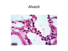
A: Alveoli (thinly walled)
B: Respiratory bronchiole (thick wall) |
|
|

|
Respiratory Bronchiole
|
|
|
|
A: RBC in capillary
B: Type 1 Pneumocyte C: Type 2 Pneumocyte (can dedifferentiate into stem cell and redifferentiate into type 1 cell) |
|
|

|
A: Macrophage
B: Type 2 Pneumocyte C: Interalveolar septa |
|
|

|
Type 2 pneumocyte: can tell because hallmark feature = osmeophilic lamellar bodies (striped things = lamellae)
|
|
|

Cartilage type? Where in respiratory system would this be find?
|
Elastic Cartilage--look at fibrous (elastin) content
Epiglottis ONLY |
|
|

Cartilage type? Where in respiratory system would this be find?
|
Elastic--notice elastic fiber content; epiglottis only
|
|
|

Cartilage type? Where in respiratory system would this be find?
|
Hyaline (not very fibrous; pretty bubbly); could find in rings of trachea, bronchi, plates of cartilage in bronchioles
|
|