Reading...
![]()
Play button
![]()
Play button
![]()
Use LEFT and RIGHT arrow keys to navigate between flashcards;
Use UP and DOWN arrow keys to flip the card;
H to show hint;
A reads text to speech;
45 Cards in this Set
- Front
- Back
|
What are the primary lymphoid organs? What occurs there?
|
Thymus and BM
Where B and T lymphocytes originate |
|
|
What are the secondary lymphoid organs? What occurs there?
|
Lympho Nodes, tonsils, appendix, Peyer's Patches, GALT, MALT, spleen
Where futher lymphocyte differentiate occur; this is where lymphocytes reside |
|
|
What is the embryonic origin of the thymus?
|
Stroma = Epithelial (endoderm of pharynx)
T Cell progenitors: Mesoderm (from BM) |
|
|
In which cavity is the thymus located?
|
Thorax: mediastinum
|
|
|
How does the cortex of the thymus differ from the medulla?
|
Cortex: hihgly populated with lymphocytes (stains intensely)
Medulla: low lymphoid cell count |
|
|
What cells comprise the thymus framework? What is their epithelial origin? What connects them to one another?
What are their other functions? |
Epithelial Reticular Cells (no reticular fibers!)
Epithelial-->Endodermal Origin Connected by desmosomes Selection, proliferation, death of thymocytes |
|
|
How can the thymus be differentiated from lymph node tissue?
|
-No nodules (no B lymphocytes)
-No afferent lymphatic vessels (no filtering) -HASSALL'S CORPUSCLES in medulla |
|
|
What is the blood-thymic barrier? What is its purpose?
|
Formed by thymic capillaries (non-fenestrated with continuous basal lamina) and sheath of reticulo-epithelial cells.
Protects developing T-cells from circulating antigens |
|
|
Where are efferent lymphatic vessels found in the thymus?
|
Near trabeculae
|
|
|
What are the stromal cells of the thymus?
|
Epithelial reticular cells
|
|
|
What are thymocytes? Describe their path in the thymus. How is this path controlled?
|
Pre-cursor T-Cells from bone marrow
Enter epithelial reticular cell network and mature. most die in cortex, about 5% make it to medulla Growth factors (thymosin) control T cell maturation Good T cells travel via medulla to bloodstream |
|
|
What role do macrophages play in the thymus?
|
Eat T cells that died trying to mature in cortex
|
|
|
What are Hassall's corpuscles?
|
HALLMARK STRUCTURE OF THYMUS!!
Found in medulla; have degenerating epithelial-reticular cells at center. Eosinic. |
|
|
How would you differentiate between areas of granulocytic maturation and areas of erythroid maturation under a microscope?
|
Erythroid sites would have much darker nuclei
Myeloid (granulocytic) sites would have lighter nuclei and granules present |
|
|
What occurs in lymph nodules/follicles?
|
Sites of antigen encounter and proliferation/differentiation of B and T cells
|
|
|
What do primary lymph follicles contain?
|
small, naive lymphocytes, no germinal center
|
|
|
What do secondary lymph follicles contain?
|
Germinal center (site of B cell proliferation)
B cells in center T cells in periphery |
|
|
Where are lymphoid follicles/nodules found unencapsulated?
|
Peyer's Patches, GALT, MALT, appendix, cortex of lymph nodes
|
|
|
Describe the structure of lymph nodes.
|
Cortex has lymphoid nodules containing B cells
Paracortical Zone houses T cells Medulla contains cords (plasma cells, macs, reticular cells, fiebrs, dendritic cells) and sinuses (lymph fluid) |
|
|
Describe the flow of lymph through lymph nodes.
|
Afferent lymphatics bring lymph to node via its convex (outer) surface
Lymph drains into sinuses Then reaches efferent lymphatics at hilum (concave/lower) |
|
|
Where do blood vessels enter and exit lymph nodes?
|
At the hilum
|
|
|
How do lymphyocytes return to the lymph node from blood?
|
Via High Endothelial Venules (cuboidal epithelium with glycoproteins and integrins that attract lymphocytes back in)
|
|
|
Where are the palatine tonsils located? Epithelial cell type?
|
Oropharynx; stratified squamous
|
|
|
If tonsils don't have efferent vessels, how are they exposed to antigens?
|
Antigens must contact epithelium that covers tonsils; tonsilar crypts facilitate this process by delving into lymphoid tissue (increased SA)
|
|
|
What are Peyer's patches and where are they found? Describe their structure.
What's their purpose? |
lymphoid aggregates in CT of ileum and appendix
B cells in germinal centers T cells in zones between follicles Peyer's Patches survey exposure to pathogens from lumen of GI tract |
|
|
What are M cells? Purpose?
|
Specialized cells in epithelium of Peyer's patches. Sample antigen in lumen and deliver it to APC's and lymphocytes located in pocket beneath them via transcytosis.
|
|
|
MALT vs GALT
Which is Peyer's patches apart of? |
MALT = mucous associated lymphoid tissue
GALT = gut-associated, ex: Peyer's Patches |
|
|
What are the two major components of the spleen?
|
Stroma = RED PULP--reticular fibers with lymphocytes, macs, APC's; AKA splenic cords or Cords of Billroth
Small masses of WHITE PULP--consists of lymphoid nodules with B cells, and Periarteriolar Lymphoid Sheaths (PALS)--sheath of T cells surrounding arteries |
|
|
Whar artery feeds into the spleen? Via?
|
Splenic artery via splenic capsule
|
|
|
Describe flow through the spleen?
|
Blood enters from splenic artery via splenic capsule
Artery divides into capsular and trabecular branches WHen reaches interior of spleen, becomes PALS and is known as CENTRAL ARTERY CENTRAL ARTER WITH PALS: sheath is location of B cell proliferation (in germinal centers) When arteriole subdivides and loses PALS, they form bundles and become PENICILLAR ARTERIOLES, which deliver blood to red pulp (splenic cords [of Bilroth]) Blood enters sinuses of red pulp and blood is drained through veins (ultimately exiting via splenic vein at splenic hilum) |
|
|
What are the functions of the spleen?
|
Lymphocyte development (as a secondary lymphoid organ)
Destruction/removal of defective or aged RBCs Phagocytosis of RBC fragments, breakdown of Hg for recycling immune defense |
|
|
Which secondary lymphoid sites are encapsulated? Unencapsulated? Partially?
|
Enc: Lymph nodes, spleen
Unencap: Lymph nodules Partial: tonsils |
|
|
Which part of the palatine tonsil is considered encapsulated?
|
Base: bordered with dense CT band that sequesters lymphoid tissue
|
|

|
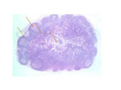
1) Lymph Nodules (lobules)
2) Interlobular septa (CT) 3) Cortex 4) Medulla – where mature T cells exit into periphery |
|

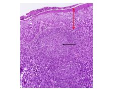
Identify.
|
Thymus: Hassall’s corpuscles!
|
|

Identify areas of erythrocytic and granulocytic development.
|
Darker nuclei clumps = erythrocytic
|
|
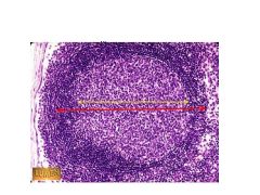
Identify.
|
Lymph follicle:
Yellow: Germinal Center—indicative of active lymph follicle Red: Margin |
|
Identify.
|
Lymph node.
Cortex= pale circle; contains B cells Paracortical zone surrounds center; houses T cells Medulla at dead center: contains cords with plasma cells, amcs, reticular cells, fibers, dendritic cells, SINUSES |
|

Identify.
|
ALL IN MEDULLA
Clumps of cells = cords Empty spaces = sinuses |
|
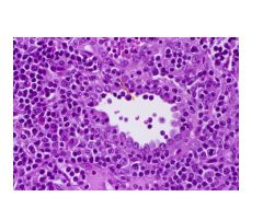
Identify.
|
Lumen of HEV: cuboidal liing that links to WBC’s for re-entry to lymphoid tissue
|
|
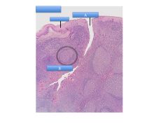
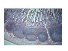
Identify structure and label.
|
Palatine Tonsil
A: Tonsilar Crypt (enhances SA) B: Lymph follicle with germinal center |
|
Identify lymphoid structure and label.
|
Palatine tonsil
Red = epithelium Black = germinal center |
|
Identify.
|
Peyer’s Patches: follicles in submucosa = SALT
Top most arrow is in mucosa; thus, it’s a MALT |
|

|
In mucosal layer; MALT
|
|

|
A: White Pulp
B: Red Pulp: sinus, cords; slow blood flow (pooling), filter BLOOD not lymph |