![]()
![]()
![]()
Use LEFT and RIGHT arrow keys to navigate between flashcards;
Use UP and DOWN arrow keys to flip the card;
H to show hint;
A reads text to speech;
301 Cards in this Set
- Front
- Back
|
What are the different types of glucose transporters, and in which tissue are they found? |
GLUT1 - RBC, brain - transport across blood-brain barrier GLUT 2 - Liver, kidney, pancreas, - Regulation on insulin release GLUT3 - Brain, placenta, others - Uptake into neurons, GLUT4 - Muscle, adipose tissue - Insulin-mediated transport GLUT5 - Kidney and gut - Absorption of fructose |
|
|
What are the main differences between hexokinase II and IV
|
Hexokinase II - Muscle cells - High affinity for glucose - Inhibited by glucose-6-phosphate Hexokinase IV - Liver cells - Low affinity for glucose - Inhibited by regulator protein, activated by either glucose or fructose-6-phosphate - Regulator protein will cause sequestration in the nucleus - Not allosteric inhibited by glucose-6-phosphate |
|
|
what are some substrate-level phosphorylations? |
Glycolysis - Phosphoglycerate kinase - Pyruvate kinase Kreb´s cycle - Succinate-CoA lyase - Phosphoenolpyruvate carboxykinase |
|
|
How is the regulation of phosphofructokinase-1 and fructose bisphosphatase 1 connected? |
They have a mutual regulator, F26BP which is an allosteric regulator of PFK-1 and FBPas-1 Insulin and glucagon is mediated by F26BP PFK-1 is inactive without the presence of F26BP regulation F26BP levels are regulated by PFK-2 and FBPase-2 When PFK2 is active - F26BP levels rise, this increases, which stimulate PFK-1, which stimulates glycolysis - Active due to stimulation of insulin on pp, and the cleaving of the phosphate When FBPase-1 is active -Levels of F26BP will decrease. This will stimulate FBPase-1, and since PFK-1 need F26BP regulation to function,it will decrease glycolysis - Stimulate gluconeogenesis - Glucagon will stimulate cAMP secretion, which stimulates PKA to phosphorylate |
|
|
What regulates PFK-1 except for F26BP? |
High ATP and citrate levels - Inhibit PFK-1 High levels of ADP and AMP - activation of PFK-1 High levels of AMP will also strongly allosterically inhibit F-1,6-bisphosphatase |
|
|
Arsenate poisining, and what is a reaction which can use arsenate? |
Arsenate AsO4, is structurally and chemically similar to phosphate. This allows many enzymes, which require phosphate to use arsenate instead, making it very dangerous. Arsenate is very toxic Glyceraldehyde-3-phosphate dehydrogenase - Will create 1-arseno-3-phosphoglycerate instead |
|
|
What are the types of pyruvate kinase? |
Pyruvate kinase L - Found in liver only, an isoenzyme of - Regulated by glucagon via PKA (cAMP) - Covalently regulated Pyruvate kinase M - Found in all other glycotic tissues, also liver - Not regulated by glucagon, or covalently - Activated by F16BP - Inhibited by alanine, long chain fatty acids, ATP, acetyl-CoA |
|
|
What are the 3 catabolic fates of pyruvate formed in glycolysis? |
1. Pyruvate -> ethanol + CO2 - Fermentation to ethanol in yeast - Hypoxic or anaerobic condition in bacteria 2. Lactate - Fermentation to lactate in very active muscles, erythrocytes - Also occurs in some cells under aerobic, as they lack mitochondria - Anaerobic conditions 3. Pyruvate -> Acetyl CoA - Aerobic condition - Enters citric acid cycle |
|
|
What is the short- time and longtime regulation of glycolysis? |
Short time - PFK1, hexokinase, pyruvate kinase Longer time - Glucagon, epinephrine, insulin * Regulate glucose transporters GLUTx |
|
|
How does acetyl-CoA regulate pyruvate dehydrogenase complex and pyruvate carboxylase? |
Acetyl-CoA is a positive allosteric modulator of pyruvate carboxylase - Creates oxaloacetate, and continues in the gluconeogenesis Acetyl-CoA is negative allosteric modulator of pyruvate dehydrogenase complex |
|
|
When does acetyl-CoA accumulate? |
When the cell energy needs are met - the oxidative phosphorylation slows - NADH/NAD+ ratio rises - Change in ratio, will inhibit the citric acid cycle - Acetyl-CoA accumulates |
|
|
How is pyruvate dehydrogenase complex regulated? |
Inhibited by ATP, acetyl-CoA, NADH and fatty acids Activated by AMP, CoA, NAD+, Ca2+ It's phosphorylated by PDK kinase and dephosphorylated by pdk phosphatase. Kinase is stimulated -> inhibited PDK - NADH - ATP Kinase is inhibited -> activation of PDC - CoA-SH - NAD+ - ADP - Pyruvate Stimulation of phosphatase -> Dephosphorylation of PDC -> activation - Ca2+ - Mg+ - Insulin |
|
|
How is citrate synthase regulated? |
Inhibited by NADH, succinyl- CoA, citrate, ATP Activated by ADP |
|
|
How is isocitrate dehydrogenase regulated? |
Activated by Ca2+ and ADP Inhibited by ATP |
|
|
How is a-ketoglutarate dehydrogenase complex regulated? |
Activated by Ca2+ Inhibited by succinyl-CoA, NADP - Succinyl-CoA is the product |
|
|
What enzyme does substrate-level phosphorylation in the citric acid cycle? |
Succinyl-CoA synthase CoA-S is removed and replaced with a Pi - Pi is donated to the enzyme and replaced by oxygen. - Pi is donated from enzyme to GDP -> GTP |
|
|
What is the role of citric acid cycle in anabolism? |
anabolism, we use energy to create a product Citrate -> Fatty acids and steroids A-ketoglutarate -> Glutamate - Glutmate is used from glutamine, proline, arginine or purines Succinyl-CoA -> porphyrins, heme Oxaloacetate -> Aspartate, asparginine - Asparagine -> pyrmidines Oxaloacetate -> phosphoenolpyruvate - PEP -> Glucose - PEP -> serine, glycine, cysteine, phenylalanine, tyrosine, tryptofan |
|
|
What are anaplerotic reactions? what are some anaplerotic reactions? |
Reactions which replenish the intermediates of citric acid cycle, as they are removed for synthesis of glucose, fatty acids, amino acids etc. Pyruvate to oxaloacetate - Pyruvate carboxylase Aspartate to oxaloacetate - Reversed reaction of aspartate transaminase Glutamate to a-ketoglutatarate - Glutamate dehydrogenase B-oxidation of odd chain fatty acid - Final oxidation forms succinyl-CoA |
|
|
How is hexokinase I, II, III and IV regulated? |
Hexokinase I, II, III - Inhibited by their own product -> Glucose-6-phosphate Hexokinase IV - Glucokinase - Not inhibited by glucose-6-phosphate - regulated by sequestration in the nucleus via regulator protein - Regulator protein is activated by -> inhibition of hexokinase IV * Glucose and fructose-6-phosphate |
|
|
How does galactose enter glycolysis? |
Galactose -> Galactose-1-p -> UDP-galactose -> UDP-glucose -> Glucose-1-phosphate -> Glucose-6-phosphate
Galactokinase -> Gal-1-p UDP-glucose:Galactose-1-phosphate uridyltransferase -> UDP-Gal UDP-glucose 4-epimerase -> UDP-Glc UDP-glucose: Galactose-1-phosphate uridyltransferase -> Gal-1-p Phosphoglucomutase -> Gal-1-p |
|
|
How does fructose enter glucolysis?
|
Fructose -> hexokinase Enters at fructose-6-phosphate -> step 3 |
|
|
How does Mannose enter? |
Mannose -> mannose-6-p - Hexokinase Mannose-6-p -> fructose-6-p - Phosphomannose isomerase |
|
|
How does fructose -1-phosphate enter glycolysis? |
F-1-p -> glyceraldehyde + dihydroxyacetonephosphate - F-1-p aldolase Glyceraldehyde -> glyceraldehyde-3-p - Triose kinase Dihydroxyacetone phosphate -> glyceraldehyde -3-p - triose phosphate isomerase Enters at 6 stage of glycolysis |
|
|
How does glycogen enter glycolysis? |
Glycogen -> Glucose-1-p - Phosphorylase
Glycogen-1-p -> glc-6-p - Phosphoglucomutase |
|
|
What is an enzyme in gluconeogenesis, found in both mitochondrial and cytosol, and why? |
Mitochondrial malate dehydrogenase and cytosolic malate dehydrogenase Pyruvate is converted to oxaloacetate by pyruvate carboxykinase Oxaloacetate can't pass the mitochondrial membrane, and is therefore converted to malate by mitochondrial malate dehydrogenase. When it has passed the mitochondrial membrane, it's converted back to oxaloacetate, and it continues the glucogeonetic pathway |
|
|
What are the activators of gluconeogenesis and glycolysis?
|
Gluconeogenesis - Acetyl-CoA, Glucagon, Epinephrine, Norepinephrine, Glucocorticoids Glycolysis - Inhibit gluconeogenesis - ADP, AMP, citrate, fructose-2,6-bp, insulin |
|
|
Cori cycle |
Lactate is formed by active skeletal muscle in anaerobic environment or by erythrocytes. Lactate is carried in the blood to the liver, where it´s converted back to pyruvic acid, then glucose via gluconeogenesis. - Convertion requires 6ATP |
|
|
Alanine cycle |
Alanine transports ammonia from skeletal muscle to the liver In the muscle, muscle protein is degraded to amino acids which inturn gives NH4+. In the alanine cycle we can transport both NH4 and pyruvate to the liver. Where NH4 enters urea cycle, and pyruvate enters gluconeogensis. We can covert glutamate to glutamine and transport it to liver. Or by alanine aminotransferase, we can transfer the amino group to pyruvate and transport both |
|
|
What is the hormonal regulation of gluconeogenesis? |
Insulin -> inhibit Glucagon -> stimulate Epinephrine, norephinephrine - > stimulate Glucocorticoids -> stimulate |
|
|
SREBP-1c How does it regulate gluconeogenesis and glycolysis? |
SREBP-1c - sterol regulatory element binding proteins Synthesis is increased by insulin and decreased by glucagon SREBP-1c will increase synthesis of - Hexokinase IV, pyruvate kinase, lipoprotein lipase, acetyl-CoA carboxylase, fatty acid synthase complex SREBP-1C will decrease - Synthesis of PEP carboxykinase, FBPase-1, glucose-6-phosphatase |
|
|
How does CREB regulate gluconeogenesis and glycolysis? |
cAMP response element binding protein Synthesis increased by glucagon via cAMP Increases synthesis of PEP carboxykinase and glucose-6-phosphate |
|
|
How does FOXO1 regulate gluconeogenesis and glycolysis? |
Insulin will have the effect of turning off expression of the genes - Insulin will activate a signalling cascade, which leads to activation of PKB. - PKB will phosphorylate FOXO1 in cytosol - FOXO1 will be attached t ubiquitin, which will mark it for destruction via proteasome. - FOXO1 which remains dephorphorylated can enter the nucleus and attach to DNA. - To trigger transcription of it's associated genes Increases synthesis of gluconeogenetic enzyme Supresses synthesis of glycolytic enzymes, pentose phosphate pathway enzymes and triacylglycerol synthetic enzymes |
|
|
How does chREBP regulate gluconeogenesis and glycolysis? |
ChREBP is found inactive in the cytosol - 2xPhosphorylated, cant move into nucleus - PP2A dephosphorylates it, allowing it to enter nucleus - In nucleus second dephosphorylation by PP2A - This allows it to be associated with mix - ChREBP-Mix complex will bind to a carbohydrate response element, and stimulate transcription PP2A is allosterically activated by xylulose-5-phosphate - xylulose-5-phosphate is a signal that other pathways which utilize glucose, has enough substrate ChREBP will therefore turn synthesis of several enzymes - Pyruvate kinase, fatty acid synthase and acetyl-CoA carboxylase |
|
|
What is the function of NADPH created by the pentose phosphate pathway? |
needed for synthesis - Fatty acid biosynthesis - Cholesterol biosynthesis - Neurotransmitter biosynthesis - Nucleotide biosynthesis Detoxification - Reduction of oxidized glutathione - Cytochrome P450 monoxygenases Also needed to counter the damaging effects of oxygen radicals |
|
|
Wha it the fat of pentose from pp pathway? |
needed in rapidly dividing cells - Bone marrow, skin, intestinal mucosa, tumors Uses ribose-5-phosphate - RNA, DNA, ATP, NADH, FADH2, CoA |
|
|
What enzymes is the oxidative phase made up of, and how are they regulated? |
Glucose-6-phosphate dehydrogenase Lactonase 6-phosphogluconate dehydrogenase Phosphopentose isomerase |
|
|
When is the non-oxidative phase active? |
In tissues which primarily require NADPH, pentose phosphates will be recycled back into glucose-6-p Transketolase and transaldolase |
|
|
What are 4 fates of glucose-6-phosphate? |
1. Ribose-5-phosphate is needed - Fructose-6-p into ribose-5-p - Glyceraldehyde-3-p into ribose-5-p 2. Ribose-5-phosphate and NADPH is needed - Glucose-6-p -> Ribulose-5-p+ NADPH -> Ribose-5 p 3. NADPH needed - Ribulose-5-p produced, but recycled 4. NADPH and ATP needed - Ribulose + NADPH is first made, then ribose is sent into glycolysis via fructose-6-p and glyceraldehyde-3-p |
|
|
How are reactive oxygen created in mitochondria? |
Created when the rate of electrons entering the respiratory chain, and electrons transferred through are mismatched. 1. Superoxide radical production will increase at complex I and III 2. Partially reduced ubiquinone radical will donate electron to O2 3. Superoxide will act as a aconitase to release F2+ 4. Fenton reaction will form reactive hydroxyl free radical -> OH |
|
|
How does NADPH and glutathione protect against ROS? |
Reduced glutathione or GSH will donate it's electrons to H2O2, reducing it to H2O and O2. GS-SG will be regenerate to oxidized form by NADPH For this we need glucose-6-phosphate dehydrogenase as it produces NADPH+H In mitochondria nicotinamid nucleotide transhydrogenase is used instead, which transfers electrons from NADH to NADP |
|
|
What are some deficiencies which prevent glutathione and NADPH from protecting against ROS? |
Glucose-6-phosphate dehydrogenase deficiency - Causes Heinz-bodies in red blood cells Wernicke-Korsakoff syndrome - Mutation in transketolase gene, which decreases affinity to TPP |
|
|
What is muscle glycogen used for, and what is liver glycogen used for? |
Muscle glycogen is used to generate ATP for muscle contraction Liver glycogen is used to maintain glucose blood levels |
|
|
What is the structure of glycogen? |
Chain structure, consisting of multiple chains with branches a-1,6 linkage - Connects branches to main chain a-1,4 linkage - Connects the glucose molecules together On the end of the main chain, and on all branches we find reducing ends |
|
|
Glycogenin and how is a primer formed? |
The primer for glycogen synthase Tyrosine residue of the protein glycogenin, will form the first glycosidic bond between glucose and UDP-glucose 1. Primer consist of a glycogen molecule of 8 glucoses, remaining from previous degradation 2. Glycogen molecule attaches to glycogenin through tyr 3. One ATP is needed to start glycogen synthesis |
|
|
How is a glucose molecule added during glycogen synthesis? |
1. Glucose enters liver or muscle cell -> Becomes phosphorylated by hexokinase II or IV 2. Phosphoglucomutase will convert G6P to G1P 3. G1P will react with UTP to form UDP-glucose, catalyzed by UDP-glucose phosphorylase 4. Glycogen synthase transfers UDP-glucose to non-reducing end of a glycogen primer 5. The UDP is released and reconverted to UTP via ATP |
|
|
How are branches formed in glycogen? |
Formation of branches starts when a chain becomes 11 or more glucose residues. At this point an oligomere of 6-8 glucose molecules is removed from the non-reducing end of the chain This is reattached to the chain via a-1.6 linkage by glycogen branching enzyme This process adds another non-reducing end to the glycogen chain |
|
|
What are the steps of glycogen degradation or glycogenolysis? |
1. Glycogen phosphorylase removes glucose residues from non-reducing ends - Release 1 glucose-1-phosphate at the time - Only remove until there are 4 glucose units until branching point 2. Debranching enzyme - Removes 3 out of 4 glucose left on the branch - Attaches them to non-reducing end of another branch - Debranching has a transfer like activity 3. Remaining glucose removed by a-1,6 glucosidase - releases free glucose 4. Glucose-1-phosphate -> glucose-6-phosphate by phosphoglucomutase - In liver G6P are converted to glucose by glucose-6-phosphatase and released into blood stream via GLUT2 Glucose-6-phosphatase is an integral membrane protein of ER - Prevents it from cleaving all G6P in the cell |
|
|
How is glycogen phosphorylase regulated? |
Glycogen phosphorylas has 2 forms
1. Glycogen phosphorylase a - More active - Activated by Phosphorylase b kinase by phosphorylation - Epinephrine, Ca2+, AMP will activate the kinase in muscle, - Glucagon will activate the kinase in liver - Will activate will activate via cAMP -> PKA -> phosphorylase kinase b 2. Glycogen phosphorylase b - Less active - Not phosphorylated - When muscle returns to rest, phosphorylase a phosphatase or PP1 removes phosphate groups, converting it Glycogen phosphorylase in liver has glucose sensor - Glucose will bind to an allosteric site of phosphorylase a, and this causes a change in conformation which exposes it's phosphorylated groups - The PP1 will convert it to phosphorylase b, - Reducing the activity, and slowing glycogen breakdown - Also stimulated by insulin indirectly |
|
|
How is glycogen synthase regulated? |
Glycogen synthase has two forms, b and a 1. Glycogen synthase a - Active form - Converted by Phosphoprotein phosphatase 1 2. Glycogen synthase b - Inactive form - Converted by glycogen synthase kinase 3 GSK-3 phosphorylates glycogen synthase b -> inhibiting glycogen synthesis - Inhibited by insulin - GSK-3 requires prior phosphorylation or priming from casein kinase II - Insulin will trigger activation of glycogen synthase b by blocking the activity of GSK3, and activating PP1 in muscles. - insulin will phosphorylate GSK3 via PKB/PKA PP1 -> dephosphorylates glycogen synthase b -> glycogen synthase a - Promotes synthesis - Removes phosphoryl groups from all 3 enzymes |
|
|
How is GSK-3 regulated? |
GSK-3 phosphorylates glycogen synthase b -> inhibiting glycogen synthesis - Inhibited by insulin - GSK-3 requires prior phosphorylation or priming from casein kinase II- Insulin will trigger activation of glycogen synthase b by blocking the activity of GSK3, and activating PP1 in muscles. - insulin will phosphorylate GSK3 via PKB/PKA |
|
|
What enzyme can remove phosphoryl groups from all three of the enzymes phosphorylated in response to glucagon in liver and epinephrine in muscle/liver? How is it regulated? |
PP1 can remove phosphate groups from phosphorylase kinase, glycogen phosphorylase and glycogen synthase PP1 is not found in free form, but tightly bound to its traget protein by glycogen-targeting proteins. - Bind glycogen, each of the three enzymes Regulated - Positive allosteric regulation by glucose-6-phosphate - Negative covalent phosphorylation by PKA |
|
|
How is glycogen-argeting protein Gm regulated? |
Gm binds glycogen to enzymes, like PP1 Gm can be phosphorylated at 2 different site, which give a different effect - in response to epinephrine and insulin 1. insulin stimulated phosphorylation - Phosphorylation at site 1 - Activates PP1, which dephosphorylates phosphorylase kinase, glycogen phosphorylase and glycogen synthase - Insulin inhibit glycogen breakdown, and stimulate glycogen synthesis 2. Epinephrine stimulated phosphorylation - Phosphorylation at site 2 - Dissociation from PP1, inactivating it - Preventing access of PP1 to glycogen phosphorylase and synthase - PP1 is also connected to phosphorylated inhibitor 1, inactivating it - Epinephrine or glucagon will stimulate glycogen breakdown, and inhibit glycogen synthesis - |
|
|
In which region is glycogen synthase kinase 3 phosphorylated, and by which kinase? |
Pseudosubstrate region, PKB |
|
|
What is the hormonal regulation of glycogen synthesis and degradation? |
Insulin - Phosphorylation of site 1 on Gm -> PP1 activated -> synthesis of glycogen Epinephrine - Stimulates PKA -> Phosphorylates site 2 on Gm -> PP1 inhibited -> degradation of glycogen |
|
|
What are the possible pathways in enzyme regulation and degradation? - 10 |
1. Extracellular signal - Hormonal - neural - Growth factos 2. Transcription of specific gene(s) 3. mRNA degradation 4. mRNA translation on ribosome 5. Protein degradation - Tagged by ubiquitin for degradation by proeasomes 6. Enzyme sequestered in subcellular organelle - Segregation of enzyme 7. Enzyme binding substrate 8. Enzyme binds ligand (allosteric) 9. Enzyme undergoes (de)phosphorylation 10. Enzymes combines with regulatory protein |
|
|
Where does fatty acid synthesis, elongation and desaturation happen? |
Fatty acid synthesis happens in cytosol
- until C16 Fatty acid elongation happens in ER and mitochondria Fatty acid desaturation in ER |
|
|
What is the function of acetyl-CoA carboxylase in fatty acid synthesis? |
Synthesis of malonyl- CoA from acetyl-CoA Acetyl-CoA har 3 regions 1. Biotin carrier protein 2. Biotin carboxylase 3. Biotin transcarboxylase This is the first step of fatty acid synthesis, as biotin carboxylase transfers HCO3 to biotin carrier protein with the help of ATP-> ADP + Pi. Then transcarboxylase transfers activated CO2 biotin to acetyl-CoA |
|
|
what are the enzymes of fatty acid synthase? |
8 enzymes in a complex
4 basic enzymes - Ketoacyl synthase - Ketoacyl reductase - Hydroxyacyl dehydratase - Enoyl reductase 4 other - Acetyl carrier protein - ACP - Acetyl-CoA-ACP acyltransferase - AT - Malonyl-CoA-ACP acyltransferase - MT - Palmitate deacylase - PD |
|
|
What are the steps of fatty acid synthesis, which enzymes, cofactors and products do we find? |
Before fatty acid synthesis we must activate Acetyl-CoA and malonyl-CoA Step 1 - Condensation - Ketoacyl synthase - Condensates the substrates, creating acetoacetyl-ACP - CO2 is released Step 2 - Reduction - Ketoacyl reductase reduces NADPH + H -> NADP+ - D-B-hydroxybutyryl-ACP is formed Step 3 - Dehydration - Hydroxyoacyl dehydratase removes water from the substrate - Water is removed Step 4 - Reduction - Enoyl reductase reduces NADPH+H -> NADP+ - Double bond is saturated -Saturated acyl group is formed, lengthened by two carbons |
|
|
What is the source of cytosolic acetyl-CoA? |
Almost all cytosolic acetyl-CoA used in fatty acid synthesis is formed in the mitochondria from pyruvate oxidation and from the catabolism of the carbon skeletons of amino acids Inner mitochondrial membrane is impermeable to acetyl-CoA - Because of this it´s converted to citrate, before shuttled into the cytosol via a citrate transporter - in cytosol citrate is reconverted to acetyl-CoA and oxaloacetate - Acetyl-CoA enters fatty acid synthesis - Oxaloacetate is reduced to malate by malate dehydrogenase, and shuttled via Malate-a-ketoglutarate transporter Cytosolic NADPH is also created as malate is converted to pyruvate via malic enzyme |
|
|
What is the sources of cytosolic NADPH? |
The malate formed from oxaloacetate during the shuttle of acetyl-CoA to cytosol can also be used for production of NADPH
Malate to pyruvate via malic enzyme will produce NADPH NADPH is also produced in the pentose phosphate pathway - Glucose-6-phoshphate dehydrogenase - 6-phosphogluconate dehydrogenase |
|
|
How is acetyl-CoA carboxylase (ACC) enzyme I regulated? |
Found in all cells, but mostly adipose and mammary glands Inactivated by AMP-activated protein kinase - Glucagon, adrenaline, AMP, palmitoyl-CoA -> Phosphorylation of ACC - Glucagon inhibits protein phosphatase A2 Activated by insulin and citrate - Insulin -> insulin-dependent protein phosphatase dephosphorylates ACC, activating it - Protein phosphatase 2A Inactive carboxylase + citrate -> partialy active carboxylase - Inhibited by palmitoyl-CoA |
|
|
What is mechanism of fatty acid elongation in animal cells? |
Takes place in ER and mitochondria Palmitate is the precursor for most longer fatty acids Similar mechanism as fatty acid synthesis, but with other enzymes, and using CoA instead of ACP Fatty acids are elongated on the head Elongation starts with donation of carbons by malonyl-CoA Then reduction, dehydration and reduction |
|
|
What is the mechanism of desaturation in vertebrates? |
Happens in smooth ER Fatty acid desaturation is needed for cell membrane fluidity in different temperatures Double bond introduced via an oxidative reaction by fatty acyl-CoA desaturase During this process 2 different substrates will at the same time undergo electron oxidations. - Electrons will flow via cytochrome and cytochrome reductase 1. NADPH will give its electrons to cytochrome reductase - FAD -> FADH2 2. Cytochrome reductase (FADH2) will give it's electrons to cytochrome - Fe3+ -> Fe 2+ 3. Cytochrome (Fe2+) will give them on to the fatty acid |
|
|
At which carbons of a fatty acid can animals add double bands, and why is this important? |
Animals can only put double bonds before 10th carbon atom Because of this, it's essential for animals to have fatty acids with double bonds after 10C in their diet These can be made into arachidonate, which can turn into prostaglandins, thromboxanes and leukotrienes |
|
|
What can be produced from arachidonate, and how is the synthesis regulated? |
Arachidonate is produced from phospholipids
And can produce prostaglands, thromboxanes and leukotrienes - By enzymes in smooth ER - COX-1 -> synthesis prostaglandins that regualte secretion of gastric mucin - COX-2 -> synthesis prostaglands that mediate inflammation, pain, fever - Thromboxanes are produced on the pathway involving COX-1 Steroids will inhibit synthesis of arachidonate - No prostaglandins, thromboxanes or leukotrienes Non-steroids like aspirin and ibuprofen will inhibit cyclooxygenase - No prostaglandins or thromboxanes |
|
|
What are essential fatty acids for mammals? |
Linoleate and alpha-lineoleanate |
|
|
How can glycerol-3-phosphate be synthesized in liver and adipose tissue? |
In liver - Glycerol kinase - Glycerol-3-phosphate dehydrogenase In adipocytes - Glycerol-3-phosphate dehydrogenase |
|
|
Phosphatidic acid |
Diacylglycerol-3-phosphate |
|
|
What are the different head groups for phosphatidic acid? what else can phosphatic acid produce? |
Head groups can be ethanolamine, serine, inostiol Triacylglycerol can be produced from phosphatidic acid - Phosphatidic acid phosphatase - Acyl transferase |
|
|
How is lipid synthesis regulated? |
Insulin - Favors synthesis of both acetyl-CoA and fatty acids -> triacylglycerol synthesis |
|
|
Diabetes mellitus - How does this effect fatty acid synthesis |
Diabetes mellitus is the lack of insulin This causes a decrease in fatty acid synthesis and causes ketone body production |
|
|
Synthesis of triacylglyceride |
1. Production of glycerol-phosphate - Either via glycerol-3-phosphate(liver and fat) or glycerol kinase (liver) 2. Production of phosphatidic acid - Acyl transferase twice 3. Production triacylglycerols and glycerophospholipids - Phosphatidic acid phosphatase + acyl transferase -> triacylglycerol - Attachment of head group -> glycerophospholipids |
|
|
What is glycerneogenesis, whats the steps? |
A shortened version of gluconeogenesis 1. Pyruvate - Pyruvate carboxylase 2. Oxaloacetate - PEP carboxykinase 3. Phosphoenolpyruvate - Multistep 4. Dihydroxyacetone phosphate - Glycerol-3-phosphate dehydrogenase 5. Glycerol-3-phosphate 6. Triacylglycerol synthesis |
|
|
What´s the role of glycerneogenesis? |
-Adipose tissue it's coupled with reesterification of free fatty acids to control the rate of fatty acid release in blood - In fasting humans, it supports the synthesis of glycerol-3-p enough to account for 65% of fatty acid reesterification to triacylglycerol |
|
|
Regulation of glyceroneneogenesis? |
Glucocorticoid hormone - Stimulates glycerneogenesis in liver - Inhibits glycerneogenesis in adipocytes Thialozolidine - Increases glyceroneogenesis -> increasing resynthesis of triacylglycerol in adipose tissue -> Reduces amount of free fatty acid in blood - Treats type II diabetes |
|
|
Triacylglycerol cycle |
Triacylglycercol cycle shows how triacylglycerols are broken down and synthesized during starvation In the liver triacylycerols are produced from glycerol-3-phosphate and transported to blood and adipose tissue In Blood lipoprotein lipase releases glycerol, and fatty acids are used for fuel for tissues In adipose tissue fatty acids are resynthesizes into triacylglycerols, and glycerol is released |
|
|
What are the basic type of complex lipids? |
1. Phospholipids Glycerophospholipids - 2 fatty acids + alcohol Sphingolipids - Sphingosine + fatty acid + choline 2. Glycolipids Sphingolipids - Sphingosine + fatty acid + mono/oligosaccharide Galactolipids - 2xfatty acids + mono +SO4 3. Aracheabacetial ether lipids |
|
|
What are the two strategies for synthesizing complex lipids? |
Strategy 1 Diacylglycerol activated with CDP and then add head group - Remove CMP Strategy 2 Diacylglycerol + head group activated with CDP. - Remove CMP |
|
|
What are the 4 steps of assembling a phospholipid?
|
1. synthesis of backbone of molecule - Glycerol or sphingosine 2. Attachment of fatty acid 3. Addition of hydrophilic head group 4. Alteration or exchange og head group - This is where the two strategies seperate |
|
|
Which lipids are precursors of CDP-Diacylglycerol? |
Phosphatidylglycerol - CDP-diacylglycerol + glycerol Cardiolipin - CDP-Diacylglycerol + phosohatidylglycerol Phosphatidylinositol - CDP-diacylglycerol + inostiol |
|
|
How do inositol phosphates function as intracellular signaling molecules |
Splitting by phospholipase C -Diacylglycerol -> DAG - Inositol-1,4,5-triphosphate -> IP3 Splitting by phophatidyl-inositol 3 kinase - Phosphatidyl-inositol 3,4,5 triphosphate or PIP3 |
|
|
What are the roles of IP3 and DAG? |
IP3 and DAg both contribute to activation of protein kinase C, by raising cytosolic Ca2+ concentration and IP3 also activates Ca2+ dependent enzymes. 1. Hormone binding - Binds to specific receptor 2. GDP-GTP exchange on G protein 3. Activation of PLC by GTP-G 4. Cleavage of PIP2 to IP3 and DAG 5. Binding of IP3 to ER IP3 -receptor evoking Ca release 6. DAG and Ca2+ activate PKC 7. PKC phosphorylates target proteins triggering an hormone response |
|
|
What is the intracellular effects of insulin mediated by? |
PIP3 |
|
|
What is HMG-CoA? |
a common precursor for both ketone bodies and cholesterol Formation of ketonebodies occur in the mitochondria, while the synthesis of cholesterol happens in the cytosol |
|
|
HMG-CoA is a branchpoint for which two products? and in which compartments and by which enzymes is this done? |
HMG-CoA functions as a branchpoint for synthesis of cholesterol in cytosol and ketone bodies in cholesterol Synthesis of cholesterol HMG-CoA -> Mevalonate via HMG-CoA reductase - 2 NADPH -> NADP+, CoA removed Synthesis of ketone bodies HMG-CoA -> Acetyl-CoA + acetoacetate via HMG-CoA lyase |
|
|
Conversion of ketone bodies? |
3 ketone bodies -> Acetoacetate, acetone, D-B-hydroxybutyrate Acetaoacetate - Created from HMG-CoA lyase, - Also created as when fumarylacetoacetate is split by fumarylacetoacetase Acetone - Acetoacetate decarboxylase, removes CO2 from acetoacetate D-B-hydroxybutyrate - D-B-hydroxybutyrate dehydrogenase from acetoacetate - Acetoacetate is reduced to form |
|
|
How are acetoacetate activated for metabolism? |
Done by B-ketoacyl-Coa transferase
Succinate-CoA -> Succinate Acetoacetate -> Acetoacetyl-CoA |
|
|
What are the steps of cholesterol synthesis? What is the rate determining step? |
1. Synthesis of mevalonate from HMG-CoA - Rate determining step 2. Mevalonate is converted into chain of activated isoprenes - ATP is utilized to attach phosphate groups activating isoprenes - Geranyl pyrophosphate -> 10 Carbons 3. After several steps squalene is formed -> 30 carbons - Precursor is farnesyl -pp -> 15C 4. cholesterol is formed from squalene after a sequence of reactions - Cholesterol is 27 C - One of the enzymes is cyclase |
|
|
Esterification of cholesterol |
cholesterol esters are formed in the liver by ACAT - Cholesterol + fatty acyl-CoA -> Cholesteryl ester + CoA ACAT catalyzes the transfer of fatty acids from CoA to the hydroxyl group of cholesterol - creates a hydrophobic form Cholesterol esters can also be formed form LCAT - LCAT catalyzes the formation of cholesteryl ester from lecthine - Cholesterol + Lecithine -> Cholesteryl ester + Lysolecithine - Lecithine -> phosphatidylcholine |
|
|
What are the types of lipoproteins? what do they contain and what is their destination? |
1. Chylomicrons - Dietary lipids - From GI to liver 2. VLDL - Endogenous lipids, cholesterol - From liver to blood 3. LDL - cholesterol - From blood to tissue 4. HDL - Excess Cholesterol - From tissue to liver |
|
|
How is cholesterol synthesis regulated in response to cholesterol levels? |
System which does transcriptional regulation of the gene encoding HMG-CoA reductase The gene coding for HMG-CoA reductase, and 20 others gene affiliated with cholesterol are controlled by sterol regulatory element binding proteins or SREBPs. Only the soluble amino terminal domain of SREBP can function as a transcriptional activator. - This domain has no access to nucleus when it's a part of SREBP molecule To activate the transcription of HMG-CoA reductase, the transcriptional active domain must be proteolytically cleaved by SCAP When cholesterol levels are high, SREBP is inactive, secured to ER via SREBP cleavage-activating protein - SCAP binds to cholesterol, acting as a sterol sesnor - When cholesterol levels decrease, conformational changes causes release of SCAP-SREBP complex from ER In golgi SREBP is cleaved twice, and the second cleavage will release the amino-terminal domain into the cytosol - Travels to nucleus and activates target genes - Short life, rapidly degraded by proteasomes |
|
|
How is HMG-CoA reductase regulated by hormones and by |
Insulin activates HMG-CoA reductase - Dephosphorylates the enzyme Glucagon inhibits HMG-CoA reductase - Phosphorylates the enzyme Phosphorylated by AMP-activated protein kinase Unidentified metabolites of cholesterol stimulate proteolysis of HMG-CoA reductase |
|
|
What is the effect of cholesterol entering the cell on cholesterol synthesis? |
1. Down regulation of receptor mediated endocytosis 2. Inhibition of cholesterol synthesis 3. Promotion of cholesteryl esters 4. Inhibition of endocytotic receptor synthesis |
|
|
What's the theory of glucose fatty acid cycle? |
If plasma glucose is decreased, fatty acids are mobilized (fasting, glucagon) and increased levels of fatty acid will decrease glucose utilization. This restores the glucose level, and maintains homeostasis
|
|
|
AMP dependent protein kinase - AMPK |
ADP produced in synthetic reactions is converted to AMP by adenylate kinase AMP will activate AMPK, which regulates the anabolic and catabolic pathways by phosphorylating the enzymes AMPK will inhibit anabolic processes - Process using ATP -> ADP AMPK will stimulate catabolic process - ADP -> ATP Stress, fasting and exercise activates AMPK |
|
|
What reactions does AMPK stimulate?
|
AMPK stimulates catabolic processes - Stimulates glucose transport (GLUT1,4) - Stimulates PFK-2 -> Cardiac glycolysis - Stimulates fatty acid oxidation and glucose uptake in skeletal muscle - Fatty acid oxidation, glucose uptake and glycolysis in heart |
|
|
What reactions are blocked by AMPK? |
HMG-CoA reductase Transcription Acetyl-CoA carboxylase - FA synthesis Synthesis of Cholesterol, triacylglycerol, glycogen, protein |
|
|
Regulation of ketone body synthesis via HMG-CoA synthase |
Liver contains two types of HMG-CoA synthase - cytosolic for cholesterol synthesis - Mitochondrial for ketone body synthesis The mitochondrial is rate limiting for ketone body synthesis The mitochondrial HMG-CoA synthase will undergo covalent modification via succinylation/desuccinylation of pre-exisiting enzyme |
|
|
What is PPAR, what's their role? |
PPAR - Peroxisome proliferator-activated receptors - Nuclear receptors - Transcription factor, regulating expression of genes - Effect on lipid transport PPAR has roles in - alpa (liver, heart, muscle, kidney) -> Regulates FA metabolism, HDL-cholesterol - beta (anywhere) -> Kartinocytes differentiation, wound healing, mediated VLDL signaling - gamma (adipocytes, macrophages) -> Adipocyte differentiation, lipid storage, glucose homeostasis |
|
|
Farber disease |
Enzyme deficiency which results in accumulation in lysosomes of PAS-positive lipids consisting of ceramide |
|
|
What is the fate of the amino group and carbon skeleton of an amino acid?
|
Amino group - Biosynthesis of amino acids, nucleotides, and biological amines - Carbamoyl phosphate -> Urea cycle -> Urea Carbon skeletons - A-keto acids -> Citric acid cycle |
|
|
How are amino groups incorporated into biomolecules? |
They are incorporated via glutamate and glutamine - Glutamate is the source of amino groups for most other amino acids, through transmination reactions - Glutamine is source of amino groups for biosynthetic process |
|
|
What are the 3 enzymes of the ATP-dependent pathway of protein degradation? |
E1: Ubiquitin-activating enzyme E2: Ubiquitin-conjugating enzyme E3: Ubiquitin-protein ligase |
|
|
What are the two pathways to protein degradation? |
1. ATP-dependet pathway used for degradation of defective proteins, and those with short half-lives - Involves ubiquitin, which becomes covalently linked to proeins slated for destruction via an ATP-dependent pathway -Involves E1, E2, E3 - Ubiquinated proteins are degraded by proteasome 2. A second system is found in lysosomes. This recycles the amino acids of membrane proteins, extracellular proteins and those with longer half-lives |
|
|
Proteasomes |
Large complex which degrades ubiquitinated proteins Complex has two main types of subcomplexes - Regulatory Caps on either side - Barrel like core Core has 4 rings - Outer rings are formed by seven alpha subunits - Inner rings are formed by seven beta subunits |
|
|
What is the role of PLP, what is it's derivative |
A derivative of vitamin B6 PLP is an intermediate amino group carrier - accepts and donates aminogroups This reactions goes from left to right, then from left to right - Ping-pong reaction mechanism PLP + amino acid -> Shiff base intermediate -> Quinonoid intermediate -> Pyroxidaxmine phosphate + keto acid |
|
|
What are some PLP requiring enzymes? |
Aminotransferase, tryptophan synthase, cysthionine synthase and lyase, glutamate decarboxylase, serine dehydratase, ornithine decarboxylase etc |
|
|
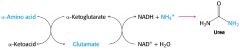
Glutamate hydrogenase reaction - Cofactor, where, regulation |
Happens in hepatocyte mitochondrial matrix Works with both NAD+ and NADP+ Allosterically regulated - ADP(GDP) stimulates - GTP (ATP) inhibits Oxidative deamination |
|
|
What is oxidative deamination? |
Release of NH4+ from glutamate, forming a-ketoglutarate - Happens in glutamate dehydrogenase |
|
|
Transdeamination pathway |
Transamination + oxidative deamination -> transdeamination |
|
|
How does NH4+ arrive to the liver? |
1. Amino acids from ingested proteins 2. Alanine from muscle - Glucose-alanine cycle via pyruvate and glutamate 3. Glutamine - From muscle and other tissues |
|
|
What are the sources of ammonia for urea cycle? |
Amino acid degradation Ammonia secretion in kidney tubules from glutamine Degradation of pyrimidine bases Produced by intestinal bacteria |
|
|
What are the ATP dependent reactions of the urea cycle, and where do they happens? |
Carbamoyl phosphate synthase I - Requires 2ATP - NH3 reacts with bicarbonate to form carbamoyl phosphate which enters cycle - Reaction takes place in the mitochondria Argininosuccinate synthetase - Requires ATP -> AMP - Happens in cytosol - Aspartate enter the urea cycle from the citric acid cycle |
|
|
aspartate-argininosuccinate shunt |
Argininosuccinate will form arginine and fumarate via argininosuccinase or lyase This fumarate can be converted to malate and enter the citric acid cycle This connection is the aspartate-argininosuccinate shunt of TCA |
|
|
Malate-aspartate shuttle What are the steps? |
shuttle of NADH in the liver, kindey and heart mitochondria 1. NADH+H is transferred to cytosolic oxaloacetate -> Malate 2. Malate crosses the inner membrane via malate-a-ketoglutarate transporter - Ketoglutarate is transported in the opposite direction 3. Malate dehydrogenase gives oxaloacetate and NADH+H 4. Oxaloacetate is first transaminated to aspartate 5. Aspartate is transported out via glutamate-aspartate transporter - Glutamate is transported in the opposite direction 6. Oxaloacetate is regenerated in cytosol |
|
|
How is the overall energetic cost of urea synthesis reduced? |
It used 3 ATP -> 2 ADP+AMP + 4Pi 1 NADH is formed in malate dehydrogenase reaction - 1 NADH -> 2.5 ATP |
|
|
How is carbamoyl phosphate synthase I regulated? |
Arginine will stimulate N-acetylglutamate synthase, and N-acetylglutamate will stimulate carbamoyl phosphate synthetase I |
|
|
What are the major goups of amino acids according to their endproducts? |
Ketogenic - Can yield ketone bodies in liver Glucogeneic - Can be converted to glucose Mixed - Both ketogenic and glucogenic |
|
|
What are the purely ketogenic amino acids? |
Leucine and lysine -> Acetyl-Coa -> ketone bodies or citrate |
|
|
What are the glucogenic amino acids? |
Aspargine Aspartate Methionine Valine Arginin Glutamine Glutmate Hisitidine Prolin They can become either pyruvate or oxaloacetate -> glucose |
|
|
What are the mixed amino acids? |
Tryptophan Phenylalanine Tyrosine Threonine Isoleucine Alanine Cysteine Glycine Serine |
|
|
What are the Coenzyme B12 dependent reactions? - What does deficiency cause? |
Methylalonyl-Coa mutase Rearrangement of L-methylmalonyl-CoA to succinyl-CoA Methionine synthase reaction Metabolic folates trapped in N5-methyl form |
|
|
What are cofactors which transfer one-carbon groups? And in which oxidation states?
|
Biotin - Most oxidized - COO- Tetrahydrofolate -> THF - Intermediate oxidized state - Methylene, methenyl, formyl, formimino groups S-adenosylmethionine - Most reduced - Methyl |
|
|
How is glutamine synthase regulated? |
Allosterically regulated - Each inhibitor will produce a partial inhibition, but all will together shut down the enzyme Glycine, alanine, Glucosamine-6-p, Histidine, CTP, carbamoyl phosphate, tryptophan and AMP |
|
|
What is the function of S-adenosylmethionine? |
AdoMet is used for methyl group transfers Synthesized from ATP and methionine by methionine adenosyl transferase Often uses methylene-THF as a methyl donor |
|
|
Reduction of N2 gas to ammonia |
N2 + 3 H2 -> 2 NH3
Triple bond between N Activation energy of N-fixation is very high |
|
|
Nitrogen fixation |
Done by nitrogenase enzyme complex Hydrolysis of 16 ATP N2 + 10H+ +8e- + 16ATP -> 2 NH4+ + 16ADP + 16Pi + H2 |
|
|
What regulates the amino acid biosynthesis? |
Allosteric regulation The enzyme regulated is often the first in the sequence, and it's inhibited by the end product of the pathway - negative feedback Regulation of the various synthetic pathways is coordinated |
|
|
What causes hyperammonemia? |
Urea cycle defects normally lead to hyperammonemia or build up of intermediates Permanent activation of glutamate dehydrogenase also causes hyperammonemia |
|
|
Congenital hyperammonemia type I
|
Carbamoyl phosphate synthetase |
|
|
Congenital hyperammonemia type II |
Ornithine transcarbamoylase |
|
|
Citrullinemia |
Argininosuccinate synthetase |
|
|
Argininosuccinate aciduria? |
Argininosuccinase |
|
|
Argininemia |
Arginase |
|
|
N-acetylglutmate synthase deficiency |
N-acetylglutamate synthase |
|
|
Maple syrup urine disease |
Defect on branched-chain a-keto acid dehydrogenase complex |
|
|
Hyperglycinemia |
Defect of the glycine cleavage enzyme |
|
|
Methylmalonic acidura |
Defect on methylmalonyl-Coa mutase |
|
|
Homocystinuria I
|
Defect of cystathionine B-synthase |
|
|
Phenylketonuria |
Defect on phenylalanine hydroxylase |
|
|
Tyrosinemia Type II |
Defect on tyrosine aminotransferase |
|
|
Tyrosinemia type III |
Defect of p-hydroxyphenylpyruvatedioxygenase |
|
|
Alkaptonuria |
Defect of homogentisate dioxygenase |
|
|
Tyrosinemia type I |
Defect of fumarylacetoacetase |
|
|
Albinism |
Defect of tyrosinase Melanine synthesis from tyrosine DOPA is a precursor of melanine Similar reaction to tyrosine hydroxylase |
|
|
Where does biosynthesis of heme happen, and how is it regulated? |
Happens in both the mitochondria and in the cytosol, and is regulated by heme as a feedback inhibitor |
|
|
What does the degradation of heme give? |
Heme degradation protects cell from oxidative damage 1. Release of iron - Fe2+ binds to ferritin - CO binds to hemoglobin - CO produced is toxic (high conc.), vasodilator (low conc.) with regulatory effects 2. Formation of bilirubin, via biliverdin - Bilirubin binds to serum albumin - Important antioxidant - Protective effect in developing brain |
|
|
What is phosphocreatine used for in muscle? When is it produced? |
Formed by creatine kinase from creatine It's an important energy reservoir in skeletal muscle - Allows for rapid regenerating ATP from ADP, by creatine kinase reaction Active contraction and glycolysis -> ATP synthesis Light activity/rest -> Phosphocreatine synthesis |
|
|
What is glutathione GSH formed from? What is it used for? |
From Glutamate, cysteine and glycine Used 2 ATP Forms reduced form GSH - redox buffer - Maintains -SH group of protein in reduced form - Maintains the iron of heme in ferrous state - Serves as reducing agent for glutaredoxin - deoxyribonucleotide synthesis - Removes toxic peroxides formed during growth or aerobic metabolism during glutathione peroxidase reaction |
|
|
What neurotransmitters are synthesized from tyrosine? Defects? |
Dopa, Dopamine, norepinephrine, Schizoprenia -> overproduction Parkinsons ->underproduction |
|
|
What neurotransmitters are synthesized from glutamate? defects? |
GABA Inhibitory neurotransmitter Underproduction can lead to epileptic seizures |
|
|
What neurotransmitters are synthesized from histidine? |
Histamine Vasodilator Released in allergic response |
|
|
What neurotransmitters are synthesized from tryptophan? |
Serotonin |
|
|
What neurotransmitters are synthesized from methionine?
|
Spermine, spermidine Involved in DNA packing |
|
|
What neurotransmitters are synthesized from arganine? |
Nitric oxide is a free radical Important biological messenger |
|
|
What are the roles of nucelotides in cells? |
1. DNA and RNA precursors 2. Carrier of chemical energy - ATP and GTP 3. Co-factor components - NAD, FAD, coenzyme A, S-adenosylmethionine 4. Activated intermediates - UDP-Glucose, CDP-diacylglycerol 5. Cellular second messenger - cAMP, cGMP |
|
|
What are the two ways of Nucleotide synthesis? |
Salvage pathway - Activated ribose (PRPP) + base -> nucleotide - Recycles free bases and nucleotides released during nucleic acid breakdown De novo pathway Activated ribose (PRPP) + amino acids + ATP+ CO2 NH3-> Nucleotide |
|
|
What are the common motives of purine and pyrimidine synthesis? |
1. Uses 5- phosphoribosyl 1- pyrophosphate 2. Uses amino acids - Purine -> glycine - Pyrmidine -> aspartate 3. N donor - Glutamine - aspartate (purine) |
|
|
Why can nucleotide synthesis limit rate of DNA replication and transcription? |
Cellular pool of nucleotides i 1% or less of the amounts required to synthesize the cell's DNA |
|
|
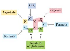
What is the origin of the different parts of purines? |
|
|
|
How is purine nucleotide biosynthesis regulated? |
It's regulated by feedback inhibition It's regulated in 4 steps by endproducts 1. AMP, GMP and IMP will inhibit the first reaction -Glutamine-PRPP amidotransferase 2. The endproducts will inhibit their own synthesis from IMP - AMP will inhibit adenylosuccinate synthetase - GMP will inhibit IMP dehydrogenase 3. Reciprocal regulation - Balance between AMP and GMP 4. AMP from endproduct will be phosphorylated to ADP, and ADP will inhibit Ribose phosphate pyrophosphokinase (PRPP synthetase) |
|
|
What are pyrmidine nucleotides made from?
|
Made from aspartate, PRPP, carbamoyl phosphate |
|
|
Where does carbamoyl phosphate synthetase II happen? |
Happens in the cytosol Will channel |
|
|
How is pyrimidine biosynthesis regulated? |
Regulated by negative feedback of the endproduct - Cytidine-5-triphosphate CTP inhibits transcarbamoylase allosterically ATP is a postive modulator of the same enzyme - Prevents change induced by CTP |
|
|
Degradation of purines and pyrimidines gives? |
Purine -> uric acid Pyrimidine -> urea |
|
|
Adenosine deaminase deficiency |
Causes immunodeficiency - T and B wont properly develop |
|
|
Salvage pathway |
Salvage pathway recycle purine and pyrmidine bases - Adenine + PRPP -> AMP + PPi - Hypoxanthine + PRPP -> IMP + PPi - Guanine + PRPP -> GMP + PPi |
|
|
Lesch-Nylan syndrome |
Prevents salvage pathway of guanin Causes - High levels of uric acid - Brain damage - Purines overproduce |
|
|
What are the 3 forms of DNA? - Helical sense - Glycosyl bond formation |
A form - Right handed - Anti glycosyl bond formation B form - Right handed - Anti glycosyl bond formation Z form - Left handed - Anti for pyrimidines, syn for purines |
|
|
How many base pairs are there between A and T versus G and C |
AT -> Double CG -> triple bond |
|
|
What is a triple helix? |
This a structure where the first half of one strands is separated and folds back on the other half of the repeat to form a triple helix with it's earlier strand |
|
|
What are some well-characterized non-enzymatic reactions of nucleotides? |
Deamination - Removes a NH2 group for cytosol, 5-methylcytosine, adenine and guanine - Creates uracil, thymine, hypoxanthine, xanithine Depurination - Add water to a guanosine residue -> Guanine and apurinic residue |
|
|
What introduces a bend or kink into the DNA? |
Formation of cyclobutane pyrimidine dimer |
|
|
What are 3 chemical agents which cause DNA damage? what is their precursor? What are some alkylating agents which cause damage to DNA? |
Sodium nitrite Sodium Nitrate Nitrosamine nitrous acid precursors Alkylating agents - S-adenosylmethionine - Dimethylnitrosamine - Dimethylsulfate - nitrogen mustard |
|
|
Nucleosomes What are the different histones? How is the nucleosome positioned? |
Histone cores connected with linker DNA H2A and H2B, H3, H4 are histone cores H1/H5 is linker DNA Its positioned in such a way to make optimal use of A=T base pairs where the histone core is in contact with the minor groove of the DNA helix |
|
|
What is the compaction of DNA in eukaryotic chromosomes? |
1. DNA 2. Beads on a string form of chromatin 3. 30 nm fiber 4. One loop 5. one rosette - Created by nuclear scaffold 6. One coil - 30 rosettes 7. Two chromatids - 10 coils each |
|
|
What are chromatin remodeling factors? |
Chromatin remodeling factors are multiprotein complexes with some subunits having helicase activity. These complexes will dissociate DNA from nucleosome, decondense chromatin, and make DNA more accessible to transcription May result in transcriptional repression, by exposing the histone tails to deactylases, or by assisting in folding |
|
|
What are some remodeling complexes, and what is their function? What does chromatin remodeling facilitate? |
SWI/SNF, ISWI, CHD, INO80 - Share similar ATPase domains, but has unique subunits SWI/SNF are master regulators of gene expression, and they also can modulate alternatic splicing They can cause eviction of histone core, or sliding of histone cores - Opens up more DNA chromatin remodeling facilitates Nucleotide excision repair - Deletion of lesions - Recruitment of NER enzymes - Stimulates repair |
|
|
What is mammalian homolog of the yeast SWi/SNF complex? |
Brm/Brg-associated factor is a chromatin remodeling complex |
|
|
ISWI family |
a chromatin remodeling family - ACF and CHRAC are reported to promote chromatin assembly -> suppressing transcription - NURF or nucleosome remodeling factor is another complex in the same family -> it activates RNA polymerase II |
|
|
CHD family |
A chromatin remodeling family Ejection of nucleosomes -> promoting transcription Also has complexes with repressive roles |
|
|
INO80 family |
A chromatin remodeling family transcriptional activation, DNA repair, telomere regulation, chromosome segregation and DNA replication SWR1, a member, has a unique ability to restructure nucleosomes, by removing H2A-H2B dimers, and replace with H2A.Z-H2B dimers - INO80 can reverse this process and stabilize chromatin - These changes inhibit genome stability |
|
|
what is DNA replication regulated by? |
Genetic and epigenetic factors |
|
|
What is the leading and lagging strand? In what direction are nucleotides synthesized, and in what direction are they read? |
Leading strand -> 3-5 strand Lagging strand -> 5-3 strand - Synthesized by polymerase III Nucleotides are synthesized in a 5-3' direction, and read in a 3-5' - Creates Okazaki fragments |
|
|
What are the steps of exonuclease activity of the DNA polymerase I? |
DNA polymerase I has 2 sites - DNA polymerase active site - 3-5 proofreading exonuclease active site 1. Mismatch is formed 2. Mispaired 3-OH end of the growing strand blocks further elongation 3. DNA polymerase slides back to position the mispaired base in the 3-5 exonuclease active site 4. Mispaired nucleotide is removed 5. DNA polymerase slides forward, and continues it's polymerization activity |
|
|
what is the exonuclease activity in DNA Pol I, II, III? - 3-5 exonuclease (proofreading) - 5-3 exonuclease |
DNA pol I - Has both 3'-5' exonuclease and 5'-3' exonuclease acitivty - Has two fragments, Large fragment (Klenow) does DNA polymerization and proofreading. The smaller fragment has the 5-3 exonuclease activity -> nick translation - RNA primers will be removed by the smaller fragment of DNA pol I, and replaced with DNA - Ligase closes the nick in the DNA strand with ATP or NAD DNA pol II - Only 3'-5' exonuclease DNA pol III - only 3'-5' exonuclease |
|
|
Which DNA pol has they highest polymerization and processivity rate?
|
Polymerization rate - Nucleotides/s - DNA pol III has the highest Processivity - nucleotides added before polymerase dissociates - DNA pol III -> above 500,000 - DNA pol II -> 1500 - DNA pol I has only 3-200 |
|
|
What are the ATP dependent stages of replication in E.Coli? |
The initiation of replication requires 3 ATP |
|
|
What are the proteins in the E. Coli replication fork? |
SSB DnaB protein - Helicase Primase DNA pol III DNA pol I DNA ligase DNA gyrase/topoisomerase II |
|
|
What is the mechanism of DNA ligase? |
1 and 2nd steps lead to the activation of the 5' phosphate in the nick - An phosphate group is first transferred to DNA ligase, then to the 5' phosphate in the nick 3. 3'-hydroxygroup will attack the newly attached phosphate and displace AMP 4. Phosphodiesterbond seals the nick |
|
|
When is it important to distinguish between parent and newly synthesized strands, and how? |
Following the replication, the template strand is methylated, but the new strand is not - Hemimethylated DNA After a few minutes DAM methylase will methylated the two strands, making them alike The cell will tag the template, to be able to distinguish the strands. |
|
|
What are the early steps of the methyl-directed mismatch repair? |
ATP-dependent mismatch repair MutH and MutS proteins will recognize the sequence - Form a complex - DNA is threaded through this complex - Scans until they meet a MutH protein bound to a hemimethylated sequence MutH will cleave the unmethylated strand - DNA licase II and several exonucleases will degrade the unmethylated DNA strand from that point towards the mismatch - Which exonuclease which is used is dependent on the location of the cleavage site relative to the mismatch - Nick is filled in by DNA polymerase III, and is sealed by DNA ligase |
|
|
What are the types of DNA repair systems in E.Coli? |
Mismatch repair - DAM methylase - MutL, MutL, MutS proteins - DNA helicase II - SSB - DNA polymerase III - Exonuclease I, VII, X - Recj nuclease - DNA ligase Base excision repair - Used for abnormal bases - DNA glycosylases - AP endonucleases - DNA polymerase I - DNA ligase Nucleotide excision repair - DNA lesions that cause large structural changes - ABC excinuclease - DNA pol I - DNA ligase Direct repair |
|
|
What are dimer pyrimidines? |
Can be caused when to adjacent thymines are exposed to UV lights - Causes linkages between the adjacent thymines Cyclobutane thymine dimer - C6 - C6 and C5-C5 linkage to adjacent pyrimidine - causes kinks in the DNA helix 6-4 photoproduct - Linkage between C6 - C4 between adjacent thymine |
|
|
Base-excision repair pathway |
1. DNA glycosylase will recognize a damaged base, and cleave it between the base and the deoxyribose in the backbone 2. AP endonuclease will cleave the phosphodiester backbone near the AP site 3. DNA pol I will repair with 5-3 exonuclease activity, and replace a portion of the strand 4. DNA ligase will close the remaining nick |
|
|
Nucleotide excision repair in E. Coli vs humans |
Repairs DNA lesions which could cause large damage The general pathway is similar in all organism 1. An exinuclease will bind to the bulky lesion, and cleave on both sides - 13 nucleotides in E coli, 29 in humans 2. The DNA segment is removed with helicase 3. GAP is filled by DNA polymerase - DNA pol I in E.Coli, DNA pol e in humans 4. Nick is sealed by DNA ligase |
|
|
Direct repair |
This a repair mechanism without removing a base or nucleotide DNA photolyase is an example of an enzyme which uses energy from absorbed light to reverse the damage to pyrimidine dimer - Folate and FADH- as cofactors Nucleotides with alkylation damage can also be repaired in a similar way - Methylguanine nucleotide can be directly repaired with methyltransferase Alkylated bases can also be reparied by AlkB - a-ketoglutarate + O2 -> succinate + CO2, OH group is attached and removed by formaldehyde in the next step |
|
|
What are the two mechanism for double stranded DNA breaks? |
Non homologous joining Homologous recombination DNA repair |
|
|
What are similatirities and differences in DNA polymerase and RNA polymerase?
|
Similarities - Direction of synthesis -> 5'-3' - Mechanism of elongation - hydrolysis of pyrophosphate - Procesivity Special features of RNA polymerase - 5'pppG or pppA (tail?) - No need for primer - No exonuclease activity - Template -> one strand of DNA |
|
|
What are the types of subunits of RNA polymerase |
rpo A - Enzyme assembly - Promoter recognition - binds some activators rpoB - Catalytic center -> Chain initiation, elongation, binds to DNA template rpoC - catalytic center rpoD - promoter specificity |
|
|
How is transcription initiation of RNA regulated? and how is the specificity controlled? |
Holoenzyme with a sigma factor will recognize one set of promoters - A substitution of sigma factor will cause the enzyme to recognize a different set of promoters - Promoters will therefore control the expression of a set of gene A sigma factor controls the specificity - Normally a core enzyme will bind to any DNA, but sigma will destabilize this binding - The holoenzyme will bind to the promoter |
|
|
What is the sigma cycle? |
RNA polymerase will bind to DNA at promoter sequence guided by a sigma subunit Once the RNA synthesis has started the sigma subunit will dissociate, and be replaced by NusA When the RNA polymerases reach the terminator sequence, the RNA synthesis will halt. - NusA dissociates, and the RNA polymerase dissociates The free polymerase can bind to any sigma subunit - |
|
|
How is RNA transcription regulated?
|
Strength of promoter - Close to consensus Activator/repressor molecules Sigmoid factor RNA degradation Antitermination attenuatiuon |
|
|
Antitermination |
Normally the RNA polymerase will stop at the terminator, only transcribing the previous region. But in this case both regions will become transcribed, as the RNA polymerase continues - RNA will represent region 1+2 |
|
|
What type of RNA synthesis terminations are there? |
Rho-dependent - Moves along the RNA, catches up with the transcription complex when it stops at a termination site - Rho portein associated with RNA and migrates in 5'-3' direction, reaching the transcription complex. - At the transcription complex is causes a release of RNA transcript via ATP-depent RNA-DNA helicase activity of Rho Rho-indepedent or intrinsic - Formation of a hairpin structure which disrupts many A=U bonds - Hairpin loop is GC-rich - Many U's at the end of 3' transcript |
|
|
What inhibits RNA polymerase? |
Actinomycin D - Both eukaryotic and prokaryotic Rifampicin - Prokaryotic RNA polymerase beta unit A-amanitin - Pol II, III - death cap |
|
|
What an eukaryotic consensus sequence? |
TATA- Box: TATAAA at -30 |
|
|
What inhibits only bacterial RNA synthesis? |
Tetracycline - Blocks binding of aminoacylt-RNA and A site of ribosome Streptomycin - Prevents transition from initiation complex to elongation - Causes miscoding Chloramphenicol - Block peptidyl transferase on ribosomes Erythromycin - Block translocation Riframpicin - Block initiation of RNA chains |
|
|
What inhibits both bacterial and eukaryotic RNA synthesis? |
Puromycin - Causes premature release Actinomycin D - Prevents RNA synthesis - Binds to DNA, and blocks RNA movement |
|
|
What inhibits only eukaryotic RNA synthesis? |
Cycloheximide - Blocks translocation reaction on ribosomes Anisomycin - Blocks peptidyl transferase a-amanitin - Blocks mRNA synthesis by binding to RNA pol |
|
|
Transcription of eukaryotes |
Seperated in time and space from translation - Different from prokaryote - Transcription happens in nucleus, translation in cytosol Has 3 RNA pol Transcription factors - By binding to promoters, and affect rate of transcription Enhancers - Increase rate of transcription mRNA 5´Cap, Poly A tail RNA editing and splicing Basal transcription apparatus, which determines starting point Upstream elements -> Frequency of initiation Inducible factors |
|
|
What are the type of RNA polymerases, and what is their location? What do they transcribe? How are they affected by a-amanitin? |
RNA pol I - Nucleolus - Transcribe rRNA - Insensitive RNA pol II - Nucleoplasm - Precursors of mRNA and snRNA - Strongly inhibited RNa pol III - Nucleoplasm - tRNA and 5s rRNA - Inhibited by high concentrations |
|
|
How is the transcription termination of RNA pol in eukaryotic cells? |
Pol I - termination factor Pol II - Termination can happen at multiple sites Pol III - Needs a run of uridins |
|
|
Ribozymes |
Group I introns Rnase P rRNA Spliceosomes Hammerhead ribozymes |
|
|
5'cap
|
7-methylguanosine is joined to the end of most eukaryotic mRNAs - GTP is methylated Cap structure is synthesizing complex is attached to RNA pol II, and will synthesize the cap early in transcription - First 20-30 ntds Function of 5'cap is to protect mRNA, and for mRNA-ribosome binding |
|
|
Poly A tail |
The addition of a poly a tail to the mRNA transcription of eukaryotes - 80-250 a residues RNA+nATP -> RNA-(AMP)n + PPi - Built by adding AMP to RNA - catalyzed by polyadenylate polymerase Function of poly A -Protection - Transport - Translation enhancement |
|
|
What are the types of introns? |
Group I
- Self splicing - G cofactor - Nuclear, mitochondrial, chloroplast Group II - Self splicing - within an intron -> lariat structure - Adenosine will have 3 phosphodiester bonds in lariat structure - Mitochondrial, chloroplast mRNA -> fungi, algea, plants Group III - Spliceosome introns - Nuclear mRNA - Spliceosomes - snRNPs - snRNAs Group IV - tRNA |
|
|
What are the steps from transcription to mature mRNA in eukaryotic cells? |
1. Transcription 2. 5'capping 2. Splicing - Introns are removed 4. cleavage - Extra RNA is remvoed 5. adenylation |
|
|
On which levels are gene expression regulated? |
Nucleus: 1. Transcriptional control 2. RNA processing control Cytosol: 3. RNA transport and localization control 4. Translation control -> protein 5. mRNA degradation control -> Inactive mRNA 6. Protein activity control |
|
|
What allows a DNA sequence to code more proteins? |
Overlapping genes - Homologous proteins Translation in different reading frames Alternative splicing - Poly A site choice - Alternative cleave and polyadenylation pattern - Gives variable domains of Ig heavy chains |
|
|
Processing of tRNA |
From the primary transcript -> intermediate - RNase D is cut (3 end) - RNase P is cut (5'end) - Base modifications are done -> methylation, deamination and reduction - CCA addition - Cytosine-cytosine-adenine sequence at the end of 3'end Intermediate -> mature tRNA - Splicing |
|
|
How can RNA be edited? |
Addition Deletion alteration or deamination |
|
|
What are the types of short RNA? |
piRNA rasiRNA qiRNA tmRNA gRNA miRNA siRNA saRNA snRNA snoRNA |
|
|
microRNA or miRNA |
Regulate the function of many eukaryotic mRNA - post transcriptional regulators Mediates the silencing of many genes Noncoding RNAs, and are complementary in sequence to particular regions of mRNA Regulate mRNA by binding to complementary sequences in 3'UTR of target mRNA - Cleaving the mRNA - Suppressing translation Protects against invading RNA viruses and to control activity of transposons Role in formation of heterochromatin |
|
|
siRNA |
Small interfering RNA Binds to mRNA -> silence it |
|
|
saRNA |
Small activating RNA induce gene activation that is long lasting |
|
|
snRNA |
Small nuclear RNA Processing of pre-mRNA in nucleus Part of spliceosome - Has different types, which have different roles |
|
|
snoRNA |
Small nucleolar - Guide chemical modification of other RNAs - Methylation or psudouridylylation |
|
|
piRNA |
piwi-interacting RNA Largest class of small RNAs in animal cells RNA-protein complexes with piwi Transcriptional gene silencing of retrotransposons in germ line cells |
|
|
rasiRNA
|
repeat associated small interfering RNA - Subclass of piRNA - in the germline - Establishing and maintaining heterochromatin structure - Controlling transcripts form repeat sequences - Silencing transposons and retrotransposons |
|
|
qiRNA |
|
|
|
tmRNA |
Transfer messenger RNA - Bacterial RNA - Properties similar to tRNA and mRNA - rescues ribosomes, recycles stalled - Facilitates degradation of messenger RNA |
|
|
qiRNA |
DNA damage induces its expression Role in DNA damage response is to inhibit protein translation |
|
|
M1 RNA |
M1 RNA is the catalytic component for ribonuclease P. It functions in the processing of tRNA molecules in prokaryotes |
|
|
gRNA |
Guided RNA gRNA will function int he editing of certain mRNA, directing where and what changes can occur |
|
|
what causes an extension of the central dogma? |
Retroviruses will extend the central dogma, to include RNA-dependet synthesis of RNA and DNA Envelope integrase Viral envelope proteins Virus structural proteins Capsid Reverse transcriptase |
|
|
Transposon/retrotransposon |
A DNA sequence which can change it's position within the genome, causing mutations and altering the genome size |
|
|
rRNA |
These form the core structure of the ribosome The ribosomes will translate the tri-nucleotides to amino acids -> translating proteins from mRNAs |
|
|
tRNA |
Used by the ribosome to translate the trinucleotide On one end of the loop a trinucleotide is displayed, on the other a corresponding amino acid |
|
|
What are the types of point mutations? |
Silent Missense - Wrong amino acid Nonsense - Unexpected stop Frameshift - Insertion or deletion |
|
|
What is interferon control? |
Virally infected cells will turn of translation to prevent the virus from spreading. This is done by interferons, a type of cytokine. The cell response by producing protein kinase R or PKR which phoshporylates elF-2 causing reduced translation PKR also indirectly induced RNAase L, which causes mRNA degradation |
|
|
What are some 3 features of the genetic code? |
Degenerate - But unambigous Conserved Universal - only slightly flexible with GMO |
|
|
What gives the accuracy of the protein synthesis? |
Wobble in codon-anticodon pairing - First two bases in codon -> strong base pairing - First base in anticodon determines how many codons can be recognized Proofreading activity of aminoacyl-tRNA synthetases -Specific interactions between tRNA and aminoacyl-tRNA synthetases Codon-anticodon pairing check on the ribosome |
|
|
What components are needed for each stage of the protein synthesis in E.Coli? |
1. Activation of amino acids - 20 amino acids + 20 aminoacyl-tRNA synthetases - 32 or more tRNA - ATP, Mg2+ 2. Initiation - mRNA - fmet - initiation codon in mRNA - 30s and 50s ribosomal subunit - Initiation factor - IF1,2,3 - GTP 3. Elongation - 70s ribosome - complex - aminoacyl-tRNA specificed by codons - Elongation factors - GTP 4. Termination and ribosome recycling - Termination coodin in mRNA - Releasing factors - EF-G - IF-3 |
|
|
Proofreading |
Aminoacyl-tRNA synthetase has two ends - Activates the amino acid for peptide bond formation - Ensure appropriate placement of amino acid in growing polypeptide The amino acid attached to the tRNA is not checked in on the ribosome, so ensuring this is the correct amino acid is important The aminoacyl-tRNA synthase will proofread after the formation of aminoacyl-AMP intermediate - If its the wrong amino acid, it's hydrolyzed to aa and AMP In addition to this most aminoacyl-tRNA synthetases can hydrolyze the ester linkage between amino acids and tRNAs in the aminoacyl-tRNA - Greatly increased hydrolysis for incorrectly charged tRNA The interaction between the aminoacyl-tRNA synthetases and tRNA is the second genetic code - Important to discriminate between the different tRNA - To discriminate we need to look at specific nucleotides which are unique - These are used as recognition site on tRNA for aa-tRNA synthetase |
|
|
Initiation complex in bacteria |
30s IF-3 -> prevents premature binding of 50s IF-1 -> A site mRNA IF-2 GTP+ fmet-tRNa 50S associates, IF's leave |
|
|
Initiation in eukaryotes |
40s elF3 PAB -> Poly a binding protein elF4F - E -> 5'cap binding - G -> PAB - A -> Helicase elF4B - scanning mRNA to find first AUG elF2 - Met-tRNA binding - Unloading it at the expense of hydrolysis of GTP |
|
|
Elongation during protein synthesis in bacteria and eukaryotes - 3 steps |
Elongation requires initiation complex, aminoacyl-tRNA , three soluble cytosolic proteins -> elongation factors and GTP - EF-Tu, EF-Ts, EF-G 1. Binding of incoming aa-tRNA - EF-Tu +GTP +aa-tRNA enter A site - GTP is hydrolyzed and EF-Tu-GDP complex is released - EF-Tu-GDP is regenerated by EF-Ts and GTP 2. Peptide bond formation - Peptidyl transferase - ribozyme 23s rRNA 3. Translocation - Ribosomes moves on codon toward 3'end of mRNA - EF-G is required for movement - EF-G binds to A site, and mimics peptidyl-tRNA |
|
|
Proofreading on ribosome |
Only the correctness of codon-anticodon pairing is checked - Mischarged aminoacyl-tRNA can't be checked |
|
|
What catalyses the peptide bond formation in protein synthesis?
|
23S rRNA, a ribozyme |
|
|
Termination of protein synthesis |
Termination is signale by presence of termination codons in mRNA When a termination codon occupies the ribosomal A site 3 termination factors are released - RF-1, RF-2, RF-3 In eukaryotes eRF recognizes all termination codons 1. These cause hydrolysis of terminal peptidyl-tRNA bond (RF1+2) 2. Release of free polypeptide and last tRNA from P site (RF3) 3. Dissociation of the ribosome complex |
|
|
What are the difference between prokaryotic and eukaryotic protein synthesis? |
Ribosome size and composition - 70 s -> 50s+30s - 80s -> 60s + 40s initiator tRNA - fMet-tRNA - Met-tRNA Initiation mRNA structure Elongation and termination is analogous, but through different factors and proteins |
|
|
What is the energy cost of protein synthesis? |
Energy cost - 2 P /aa-tRNA + 1 P for proofreading - 1 GTP /First step of elongation - 1 GTP/Translocation - not necessary > 4 NTP/peptide bond in prokaryotes The same in eukaryotes, but + indefinite P for mRNA scranning |
|
|
What are the optimal regulation points of protein synthesis? |
elF2 - Before investing too much energy elF4E - the lowest amount of inhibitor cuases the largest relative extent of inhibition miRNA and siRNA - partial or total inactivation of mRNA |
|
|
How is elF-4E regulated? |
Transcription Phosphorylation Inhibited by interactions with binding proteins |
|
|
Antibiotics |
Molecules or substances used to kill microorganisms Act against protein synthesis, mostly on the ribosome Ribosome is highly conserved, making it hard to find a species specific inhibitor Microorganisms tend to become resistant |
|
|
Protein targeting |
Directs the structures to specific destinations by synthesizing a signal sequence of amino acids on the end Signal recognition particle binds to ribosome, and direct it to SRP receptor on ER. - Binds GTP Ribosome receptor and SRP receptor are found on the outside of ER On the inside peptide translocase complex is found The product is guided into the ER, and signal peptidase will cleave the signal sequence |
|
|
Where does N/O-linked oligosaccharides happen? |
N-linked in ER O-linked in golgi |
|
|
Transported into mitochondria |
Tom receptor + pore - Outer mitochondrial membrane Tim receptor + pore - Inner mitochondrial membrane |
|
|
Transported into nucleus |
Nuclear localization sequence attached to protein Protein with NLS binds to importin a and B Translocated to the nucleus through a nuclear pore complex - RAN |
|
|
Which chemical bonds participate in stabilizing protein structure? |
Disulfide bridges H-Bonds Ionic interactions Hydrophobic interactions |
|
|
Folding |
A spontaneous process, which sometimes proteins cannot do on their own - Cooperative process -> entire protein is folded or nothing |
|
|
What are chaperones? |
Proteins which assist in the correct folding of polypeptides - Proteins assisting in the correct folding of polypeptide - Does this by preventing wrong folding They catalyze the formation of functionally active proteins, but are not a part of the product Heat shock proteins - Chaperons induced by stress, such as heat |
|
|
What induces heat shock proteins? |
Heat shock proteins are induced by stress, such as heat. During heat-stress the probability of incorrect folding is much higher |
|
|
What is the role of nucleoplasmins? |
Assembly of chromatin DNA+histone -> aggregate DNA+nucleoplasmin+histone -> nucleosome + nucleoplasmin |
|
|
Degradation in bacteria |
1. Lon, ATP dependent protease 2. Other proteases |
|
|
Degradation in eukaryotes |
1. Ubiquitination - Signal death for proteins - Needs 3 enzymes -> E1, E2, E3 2. Proteasome - Enzyme where the proteins are degraded - 26s with a core and regulatory particles(caps) |
|
|
What can the UPS regulate?
|
Ubiquitin-proteasome system regulate - Gene transcription - cell cycle - Apoptosis |
|
|
What are the 7 process that affect the steady-state concentration of a protein? |
1. Transcription 2. Post-transcriptional processing 3. mRNA degradation 4. Translation 5. Post-translationsal processing 6. Protein degradation 7. Protein targeting and transport |
|
|
How is gene expression regulated in eukaryotes? |
By changing the structure of chromatin we change the access to the eukaryotic promoters. - Heterochromatin - Euchromatin - Hypersensitive site This is mostly a positive mechanism |
|
|
what is the process of gene expression? |
Combined process of - Transcribing a gene into mRNA - Processing of that mRNA, and it's translation into protein |
|
|
What are 4 gene regulatory mechanisms? |
1. Transcriptional mechanism - Type of promoters and RNA polymerases - Control of transcription by transcription factors and TFBS 2. RNA processing - 5' capping and 3' poly-adenylation - RNA degradation rates - Splicing and alternative splicing 3. Translational mechanism - MicroRNA (miRNA) inhibit translation and degrade mRNA - silencer RNA or siRNA degrade mRNA 4. Epigenetic mechanism - DNA methylation - Histone modifications -> Acetylations, methylations, phosphorylation - Chromatin remodeling |
|
|
What are enhancer elements, and how d they affect the transcription? |
Enhancer elements are DNA sequences No promoter activity of their own, but greatly increase activity of many promoters in eukaryotes Functions as binding site for specific regulatory proteins Only effect in specific cell type, with appropriate regulatory proteins Can work both as an upstream and downstream promoter They can stimulate any any promoter in the vicinity and may act on more than one |
|
|
HIF-1 alpha |
HIF-1 is synthesized during conditions of low oxygen concentration - Hypoxia inducible factor HIF-1 regulates gene expression to reduce ROS formation HIF-1 acts as a transcription factor, and increases synthesis of - Glucose transporter - Glycolytic enzymes - PDH-kinase - Lactate dehydrogenase - Protease which degrades cytochrome oxidase These changes will all counter the formation of ROS, by decreasing the supply of NADH and FADH2, and by making cytochrome oxidase of complex IV more effective. when electron flow of FADH2 and NADH to respiratory chain decrease, the formation of ROS is reduced. |
|
|
What activates the HIF-1a domain in the nucleus? |
Interaction with p300 |
|
|
Name 4 enzyme that FoxO upregulate? |
G6Pase G6Pase transporter HMG-CoA synthase PDK4 |
|
|
What is the function of SIRT1 deacetylase on transcription factors? |
It inhibits p53, foxo and PPAR Stimulates PGC1a and LXR |
|
|
DNA replication in mitochondria |
Happens in matrix Replication not limited to S-phase Replication at random Double stranded ring, nucleoid Has no histones, no introns Polycistronic transcription Attached to inner membrane |
|
|
Which proteins are encoded into the mtDNA? |
Components of the respiratory chain - Complex I, III, IV, ATP synthase, transfer RNA, ribosomal RNA, control region of DNA |
|
|
Dense packing in mtDNA |
Almost every nucleotide is part of a coding sequence, very limited regulatory sequence |
|
|
Relaxed codon usage |
Only 22 tRNA for protein synthesis Normal codon-anticodon pairing rules are relaxed |
|
|
Variable genetic code |
4 codons have different meaning |
|
|
What is required for replication of mtDNA? |
Replisome - mtDNA polymerase y - Twinkle -> 5'-3' helicase - SSB protein - TFAM - transcription activator mitochondria very reduced accuracy |
|
|
Transcription of human mtDNA |
POLRMT - mitochondrial RNA polymerase TFAM, TFB1M, TFB2M Both mtDNA strands are transcribed at the same rate Each strand has a single promoter -> Two large polycistronic precursor RNA - Completely symmetric transcription - Strand 1: 2 rRNAs, most tRNAs, 10 poly-A-RNAs - Strand 2: 8 tRNAs, 1 small poly-A-RNA -> no useful info No cap at 5'end, poly-A at 3'end added post-transcriptionally |
|
|
How is mtDNA inherited? |
It's inherited maternally Non-mendelian, cytoplasmic inheritance |
|
|
What are hormones? |
Chemical messengers of the endocrine system |
|
|
What are the 6 steps a hormone goes through as a signal? |
1. Synthesis 2. Storage and secretion 3. Transport to target cell - Endocrine, paracrine and autocrine 4. Recognition by receptor 5. Relay and amplification of hormonal signal - Leads to cellular response 6. Degradation of hormone |
|
|
Endocrine vs paracrine vs autocrine |
Endocrine action - Hormone diffuses to distant regions of the body carried by blood Paracrine action - Hormon is released into the extracellular space and diffuses to neighboring cells Autocrine - Hormone acts on same cell that produced it |
|
|
What are peptide hormone, how are they made and what do they bind to? |
Insulin, glucagon, somatostatin etc made by proteolytic cleavage of precursor proteins Bind to receptors in plasma membrane |
|
|
Catecholamine hormones |
Epinephrine, norepinephrine Plasma membrane receptors |
|
|
Steroid hormones |
Nuclear receptors Derived from cholesterole Cortisol, progesterone etc |
|
|
Vitamin D
|
Nuclear receptors Calcitrol |
|
|
Retinoid |
From vit A Nuclear receptors |
|
|
Thyroid hormones |
Nuclear receptors T3 from tyr in thyroglobulin |
|
|
How does epinephrine desensitize B-adrenergic receptors?
|
The binding to BARK causes it to dissociate It becomes phosphorylated on the carboxyl terminus of the receptor The complex enters the cel by endocytosis, and arrestin dissociates Receptor is then dephosphorylated and returns to surface |
|
|
JAK |
Janus kinase |
|
|
STAT |
Signal transducer and activator of transcription |
|
|
JAK-STAT pathway |
1. Ligands bind to receptor, activating JAK, increasing its kinase activity 2. Activated JAK phosphorylates receptor, creating a binding site for STAT 3. STAT is recruited tor receptor, and phosphorylated by JAK 4. Activated STATs form dinners and move into nucleus where they induce transcription of target genes |
|
|
How is the JAK-STAT pathway regulated? |
Protein tyrosine phosphatase will remove phosphates from receptors and STAT - Deactivates SOCS inhibit stat phosphorylation Protein inhibitors of activated STAT or PIAS works in nucleus to inhibit transcriptional activation by STAT |
|
|
What 2 mechanism does erythropoietin receptor work on? |
JAK-STAT pathway via GBR2 and MAPK cascade |