Reading...
![]()
Play button
![]()
Play button
![]()
Use LEFT and RIGHT arrow keys to navigate between flashcards;
Use UP and DOWN arrow keys to flip the card;
H to show hint;
A reads text to speech;
107 Cards in this Set
- Front
- Back
|
what connects biceps brachii m. and extensor carpi radialis m.?
|
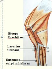
Lacertus fibrosus
|
|
|
Which ridge of the trochlea of the femur is most important in patellar lock?
|
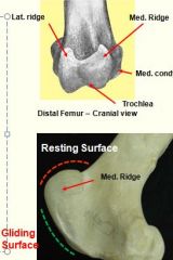
The larger medial ridge
|
|
|
What goes through tarsal canal?
|
DDF
|
|
|
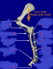
Name continuations of femoral artery?
|
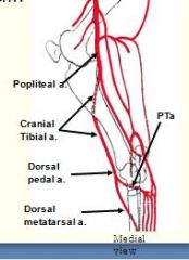
1. femoral a.
2. popliteal a. - after caudal femoral a. splits off on dorsomedial side of stifle; passes thr. two heads of gastronmeius m. 3. cranial tibial a. - goes btw. tibia and fibula 4. dorsal pedal a. - passes over flexor surface of tarsus 5. great metatarsal a. |
|
|
Main blood supply to carpus/manus?
|
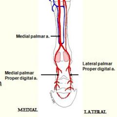
medial palmar artery
|
|
|
1. provides main innervation to structures of the manus ?
2. divides above carpus into ____ which descend the limb |
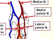
The median n.
divides above carpus into medial and lateral palmar nerves |
|
|
When does dorsal pedal a. become great metatarsal a.?
|
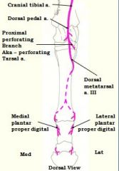
after perforating tarsal a. branches off and goes thr. tarsus
|
|
|
Where does saphenous arise from?
|
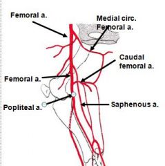
femoral a. (in femoral triangle); travels caudomedially.
|
|
|
Nerves supplying the dorsal aspect of the distal forelimb (antebrachium and on) are: (name four)
|
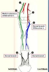
1. Dorsal branch of the ulnar nerve
2. medial cutaneous antebrachial nerve (branch of musculocutaneous n.) 3&4. dorsal branches from medial and lateral palmar (proper) digital nn. ~last couple are only nerves to dorsal aspect of digits |
|
identify
|

see picture
|
|
|
Which accessory ligaments present in hind limb?
In forelimb? |
; DDF, not for SDF b/c inserts on calcaneus according to Pasquini (p.191)
both in forelimb |
|
|
Does SDF insert on calcaneus bone?
Does DDF insert on calcaneus bone? |

Does SDF insert on calcaneus bone?
YES (that is does not have proximal check ligament / accessory.tendon like in forelimb) NO, instead it goes thr. tarsal canal created flexor retinaculum |
|
|
just above fetlock, medial palmar n. becomes the _______..
|

just above fetlock, medial palmar n. becomes the *medial (palmar proper) digital n.*
(runs along side of lateral palmar proper digital n. on abaxial side) |
|
|
Calcanean tendon is primarily* formed by what two muscles?
|
SDF, gastrocneius m.
|
|
|
Where do peroneus tertius m. and long digital extensor m. originate?
|

extensor fossa of femur
|
|
|
when/where does axillary a. start?
|
Subclavian a. becomes axillary a. after subscapular a. splits off
|
|
|
when/where does axillary a. become brachial a.?
|
axillary a. becomes brachial a. ,,,,after common interosseus a. breaks off
|
|
|
Name 3 annular ligaments:
|
1. plantar/palmer annular ligament
2. proximal digital annular ligament 3. distal digital annular ligament |
|

Identify
|

long plantar ligament
occupies lateroplantar aspect of hock (from calcaneus to 4th tarsal and metatarsals). Stabilizes calcaneus when under force of common calcaneal tendon. |
|
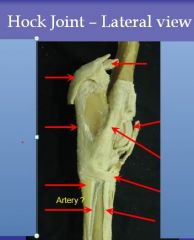
identify structure in image of hock
(What artery runs lateral to metatarsal IV?) |

see pic
|
|

identify 1-8
note: #7 is ligament, #6 and #8 are impressions, also impression to right of #5 |
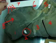
1 left lateral lobe of liver
2. left medial lobe of liver 3. quadrate lobe 4. right lobe 5. portal vein w/duodenal impression below it 6. esoph.impression / hilus 7. triangular lig. 8. gastric impression |
|
|
Lateral and medial palmar proper digital arteries are branches of -------
|
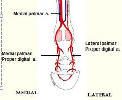
Medial palmar artery
|
|
|
is front limb where accessory (check) ligament of DDF attach?
|

back of carpus
|
|
|
cranial most muscle on forelimb ?
|
extensor carpi radialis
(common digital extensor is slightly lateral, w/LDE behind that) |
|
|
caudal most muscle on forelimb ?
|
lateral ulnar m.
|
|
|
what two tricep heads visible on medial side?
(deep to what???????????????????) |
* long head (deep to tensor fascia antebrachii) <--largest
* medial head (more cranial. tow. middle) |
|
|
what two tricep head visible on lateral side?
|
*LONG head <--larger, more caudal tricep head
* lateral head |
|
|
Digital veins ->_____ -> cephalic v.
|
palmar v.
|
|
|
flexors of digits and carpus are innervated by?
|
median and ulnar n.
therefore these nerves must pass thru caudolateral part of antebrachium |
|
|
what muscles are innervated by radial n.?
|
Extensors of arm (triceps), and Extensors of carpus/digits! Although does not extend to carpus, musc.parts of extensors (CDE, LDE) are more proximal.
|
|
|
Which muscle is present in extensor retinaculum and extensor groove (on Cr/L aspect) but is flexor of carpus?
|
Ulnaris lateralis m.
(also innervated by radial n.) |
|
|
Where does ulnaris lateralis m. insert?
|
accessory bone
(extensor ulnaris lateralis m. and flexor ulnaris lateralis m. both insert on accessory bone) |
|
|
What is path of radial n. and where does it extend to?
|
* starts caudal to humerus, goes around lateral side (innervating tricep muscles)
* Arise on Cr/L aspect of the distal humerus. * running deep along cranial side * stops before getting to carpus/hock! on very front of distal humerus |
|

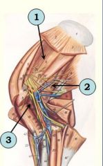
identify
|
2. Infraspinatus m.
3. Deltoideus m |
|
|
Where on the patella is the parapatellar fibrocartilage found?
|
Medial angle
|
|
identify
|

Medial group
1. Subscapularis m. 2. Teres major m. 3. Coracobrachialis m. |
|

identify muscles
|
1. triceps brachii m.
2. tensor fascia antebrachii m. |
|
|
what nerve is at risk if humerus breaks?
|
radial n., runs right on bone
|
|
|
Where are flexors of carpus and digits located on forearm?
Which flexor has 3 heads? |
caudomedial aspect, in "flexor groove"
flexor carpi radialis, FCU, SDF, DDF (3 heads) FYI: 1 of 3 heads of DDF is actually superficial to SDF? |
|
|
where do common and lateral digital extensors originate?
|
lateral epicondyle of humerus
|
|
identify
|

ulnar n.
|
|
identify
|

medial n.
|
|
|
what muscle is superficial cervical lymph node attached to or deep to?
|
brachiocephalicus m. w/omohyoideus m. attached also on bottom
|
|
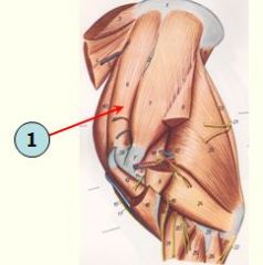
identify muscle
what is major function? |

Supraspinatus m.
Important function is stabilization of the shoulder joint Extends shoulder joint |
|
|
what nerve is at risk if humerus breaks?
|
radial n., runs right on bone
|
|
|
ulnar nerve divides into ?
|
dorsal br. of ulnar nerve
palmar br. of ulnar nerve |
|
|
Where do br. of ulnar nerve meet br. of median nerve ?
Where nerve does this form? |
Palmar br. of ULNAR nerve joins with lateral branch of MEDIAN nerve to form:
LATERAL PALMER NERVE |
|
|
What bones are located in the proximal row of the hock joint?
|

Talus
Calcaneus |
|
|
What bones are located in the middle and distal rows of the hock joint?
Which two are fused? Which is two stories? |

T1 and T2 (fused)
T3 and T4 (T4 is two story bone) |
|
|
Oviduct/uterine tube parts cranio-caudally:
|
Infundibulum - Ampulla - Isthmus
|
|
|
part of the cervix that projects into the vagina is called portio vaginalis, it _______ in the horse.
|
forms the annular fornix
|
|

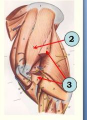
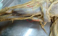
identify muscles in lateral shoulder shown
|

see picture
|
|
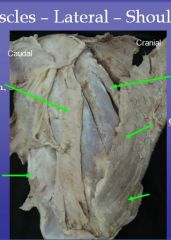
identify muscles in lateral shoulder shown
|

see picture
|
|

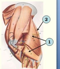
identify muscles in medial shoulder shown
|
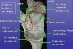
see pic
|
|
|
Where does the dorsal limb of the cunean tendon insert on?
|

Metatarsal 3
|
|
|
What bursa lies under the cunean tendon and over the medial collateral ligament of the hock?
|
Subtendinous or cunean bursa
|
|
|
Above the fetlock the medial palmar a. divides into ____ which course on the abaxial surfaces of the proximal sesamoid bones
|

medial/lateral palmar proper digital aa.
|
|

identify structures from stifle - medial view
|
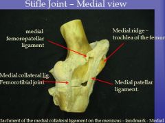
see pic
|
|
|
What structure does the medial cutaneous antebrachial n. (from musculocutaneous) course over?
|
Lacertus fibrosus
|
|

identify structures in image of hock
|

see pic
|
|
|
What is located between insertion of accessory gluteal muscle
AND cranial part of greater trochanter of the femur? |
trochanteric bursa
|
|
|
Where does the medial limb of the cunean tendon insert on?
|
Tarsals 1 and 2
|
|
|
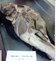
Which patellar ligaments is attached to the parapatellar fibrocartilage/parapatellar ligament of patella?
|
medial patellar ligament
|
|
identify structure shown from forelimb
|
dorsoscapular ligament
|
|

identify structure on hindlimb:
|
see pic
|
|
|
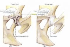
what ligaments of hip joint insert on fovea capititis ?
what are their origins? |
accessory ligament - prepubic tendon
ligament of head of femur - acetabular fossa |
|
|
What bones are involved in the hip joint?
|

Ilium
Pubis Ischium Head of Femur |
|

identify these 3 ligaments of hip joint
|

see pic
|
|
|
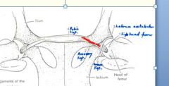
List ligaments of the hip joint.
|
Lig. of head of the femur, the accessory lig., and transverse acetabular lig. and labrum acetabulum (acetabular lip) which deepens acet.cavity
|
|
|
identify joint cavities shown on lateral stifle:
(what's cavity on medial side called?) |

femoropatellar joint cavity
lateral femorotibial joint cavity medial cavity (just caudal to medial collatoral ligament, where meniscus is) |
|
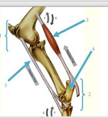
identify parts of stay aparatus
|

see picture
|
|
|
what are branches of sciatic nerve?
what's their relation to each other? Where does split happen? |

tibial n. <- caudal
common peroneal n. <- more cranial ~split just before stifle joint |
|
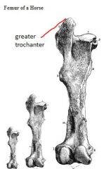
what are bony process on proximal femur called?
What is major impression on shaft called? |

trochanters
greater trochanter (distinct cranial and caudal parts) Third trochanter Lesser trochanter / trochanter minor is in the form of ridge |
|

accommodate
terminal branches of the Plantar proper digital arteries, which form the terminal arch inside the bone |

Solar foramina (red arrows) – accommodate
terminal branches of the Plantar proper digital arteries (along proximal edge of P3, closer to P2, are extensor processes) |
|
identify bony processes
|

see pic
|
|
|
which has rounder shaft 3rd metatarsal or 3rd metacarpal?
|
metatarsal
|
|
|
• Dorsal branch of accessory n.
|
Dorsal branch innervates the trapezius m.
|
|
|
• Ventral branch of accessory n. dives into ?
|
Ventral branch of accessory n. dives into sternocephalicus m.
|
|

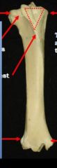
identify structure shown
|

1. caudate lobe
2. unknown 3. caudal vena cava 4. triangular ligament 5. caudate process of caudate lobe |
|
|
what lung has accessory lobe?
|
RIGHT
|
|

identify structures on humerus
|
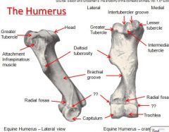
see pic
|
|
|
list accessory glands (3) in order (cr. to cd.) based on location to root of penis:
|

1. vesicular gland
2. bulbourethral gland 3. prostate gland |
|
identify structures
|

see pic
|
|

identify
|

ampulla of DD
|
|
|
surrounding external urethral process
|
Fossa glandis of penis
|
|

|
proper lig. of ovary
|
|
|
when penis erect which is further back on shaft: preputial ring or preputial oriface?
|
preputial ring is front of preputial orifice
|
|
|
saphenous n. originates from ?
|
femoral n. (medial side of leg)
|
|
|
is saccus cecus closer to cardiac/esoph. or pyloric sphincter? (edit response)
|
*cranial portion?, closer to cardiac sphincter w/esoph.
* assoc. w/fundus and nonglandular portion * appears white, with internal folds |
|
|
cardiac groove is bigger on which lung?
|
LEFT lung has bigger cardiac groove
|
|
|
which nerve wraps (makes U-turn) around aorta and to go back cranially?
|
LEFT recurrent laryngeal n.
|
|
|
which lung has accessory lobe?
|
RIGHT lung has acc. lobe
|
|
|
describe path of caudal vena cava and its relation to accessory lobe of ___lung:
|
- caudal vena cava extends to RIGHT of mediastinum along w/R.phrenic n. (inside caval fold)
- space betw. caval fold and caudal mediastinum (mediastinal recess) houses acc.lobe of R lung |
|
|
Which side is azygous v. present on?
|
RIGHT side only
|
|
|
External jugular v. located inside?
|
jugular groove
|
|
|
two parts of brachiocephalicus?
|
cleidomastoideus m.
cleidobracialus m. |
|
|
contains carotid sheath?
|
visceral space
|
|
|
trick for remembering colon
|
r's v. sm. funny like very put. fsh
L D don't ffL R dsr 444-1322 |
|
|
what part of asc.colon has largest diameter?
|
Right dorsal colon
|
|
|
narrowest part of colon?
|
left dorsal colon
|
|
|
which has more taeni coli, ventral or dorsal loop of asc.colon?
|
ventral loop has 4 TC
LD has 1 RD has 3 transv. has 2 desc. has 2 |
|
|
which kidney is heart shaped
|
RIGHT
|
|
|
how do u tell Cr&Cd. terminal recess of kidney apart?
|
urethra always to caudal side
(and in penis shaped left kidney, pointy end is cranial) |
|
|
coronary sinus is below ?
what is below CS? |
CS is below caudal vena cava, on R. side of heart
subsinuosal IV groove |
|
|
what is muscle between external jugular vein and carotid sheath?
|
omohydoideus m.
|
|

Identify structure here
|

see pic
|
|

What arteries does Great M/t artery give off?
Where? |

Medial/Lateral plantar (proper) digital arteries
- branch off just proximal to fetlock joint (where dr. aires is pointing) |