Reading...
![]()
Play button
![]()
Play button
![]()
Use LEFT and RIGHT arrow keys to navigate between flashcards;
Use UP and DOWN arrow keys to flip the card;
H to show hint;
A reads text to speech;
55 Cards in this Set
- Front
- Back
|
Purpose of the thorax
|
•
To provide bony protection to the heart, lungs, and great vessels. • To assist in breathing (air exchange). • To provide protection to abdominal viscera. • Act as conduit for structures that pass through the thorax from one body region to another & for structures that connect organs in the thorax to other body regions. |
|
|
____separates the thorax from the abdomen
|
diaphragm
|
|
|
–
Serves to attach the vertebral column posteriorly to the sternum anteriorly. – The internal organs –heart & lungs-are housed and protected within |
rib cage
|
|
|
–
Manubrium – Body – Xiphoid process |
parts of the sternum
|
|
|
Anterior cartilaginous ends of the first seven ribs
|
•
Sternocostal joints – |
|
|
–
Manubrium fuses with the body of the sternum |
•
Manubriosternal joint |
|
|
–
Cartilages of rib 6-10 form small synovial-lined joints |
•
Interchondral joints |
|
|
–
Attachment of ribs to the vertebral column – Facet & demifacets: articulating surfaces on the vertebral body |
Costovertebral & costotransverse joints
|
|
|
Movements of Costovertebral & costotransverse joints
– Nonaxial, diarthrodial, gliding joints – Elevation & depression of the rib cage |
Movements of Costovertebral & costotransverse joints
|
|
|
Innervation/nerves
• Dermatomes • Thoracic spinal nerves • Sympathetic nervous system • Phrenicnerves (C3-C5) – Controls the diaphram |
(T2-T12)
|
|
|
Innervation/nerves
• Phrenicnerves |
–
Controls the diaphram (C3-C5) |
|
|
Lie on either side of the mediastinum surrounded by the right and left pleural cavities
|
lungs
|
|
|
Originate from the pulmonary trunk
– Carry deoxygenated blood to the lungs from the right ventricle of the heart. • |
Pulmonary arteries
|
|
|
Superior & inferior veins carry oxygenatedblood from the lungs back to the heart.
|
Pulmonary veins
|
|
|
Occurs when people hold their breath and exhale.
|
Valsalva’s Maneuver
|
|
|
Innervation
– Diaphragm: |
phrenic nerve (C3, C4, C5)
|
|
|
Innervation
– Intercostal muscles: |
intercostal nerve (T2-T6)
|
|
|
Nose, oral cavity, pharynx, & larynx
|
upper respiratory tract
|
|
|
Trachea, bronchial tree
|
lower respiratory tract
|
|
|
What phases of respiration
– Elevation (raising) of ribs & increase in size of thoracic cavity via descent of the diaphragm muscle & expiration of the thoracic wall – Occurs when an individual is resting or sitting quietly – |
•
Inspiration |
|
|
Diaphragm muscle
• External intercostal muscles |
Quiet inspiration phase
|
|
|
What respiration phase uses:
Diaphragm muscle • External intercostal muscles Sternocleidomastoid • Scalenes • Pectoralis major • Levator costarum • Serratus posterior superior |
Deep inspiration phase:
• |
|
|
Occurs when an individual is breathing harder
|
Deep inspiration phase:
• |
|
|
What respiration phase uses:
Diaphragm muscle • External intercostal muscles Sternocleidomastoid • Scalenes • Pectoralis major • Levator costarum • Serratus posterior superior• Levator scapula • Upper trapezius • Rhomboids • Pectoralis minor |
Forced inspiration phase
|
|
|
Occurs when an individual is working very hard; in a state of “air hunger.”
|
Forced inspiration phase
|
|
|
Occurs through relaxation of the external intercostal muscles,
• Gravity pulling the rib cage downward, • Elastic recoil of the thoracic wall & tissue of the lung & bronchi. • (internal intercostals) • No muscle action is occurring. |
Quiet expiration phase
|
|
|
Muscles that can pull down on the rib & muscles that can compress the abdomen, forcing the diaphragm upward.
|
Forced expiration phase:
|
|
|
What expiration phase uses:
Internal intercostals +: • Rectus abdominis • External oblique • Internal oblique • Quadratus lumborum • Transverse abdominis • Serratus posterior inferior |
Forced expiration phase:
|
|
|
Common pathologies of the respiratory system
|
Upper respiratory infection (URI)
Lower respiratory infection (LRI) Bronchitis Emphysema Asthma Hyperventilation Stitch Hiccups Pleurisy Pneumothorax Rib separation Rib dislocation Flail chest |
|
|
What system?
heart pumps blood, which creates blood pressure, and circulates oxygen, nutrients, and other substances |
circulatory system
|
|
|
Fibroserous sac surrounding the heart & the roots of the great vessels
|
pericardium
|
|
|
receives blood from the upper body by way of the superior vena cava & from the lower body via the inferior vena cava
|
right atrium
|
|
|
receives blood from the lungs by way of the four pulmonary veins.
|
left atrium
|
|
|
has relatively thin walls & pumps blood to the lungs through the pulmonary artery.
|
right ventricle
|
|
|
has thicker walls & pumps blood to the body through the aorta
|
left ventricle
|
|
|
More force is required to pump the blood through the body than through the lungs, so which muscular wall is thicker?
|
left ventricle
|
|
|
Interatrial, interventricular, & atrioventricular septa separate-----.
|
the four chambers of the heart.
|
|
|
means contraction
|
systole
|
|
|
means relaxation
|
diastole
|
|
|
When the ventricles are in systole, the atria are in diastole
|
cardiac cycle
|
|
|
–
Heart sounds –two sounds per heartbeat: lub-dup created by the closure of the valves • First sound – |
closure of the AV valves(lub)
|
|
|
–
Heart sounds –two sounds per heartbeat: lub-dup created by the closure of the valves • Second sound – |
closure of the aortic & pulmonary semilunarvalves (dup)
|
|
|
heart rates:
normal adult? child? physically fit? |
Adults 60 to 80 beats per minute
– Children & infants have faster pulses – Person excellent physical condition has a slow resting pulse |
|
|
amount of blood pumped out of the ventricle in 1 minute
|
cardiac output
|
|
|
average resting cardiac output
|
5 to 6 Liters
|
|
|
•
Decreases heart rate • Reduces force of contraction • Constricts the coronary arteries |
–
Parasympathetic |
|
|
Increases heart rate
• Increases the force of contraction |
Sympathetic
|
|
|
•
Form the passageway for the blood • Help maintain blood pressure & provide the capillaries sites for the exchange of materials between the blood & the tissues. |
Vascular system:
– Arteries, capillaries, & veins |
|
|
–
Return tissue fluid to the blood to maintain blood volume; – To protect the body against pathogens & other foreign material |
Lymphatic System
|
|
|
Parts of the lymphatic system?
|
Parts
• Lymph & lymph vessels • Lymphatic tissue: lymph nodes & nodules, spleen, & thymus |
|
|
Purpose of Abdomen
|
•
To house and protect major viscera To assist with breathing |
|
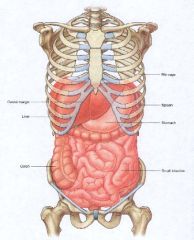
What is housed in the abdominal cavity?
|
Small intestineColonLiverStomachSpleenRib cage
|
|
|
To break down food into simple chemicals that can be absorbed into the blood & lymph and utilized by cells
|
digestive system
|
|
|
Organs of the digestive system
|
esophagus
• Stomach • Small intestine – Duodenum, Jejunum, ileum • Large intestine – Ascending colon, transverse colon, descending colon, sigmoid colon • Rectum & Anal canal • Nerve Supply: Vagus Nerves |
|
|
Regulators of the internal environment of the body
|
Urinary & Endocrine System
|