![]()
![]()
![]()
Use LEFT and RIGHT arrow keys to navigate between flashcards;
Use UP and DOWN arrow keys to flip the card;
H to show hint;
A reads text to speech;
30 Cards in this Set
- Front
- Back
|
Male Reproductive System Overview |

1. Paired Testes 2. Set of Tubules form Testes to Penis 3. Glands That contirbute semen 4. Penis (Muscles) |
|
|
Glands that Contribute Semen |
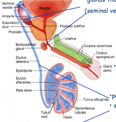
1. Seminal Vesicle 2. Prostate 3. Bulbourenthral Gland |
|
|
Tubules From the Testes |

1. Tubuli Recti 2. Rete Testes 3. ductuli Efferentes 4. Epididymis 5. Ductus Deferens |
|
|
Testes |
1. Seminiferous Epithelium 2. Spermatogenesis 3. Sertoli and Leydig Cells 4. Ducts in Testes |
|
|
Testis |
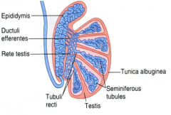
Primary Male Sex Organ 1. Produce Spermatozoa 2. Produce Testosterone |
|
|
Tunica Albuginea |
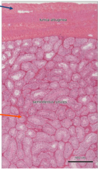
1. Dense Fibrous CT 2. Posteriorly Mediastinum Testis |
|
|
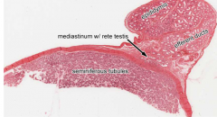
Mediastinum Testis |
1. Septa subdivide testis into 250 lobules |
|
|
Seminiferous Tubules |
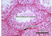
Site of spermatogenesis |
|
|
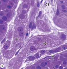
Seminiferous Epithelium |
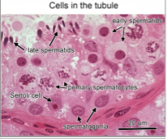
1. Sertoli Cells: Physically and metabolically support developing sperm, form blood testis barrier (Prevent autoimmune attacks) 2. Spermatogenic Cells: Produce Sperm |
|
|
Interstitial Tissue |
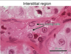
1. Leydig Cells: produce androgens (testosterone) |
|
|
Sertoli Cells |
1. Produce androgen binding protein, inhibin also phagocytose excess cytoplasm shed during spermatogenesis. |
|
|
Leydig Cells |
1. Characteristic lipid droplets of steroid producing cells. 2. Produce Androgens (Testosterone) |
|
|
Spermatogenesis Diagram |
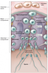
1. Spermatogonia 2. Primary Spermatocyte 3. Secondary Spermatocyte 4. Spermatid |
|
|
Spermatogenesis Histology |
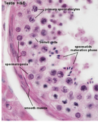
1. Sertoli= Support and Blood Barrier 2. Spermatogonia-Primary Spermatocyte-Secondary Spermatocyte-Spermatid |
|
|
Intratesticular Ducts |
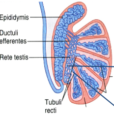
Carry spermatozoa and liquid from seminiferous tubules to the duct of the epididymis 1. Tubuli Recti 2. Rete Testes |
|
|
Tubuli Recti |
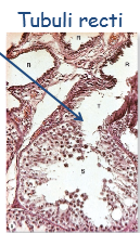
1. Connect Seminiferous Tubules to Rete Testes |
|
|
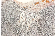
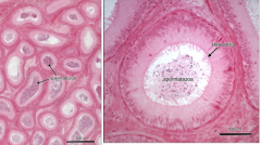
Rete Testis |
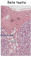
1. Drain into 20ish ductuli efferentes 2. Simple Cuboidal ET |
|
|
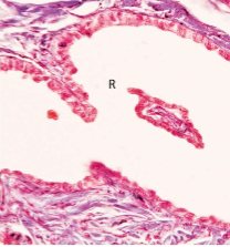
Rete Testis High Mag |
|
|
|
Ductuli efferentes |
1. Connect Rete Testis to Epididymis 2. Scalloped or Festooned Epithelium 3. Alternates between non-ciliated cuboidal cells with microvilli and taller ciliated cells |
|
|
Epididymis |
1. Connects Ductuli Efferentes to Ductus Deferens 2. Single tubule 4-5 meters long, highly tortuos 3. Lined with pseudostratified columnar ET 4. Sperm Undergo Final Maturation here, become motile |
|
|
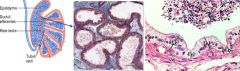
Testis to Epididymis Overview |
|
|
|
Ductus Deferens |
1. Connects Epididymis to the urethra via the ejaculatory duct in the prostate Gland 2. Long straight tube 3. Lined with pseudostratified columnar ET, some stereocilia 4. THick Muscularis (Longitudinal inner and outer with middle circular) |
|
|
Accessory Glands |

Contribute to the Semen 1. Seminal Vesicle: Exocrine Gland 2. Bulbourethral Glands Prostate Gland |
|
|
Seminal Vesicle |
1. 70% of Ejaculate -Fructose -Prostaglandins= Stimulate acitivty in female reproductive tract - Firbrinogen |
|
|
Bulbourethral Glands |
Release clear mucus like secretion to coat urethra. Make ejaculation smoother |
|
|
Prostate Gland |
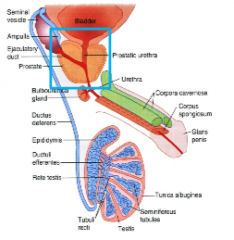
1. 3 Layers of Tubuloacinar Glands 2. Secrete Proteolytic enzymes, high levels of zinc, citric acid, and acid phosphatase 3. Produces prostate specific Antigen which helps to slow release of sperm |
|
|
Layers of Prostate |
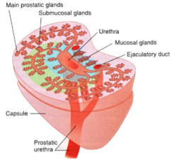
1. Transition Zone= Mucosal Glands 2. Central Zone= Submucosal Glands 3. Peripheral Zone= Main Glands (primary area of prostate cancer) ***All ducts empty into prostatic urethra |
|
|
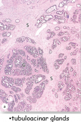
Prostate Gland: Tubuloacinar Glands |
|
|
|
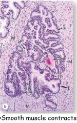
Prostate Gland: Smooth Muscle Contraction |
|
|
|
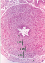
Prostate Gland: Corpus Amylaceum |
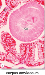
Concretions: Corpora Amylacea become numerous with age |