![]()
![]()
![]()
Use LEFT and RIGHT arrow keys to navigate between flashcards;
Use UP and DOWN arrow keys to flip the card;
H to show hint;
A reads text to speech;
36 Cards in this Set
- Front
- Back
|
ABCDs of Melanoma |
A= Asymmetry B = Borders irregular C = Colour variation D = Diameter >0.6cm Dark colour |
|
|
Allen's test: 1) What for 2) When 3) How |
1) Clinical test to test patency of ulnar artery 2) Before ABG 3) Patient elevates hand and makes a fist Occlude both radial and ulnar arteries with thumbs x 30 secs Tell patient to open hand Hand should appear blanched Un-occlude ULNAR artery. Colour should return in 5-15 secs |
|
|
Ballance's sign |
Constant dullness in the Left flank with resonance in the Right flank Sign of: Splenic rupture Haematoma |
|
|
Barrett's oesophagus What cells usually line oesophagus? |
Columnar metaplasia, with goblet cells, of the distal oesophagus, assoc with GORD Normally: Stratified squamous cells |
|
|
Battle's sign |
Ecchymosis over mastoid processes Due to basilar skull fracture |
|

Beck's triad |
(Beck becoming muffled as he grabs his chest on stage) Seen in Cardiac Tamponade 1) JVD 2) Decreased BP 3) Decreased/muffled heart sounds |
|
|
Bergman's triad |
(iceBERG made of FAT - a FatBerg) Seen in fat embolus syndrome 1) Mental status changes 2) Petechiae (axilla/thorax espec) 3) Dyspnoea |
|
|
Blumer's shelf |
Metastatic disease of rectouterine/rectovesical pouch Creates a 'shelf' palpable on DRE |
|
|
Boa's sign |
(BOA constricting your CBD) Right subscapular pain Seen in cholelithiasis |
|
|
Borchardt's triad |
(B for belly) Seen in gastric volvulus 1) Emesis, followed by retching 2) Epigastric distension 3) Failure to pass NG tube |
|
|
Carcinoid triad |
(FDR in his Car) 1) Flushing 2) Diarrhoea 3) Right heart failure |
|
|
Charcot's triad |
(CHarcot's for CHolangitis - sCHARCs find bile ducts particularly delicious, especially when cooked in a FLAME) Sign of cholangitis 1) Fever (chills) 2) Jaundice 3) RUQ pain |
|
|
Chvostek's sign |
(CHeek for CHvostek) Twitching of facial muscles As you tap the cheek Sign of HyPOcalcaemia |
|
|
Courvoisier's Law |
GB nontender + enlarged = NOT chronic cholelithiasis BUT obstructed CBD, MC due to pancreatic cancer Chronic cholelithiasis ==> scarring and tenderness |
|
|
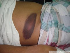
Cullen's sign |
Blueish discolouration of the periumbilical area due to retroperitoneal haemorrhage tracking to anterior abdominal wall through fascial planes. Common cause = acute haemorrhagic pancreatitis |
|
|
Cushing's triad |
Signs of increased intracranial pressure 1) Hypertension 2) Bradycardia 3) Irregular respirations |
|
|
Dance's sign |
Empty RLQ in children with ileocaecal intussusception |
|
|
Fothergill's sign |
Used to differentiate intra-abdominal masses from masses in abdominal wall If mass can still be felt with tension on the musculature of the abdominal wall (e.g. half sitting upright position), the mass is in the abdo wall. |
|
|
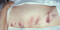
Fox's sign |
Ecchymosis of the inguinal ligament Due to retroperitoneal bleeding tracking |
|
|
Goodsall's rule |
Anal fistulae Course in a straight path anteriorly and A curved path posteriorly |
|
Clinical sign? |
Cullen's sign (see the vampire bites!?) Blueish discolouration of the periumbilical region due to retroperitoneal haemorrhage tracking through fascial planes to anterior abdo cavity Common cause = acute haemorrhagic pancreatitis |
|
Clinical sign? |
Fox's sign Ecchymosis of the inguinal ligament due to retroperitoneal bleeding tracking |
|
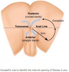
What is this diagram trying to demonstrate? |
Goodsall's rule Anal fistulae Course a straight path anteriorly and a Curved path posteriorly |
|
|
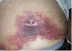
Grey Turner's Sign |
Bluish discolouration of the flank due to retroperitoneal haemorrhage with blood dissecting to flanks |
|
|
Hamman's sign |
(The heart is a piece of ham) Sign of emphysematous mediastinum Crunching sound heard on auscultation of the heart Commonly due to Boerhaav's syndrome, pneumomediastinum etc |
|
|
Howship Romberg's Sign |
(Ship sailing out of your back passage onto your thigh) Obturator hernia sign Pain along the inner aspect of the thigh due to nerve compression by obturator hernia |
|
|
Kehr's sign |
(Splenic rupture sign) Severe Left shoulder pain Due splenic rupture Causing blood to irritate diaphragm ==> referred pain to Left shoulder |
|
|
Homan's sign |
DVT sign Forced dorsiflexion of the foot ==> Calf pain |
|
Clinical sign? |
Grey Turners A sign of retroperitoneal haemorrhage Blueish discolouration of the flank due to dissecting of blood from the retrperitoneum |
|
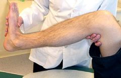
What sign is being tested? |
Homan's sign Sign of DVT Forced dorsiflexion ==> Calf pain |
|
Clinical sign? |
Kehr's sign Sign of splenic rupture Left dhoulder pain due to diaphragmatic irritation |
|
|
Parkland formula |
Formula used for calculating how much IV fluids needed by burn victim in first 24hrs of Rx 4 x weight of patient in kg x % body burned in mL First half of fluids is given in first 8hrs. Next half is given over the next 16hrs |
|
|
Jenkin's rule |
Amt of suture needs to be 4 times the length of the wound |
|
|
Hilton's Method |
Abscess exploration method prior to drainage Incision made over abscess Wound is explored bluntly using a forceps |
|
|
Sebaceous cyst: How do you clinically diagnose? |
1) Fluctuation: Test twice. at different right angles, minimum 2) Transillumination |
|
|
Wrist ganglion: How do you clinically diagnose |
1) Location: At the wrist 2) Can move skin freely over the ganglion 3) Fluctuation in 2 directions 4) Transilluminates brilliantly |