![]()
![]()
![]()
Use LEFT and RIGHT arrow keys to navigate between flashcards;
Use UP and DOWN arrow keys to flip the card;
H to show hint;
A reads text to speech;
20 Cards in this Set
- Front
- Back
|
Sensation |
The act of receiving a stimulus |
|
|
Perception |
The act of interpreting a sensation |
|
|
Simple Unencapsulated receptors |
These receptors have free dendritic nerve endings that often sense pain and temperature. Examples include Merkel discs, root hair plexi, and itch receptors. |
|
|
Simple encapsulated receptors |
These receptors function largely as mechanoreceptors and consist of fiber terminals enclosed in connective tissue. Examples include Pacinian corpuscles, Ruffini's corpuscles, and golgi tendon organs. |
|
|
Sweet, salty, sour, bitter, umami, fat |
These are the six taste sensations we can perceive. |
|
|
Chemorecptors |
These receptors respond to chemicals that dissolve in solution. Examples are olfactory receptors and taste buds |
|
|
Diplopia and strabismus |
Two disorders of the eye caused by homeostatic imbalances of the extrinsic eye muscles. |
|
|
Fibrous tunic |
This forms the outer coat of the eye and consists of both the sclera (white of the eye) and the cornea. |
|
|
Choroid region |
Part of the vascular tunic (uvea), this region supplies blood to the tunics of the eye. |
|
|
Iris |
Part of the vascular tunic, this region contracts (parasympathetic action) and dilates (sympathetic reaction) to regulate the amount of light that stimulates the retina. |
|
|
Retina |
Part of the sensory tunic, this region can be divided into two layers; the pigmented layer and the neuronal layer. |
|
|
Cones |
Photoreceptors that respond only to bright light and are responsible for high-acuity color vision. Each of these receptors synapse with only one ganglionic cell (1:1 ratio). |
|
|
Rods |
Photoreceptors that respond to very low levels of light and are concentrated along the periphery of the retina. These receptors synapse with ganglionic cells at a 10:1 ration (10 receptors for each ganglionic cell). |
|
|
Organ of Corti |
This organ is responsible for hearing and is located in the cochlear duct. Afferent fibers of the cochlear nerve synapse with outer hair cells that have stereocilia. These stereocilia allow for the sensation of vibrations within the tectorial membrane. |
|
|
Semicircular canals |
These structures detect angular acceleration along three perpendicular axes. Stereocilia encapsulated in capulae allow for acceleration sensation. |
|
|
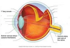
eye diagram (counterclockwise from top left): ciliary body, cornea, iris, pupil, aqueous humor, lens, vitreous humor, optic disc, optic nerve, fovea centralis, retina, choroid, sclera. |
|
|
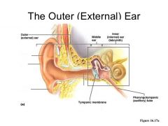
Outer ear (counterclockwise from top left): Auricle (pinna), helix, lobule, external auditory meatus |
|
|
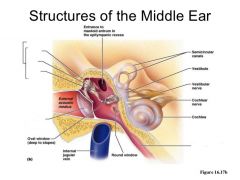
Middle ear (counterclockwise from top left): Ossicles, malleus, incus, stapes, tympanic membrane, pharyngotympanic tube |
|
|
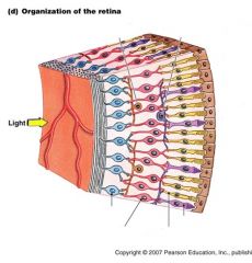
Organization of the retina (counterclockwise from top right): horizontal cell, amacrine cell, ganglion, bipolar cell, rod, cone |
|
Why do we sense sound of different frequencies at different segments of the basilar membrane |
The range of frequencies we can sense is due to the length of basilar fibers along the basilar membrane. vibrations of higher frequencies localize around the base of the membrane while vibrations of lower frequencies localize around the apex. |