![]()
![]()
![]()
Use LEFT and RIGHT arrow keys to navigate between flashcards;
Use UP and DOWN arrow keys to flip the card;
H to show hint;
A reads text to speech;
122 Cards in this Set
- Front
- Back
|
What do the first order neuron of the sensory pathways starting at receptor do? |
(PNS) Sensory fiber carry impulses to spinal cord/brainstem |
|
|
What do the second order neuron of the sensory pathway starting at the receptor do? |
(CNS) Sensory fibers carry impulses up spinal cord/brainstem to thalamus |
|
|
Where do the second order neuron sensory fibers carry impulses to? |
Thalamus |
|
|
What do third order neuron of the sensory pathway starting at the receptors do? |
(CNS) Sensory fibers carry impulses from thalamus to appropriate lobe of cerebrum |
|
|
What is sensory adaptation? |
Threshold changes for a stimulus to create an action potential in receptors. Tolerance occurs and greater stimulus is needed. |
|
|
General receptors are spread throughout ___. |
the body (skin, muscle, organs, etc.) |
|
|
Special sense receptors are found ____ |
concentrated in complex organs located in head. |
|
|
What is a receptive field? |
The area monitored by a single receptor cell is its receptive field. |
|
|
The ___ a receptive field the more accurate the area of stimulus |
smaller |
|
|
What are the five receptor types? |
1. Mechanoreceptors 2. Thermoreceptors 3. Chemoreceptors 4. Photoreceptors 5. Nocireceptors |
|
|
What are mechanoreceptors? |
Detect mechanical or physical changes such as touch, vibration, pressure, or stretch |
|
|
what is proprioceptor? |
Detects position of body in space |
|
|
What is thermoreceptors? |
Detect heat or cold within range |
|
|
What are chemoreceptors? |
Detects chemical changes (molecules dissolved in fluid). |
|
|
What are chemoreceptors used for? |
Taste, smell |
|
|
What are photoreceptors? |
detects photons (light) |
|
|
What are nociceptors? |
Detects tissue damage and pain |
|
|
What is the most complex special sense organ? |
Vision |
|
|
What are the two photoreceptors? |
Rods and cones |
|
|
What do cones detect? |
Color (red, green, blue) |
|
|
What do rods detect? |
Light/dark |
|
|
Impulses for vision are sent to ___ lobe |
occipital |
|
|
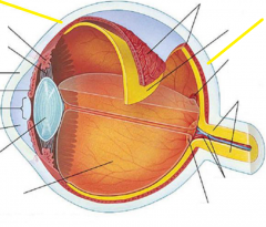
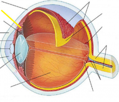
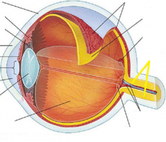
What are the three layers of the eye? |
1. Fibrous tunic 2. Vascular tunic 3. Nervous tunic |
|
|
What are the two layers of the fibrous tunic? |
Cornea and sclera |
|
|
What inserts into the sclera? |
Sex extra-ocular muscles |
|
|
The sclera contains ___ and ___ |
small blood vessels, nerves |
|
|
The cornea is made up of ____ |
stratified squamos epithelium and dense matrix of multiple layers of collagen fibers. |
|
|
The sclera appears ___ the cornea appears ___ |
white (white of the eye), transparent |
|
|
What are the three components of the vascular tunic? |
1. Choroid 2. Lens 3. Ciliary body |
|
|
What is the choroid? |
Pigmented layer that absorbs light |
|
|
What is the iris? |
The colored, muscular ball; focuses image on retina |
|
|
What is the ciliary body? |
Muscular ring used to adjust shape of lens; secretes aqueous humor |
|
|
What does the canal of Schlemm do? |
Absorbs aqueous humor |
|
|
What happens in glaucoma? |
Due to increased ocular pressure. The fluid in the eye (aqueous humor) does not reabsorb adequately. |
|
|
What may cause glaucoma to occur? |
High blood pressure, diabetes, surgery, family history |
|
|
What may glaucoma damage? |
Optic nerve and retina (loss of peripheral vision, or blindness) |
|
|
What is the nervous tunic also known as? |
Retina |
|
|
What is found in the nervous tunic? |
Photoreceptors, optic disc, and fovea |
|
|
What is the optic disc? |
"blindspot" optic nerve axons exit orbit; no photo receptors here. |
|
|
What is the fovea? |
Focal point. Concentration of cones at back of eye |
|

|
Palpabrae |
|

|
Palpabrae fissure |
|

|
Cornea |
|

|
Lacrimal gland |
|

|
Nasolacrimal duct |
|
|
Sclera |
|

|
Lens |
|

|
Iris |
|

|
Pupil |
|
|
Ciliary body |
|

|
Ora serrata |
|

|
Vitreous humor |
|

|
Aqueous humor |
|

|
Posterior segment |
|

|
Anterior segment |
|

|
Choroid |
|

|
Retina |
|

|
Optic disc |
|
|
Optic nerve |
|
|
What does the lacrimal apparatus do? |
Keeps a constant flow of tears that keep surface moist and clean |
|
|
How do the sclera and cornea differ? |
Sclera is vascular, dense fibrous CT containing collagen and elastic fibers. All the extra ocular muscles insert into sclera Cornea is composed of dense matrix of collagen fibers, avascular, transparent and continous with the sclera. Is covered by stratified squamos epithelium. |
|
|
What is the functional significance of the macula lutea? |
Most of the cones are concentrated in macula lutea (area where visual image arrives after passing through cornea and lens). |
|
|
What is the functional significance of the fovea centralis? |
Central portion of macula lutea where highest concentration of cones are found |
|
|
What is the action of the inferior rectus? |
Move eys inferiorly |
|
|
What is the action of the medial rectus? |
Move eyes medially |
|
|
What is the action of the superior rectus? |
Move eyes superiorly |
|
|
What is the action of the lateral rectus? |
move eyes laterally |
|
|
What is the action of the inferior oblique? |
Move eyes superiorly and laterally |
|
|
What is the action of the superior oblique? |
Move eyes inferiorly and laterally |
|
|
What muscle is involved when the pupil dilates (gets bigger)? |
Pupillary dilator muscle (radial) |
|
|
What muscle is involved when the pupil constricts? |
Pupillary constrictor muscle |
|
|
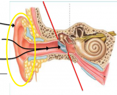
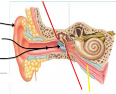
Auricle |
|

|
External acoustic meatus |
|

|
Tympanic membrane |
|

|
Ossicles |
|

|
Malleus |
|

|
Incus |
|

|
Stapes |
|

|
Oval window |
|

|
Round window |
|
|
Pharyngotympanic tube |
|
|
Describe the action of the tensor tympani |
When it contracts, the malleus is pulled medially, stiffening the tympanium and reduces amount of movement |
|
|
Describe the action of the stapedius muscle. |
Contraction pulls the stapes, reducing movement of stapes at oral window. |
|

|
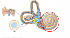
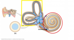
Vestibule |
|
|
Utricle |
|

|
Saccule |
|

|
Maculae |
|
|
Semicircular canal |
|

|
Ampullae |
|

|
Cristae ampullaris |
|
|
What type of sensory information stimulates the vestibule? |
Gravity and linear acceleration |
|
|
What type of sensory information stimulates the semicircular canals? |
Rotation of the head |
|
|
What structures make up the external ear? |
The auricle and the external acoustic meatus |
|
|
What structures make up the middle ear? |
Tympanic membrane and cavity, the auditory ossicles (malleous, incus, stapes), and the oval and round window. |
|
|
The inner ear is made of canals embedded in what bone? |
Temporal |
|
|
What is the structure in the inner ear responsible for hearing? |
Cochlea |
|
|
What structure in the inner ear is responsible for equilibrium? |
Vestibule |
|
|
What type of receptors are found in the cochlea? |
Mechanoreceptors |
|
|
How are the (auditory) vibrations transmitted? |
Outer ear (ear drum) -> middle ear bones -> fluid in inner ear -> bend receptors in cochlea
|
|
|
Where are the hearing impulses sent to? |
Temporal bone |
|
|
Where type of receptors are located in the inner ear responsible for equilibrium and where are those receptors located in? |
Mechanoreceptors located in semicircular canals and vestibule |
|
|
What happens within the inner ear when head or body position moves? |
Fluid moves |
|
|
Where are the impulses sent from the mechanoreceptor in the vestibules and semicircular canals? |
Cerebellum |
|
|
What is held within cochlear duct? |
Spiral organs/receptor cells |
|
|
What part of the ear begins to amplify sounds? |
Middle ear |
|
|
What part of the inner ear is responsible for dynamic equilibrium? |
Semicircular duct |
|
|
What part of the inner ear is responsible for static equilibrium? |
Utricle and saccule |
|
|
Utricle and saccule are responsible for ___ equilibrum |
Static |
|
|
Semicircular ducts are responsible for ___ equilibrium |
dynamic |
|
|
What type of receptors does the olfactory system use? |
Chemoreceptors |
|
|
Where are the receptors found for the olfactory system? |
Mucus membrane, specifically found in 1-inch path at roof of nasal cavity |
|
|
Where are the impulses sent from the olfactory system? |
Temporal lobe |
|
|
What type of receptors are used for gustation? |
Chemoreceptors |
|
|
Where are the receptors found for gustation? |
Most on tongue papillae and some on cheeks, palate, throat, and larynx |
|
|
How many different cranial nerves detect taste? |
3 |
|
|
What other special sense does the gustation system work in conjunction with? |
Olfactory |
|
|
Where are the gustation impulses sent to? |
Parietal lobe |
|
|
What are the three cranial nerves involved with gustation? |
Facial nerve (N VII), Glossopharyngeal nerve (N IX), and the Vagus nerve (N X) |
|
|
What are the three cranial nerves involved with gustation?
|
Facial nerve, glossopharyngeal nerve, and vagus nerve
|
|
|
Describe the arrangement of cells in the retina from where the light hits to the deeper layers
|
1. Light hits Ganglion cells that face the vitreous chamber
2. Bipolar cells 3. Rods and cones band around periphery posterior retinal surface |
|
|
What fluid is found in the vestibular ducts?
|
Perilymph
|
|
|
What fluid is found in the cochlear duct?
|
Endolymph
|