![]()
![]()
![]()
Use LEFT and RIGHT arrow keys to navigate between flashcards;
Use UP and DOWN arrow keys to flip the card;
H to show hint;
A reads text to speech;
444 Cards in this Set
- Front
- Back
|
name 2 immunomodulatory antibiotics |
vincristine Cyclosporine A |
|
|
name the types of hypersensitivities
|
type I: |
|
|
list the effects of glucocorticoids (i.e. pred) on the immune system: |
1. apoptosis of activated cells 2. decreases expression of cytokines , chemotaxins and surface molecules i.e. costimulators, and when APCs present to T cells without costimulators immunotolerance results 3. inhibit Fc receptor mediated phagocytosis of antibody coated cells 4. inhibits CD4+ and CD8+ T cell functions 5. reduce platelet activity and capillary blood flow
outcome = broad spectrum immunosuppression that crosses BBB
|
|
|
what are the side effects of glucorticoid use? |
1. hypoadrenocorticism 2. hypothyroidism 3. hyperparathyroidism 4. insulin resistance (diabetes) 5. GI ulceration (inhibits prostaglandins) 6. recrudescence of viral and protozoal infections especially in cats 7. infections due to immunosuppression 8. myopathy- common in large breed dogs,less activity - predisposes to UTI and aspiration pneumonia, can also cause glomerulosclerosis and precipitation of CHF |
|
|
cytotoxic agents are used to treat what specific neoplasia? |
lymphoma |
|
|
what is the onset of action for azathioprine? |
lag phase of a few weeks before start to get immunosuppression |
|
|
azathioprine is useful in both the induction and maintenance phase for treating lymphoma. True or false? |
false, it is only weakly immunosuppressive and takes a few weeks to have immunosuppressive effect so only useful in maintenance phase, cant use in induction when need something stronger is pred sparing in maintenance phase |
|
|
what are the potential side effects of azathioprine? |
- myelosuppression - non regenerative anaemia - hepatotoxicity - pancreatitis - GI effects -* also a teratogen/mutagen - important to warn owners |
|
|
name 3 cytotoxic agents |
azathioprine chlorambucil cytarabine |
|
|
which drug is recommended for treating meningitis of unknown origin (MUO)? |
cytarabine |
|
|
why is vincristine good for acute management of IMT? |
Because it binds tubulins and so concentrates in platelets - have vincristine on them and then this is exposed to splenic macrophages |
|
|
describe MOA of cyclosporine A |
interrupts signalling pathway by which T cells are triggered to proliferate, suppression of cytokines and costimulators and failure to activate expression genes for IL-2 the most important cytokine for T cell proliferation CsA is a potent T cell inhibitor but has broader anti-inflammatory and immunosuppressive effects and at higher doses can affect all immune cells |
|
|
cyclosporine A is mainly used in maintenance phase. True or false |
true |
|
|
cyclosporine A is cytotoxic. True or false? |
false |
|
|
cyclosporine A crosses the BBB. True or false? |
false |
|
|
what are the potential side effects of CsA? |
1. GIT signs - most common 2.gingival hyperplasia- common 3. hepatic, renal 4. hair growth 5. hypertension 6. Toxoplasmosis- cats 7. neosporosis- dogs 8. many drug interactions - affects their absorption i.e. pred clearance is slowed and is influences by other drugs |
|
|
describe treatment for IMHA and IMT, including induction and maintenance as well as supportive therapy |
induction: - pred + vincristine or - pred + human IgG ($$$)
maintenance: - pred + azathioprine (dogs) or chlorambucil (cats) - cyclosporine A ( T cell suppression to reduce B cell activation) - Fc receptor antagonists i.e. danazol, pred
supportive: - transfusion, circulatory support - antithrombotic i.e. clopidogrel, aspirin - GI protection - best GI protection is food |
|
|
list some reasons for loss of remission when using immunomodulatory agents |
1. drug tapered too early i.e. moved from induction to maintenance too quickly 2.higher than expected dose needed to maintain immunotolerance 3. adjuvant is present i.e. costimulatory molecule (need to look for infection that may be causative or secondary to immunosuppression i.e. UTI, asp pneumonia) 4. infection - primary or secondary i.e. UTI, aspiration pneumonia 5. neoplasia 6. prednisolone resistance |
|
|
which type of vaccines are okay to use in immunosuppressed animals? a) live b) killed c) recombinant d) none |
c) recombinant recombinant are okay because they don't have adjuvants killed vaccines should be avoided because they are given with adjuvants to stimulate expression of cosstimulatory molecules |
|
|
what should you suspect if you are presented with a young puppy with recurring infections |
primary immunodeficiency |
|
|
define primary autoimmunity |
immune response directed at self antigen with no recognisable exogenous trigger
*these diseases are more commonly linked to MHC II molecular mimicry |
|
|
list examples of pyrogens |
- endotoxin - exotoxin - viral nucleic acids - IFNs - TNF-alpha - bile acids |
|
|
what are the clinical signs of chronic PUO? |
- fever > 1wk - lethargic - malaise - vague pain responses |
|
|
list 3 primary pathogens that commonly cause chronic infections in cats |
- FIV - FIP (FCoV) - FeLV |
|
|
what is the first thing you should do when investigating a dog with PUO? |
investigate the urinary tract (including prostate in an entire male) |
|
|
describe the diagnostic investigation for PUO |
step 1: screening 1. history and PE - esp investigate urinary tract in dogs + prostate 2. CBC, biochem, UA, urine C+S and UPCR - moderate proteinuria likely due to immunoreactive GN which is why do UPCR* - UTI dogs step 2: search for specific lesion somewhere 1. imaging - thoracic rads - abdominal US - spinal and joint rads - echo (27% of dogs with endocarditis have no murmur on initial presentation) step 3: specific and invasive tests 1. joint taps 2. CSF 3. blood culture 4. skin biopsy 5. PCR for unusual microbes i.e. Bartonella in dogs 6. serology - cats |
|
|
what is the most common immune mediated polyarthropathy in dogs? |
type 1 - idiopathic |
|
|
what are the clinical signs of immune mediated polyarthropathies? |
- pyrexia - lethargy - asthenia (unwilling to move) - shifting/compound lameness - back pain if facet joints are area of most pain - distal joints i.e. carpi/tarsi are hot, have periarticular swelling and reduced ROM - collapse/deformity of the joints in severe cases (erosive forms) +/- CS related to underlying causes |
|
|
describe diagnostic work up for immune mediated polyarthropathies |
1. PE 2. rads - may not see changes in early stages, if has been present for 1 month + will see changes and should be able to distinguish between erosive and non-erosive 3. arthrocentesis - confirms diagnosis - tap 4-6 joints to confirm polyarthritic - carpi and tarsi easiest - non septic neutrophilic inflammation ** if the patient is young large breed prone to SRMA should also do CSF tap to identify any concurrent meningitis 4. exploratory arthrotomy and bone biopsy - rarely done
tests to assess whole patient and look for cause: 1. CBC, biochem, UA, C+S, UPCR 2. chest rads 3. abdominal US 4. ANA - SLE potential cause 5. rheumatoid factor - will be more lymphocytes in joint fluid if RA |
|
|
what are the 4 types of IMPAs? |
type 1- idiopathic - most common - just tx the PA type 2- reactive - secondary to an infection elsewhere in body - UTI, endocarditis type 3- enteropathic arthritis - secondary to underlying enteropathy i.e. IBD type 4 - paraneoplastic - neoplasia remote from joints |
|
|
describe treatment for immune mediated polyarthropathies |
1. treat underlying cause - i.e. GN 2. immunosuppresive tx - pred + azathioprine dogs - pred + chlorambucil cats 3. analgesia 4. environmental modification |
|
|
how can diagnosis of SLE be confirmed |
positive ANA + positive for at least 2 immune mediated diseases |
|
|
list 2 ddx for UPCR > 2 |
1. renal failure 2. glomerulopathy |
|
|
describe diagnostic work up for glomerulopathy |
1. PE 2. CBC, biochem, UA including culture and UPCR - UPCR > 2 highly suggestive (check sediment) 3. abdominal imaging 4. +/- renal biopsy - histopath, electron microscopy and IHC = definitve diagnosis but very invasive test and won't really alter outcome |
|
|
prednisolone is the mainstay of therapy for immune mediated glomerulopathies. True or false? |
false. immunosuppressive therapy doesn't seem to help tx GN but may be needed to tx underlying cause generally GN just need monitoring and will go away when primary disease resolves |
|
|
outline therapies for glomerulonephropathies |
1. ACE inhibitors - reduce glomerular filtration pressure due to reducing proteinuria 2. diet modification - low protein, low salt 3. omega 3 supplementation 4. +/- amlodipine if hypertension 5. +/- spironolactone if nephrotic syndrome 6. +/- low dose aspirin if hypoalbuminaemia - to reduce thrombotic risk 7. tx CRF (^^) |
|
|
what are the different presentations of reactive amyloidosis? |
1. proteinuria - sharpeis 2. CRF- absynnians and somalis 3. haemabdomen/liver rupture- devon rex, orientals, siamese |
|
|
list the components involved in: a) primary haemostasis b) secondary haemostasis |
a) primary haemostasis - platelets - vWF - endothelium
b) secondary haemostasis - coagulation factors - vitamin K |
|
|
what are the clinical signs of primary haemostatic disorders? |
- petecchiae - ecchymoses on skin and mucosae - mucosal bleeding- nose, gingiva, gut, lower UT |
|
|
what are the clinical signs of secondary haemostatic disorders? |
- bleeding into body cavities- haemothorax, haemabdomen, haemarthroses - haematomas - nose bleeds (more common with primary) |
|
|
what is the first step in diagnosing a bleeding disorder |
work out if is primary or secondary haemostatic disorder based on clinical signs and screening tests |
|
|
describe how to distinguish between primary and secondary bleeding disorders |
1. clinical signs - primary: petecchiae, ecchymoses, mucosal bleeding - secondary: bleeding into body cavities, haematomas, +/- nose bleeds 2. screening tests i. BMBT - most readily available screening test for primary haemostatic disorders, is redundant if obvious petecchiation* ii. PT and APTT - screening for secondary haemostatic disorders |
|
|
list the tests for diagnosing primary haemostatic disorders |
1. BMBT 2. Platelet count 3. PFA 100 - tests platelet aggregation +/- adhesion - bench top analyser 4. vWF 5. bone marrow aspirate (BMA) 6. (TEG) |
|
|
list the tests for diagnosing secondary haemostatic disorders |
1. PT and APTT 2. ACT 3. individual factor assays 4. (TEG) |
|
|
At what platelet count (functional platelets) does spontaneous bleeding occur? |
< 20-50,000/uL |
|
|
what are the causes of thrombocytopaenia? |
1. decreased production - bone marrow neoplasia - bone marrow toxicity 2. sequestration - splenomegaly - vascular damage - DIC 3. destruction - primary IMT - idiopathic - secondary IMT (most common*) i.e. drugs, haemotrophic infections, other immune mediated dz, neoplasia |
|
|
IMT is a : a) Type I HS b) type II HS c) type III HS d) type IV HS |
b) type II HS |
|
|
in IMT what are the antibodies directed against? |
glycoprotein 2b3a |
|
|
what are the clinical signs of IMT? |
- petecchia on skin and in mouth - commonly bleeding - melaena, haematuria - losing whole blood so become hypovolaemia- poor ability to oxygenate tissues |
|
|
describe diagnosis of IMT |
1. clinical signs 2. bloods: smear, CBC, biochem + UA 3. chest rads 4. abdominal US - look for underlying cause i.e. splenomegaly - neoplasia 5. serology for haemotropic infections in cats 6. PCR for Anaplasma platis if in Nthn Aus |
|
|
how can a thrombopathia be ruled in |
platelet count and PFA 100 |
|
|
describe treatment for IMT |
1. treat underlying cause 2. immunosuppressive therapy - pred +/- azathioprine - monitor platelet count every 3-4wks and taper dose accordingly 3. platelet rich plasma or fresh whole blood if actively bleeding 4. IgG therapy ($$$) 5. vincristine - adheres to platelets hence protects platelets when they are ingested by spleen - a single dose can increase platelet counts out of danger zone more rapidly than immunosuppresive therapy 6. supportive care - crystalloid +/- colloid - rest, nutrition, soft food - gut protectants- nutrition |
|
|
what are the causes of thrombopathias? |
primary - boxers, american cocker spaniel, GSD, basset hound secondary - drugs most common- NSAIDs, Ca channel blockers, beta blockers - liver diseases - neoplasia |
|
|
what kinds of drugs can cause thrombopathias? |
NSAIDs Ca channel blockers beta blockers |
|
|
how are thrombopathias diagnosed? |
1. BMBT 2. platelet count 3. PFA 100 - do before liver biopsy - confirms diagnosis |
|
|
name a drug that activates platelets |
desmopressin (synthetic ADH) |
|
|
what type of HS is acute vasculitis? |
type III HS |
|
|
list the triggers of acute vasculitis |
1. drugs - sulphonamides 2. infections 3. neoplasia 4. idiopathic - sharpei, beagles |
|
|
what are the clinical signs of acute vasculitis? |
- petecchiae - swelling - necrosis of tail tips, ear tips (chronic) - effusions into body cavities |
|
|
describe diagnosis and treatment of acute vasculitis |
diagnosis 1. CS 2. BMBT - prolonged 3. platelet count - normal 4. PFA 100 - normal 5. deep skin biopsy 6. UPCR - investigate for concurrent GN
treatment 1. tx underlying cause - remove all drugs and wait 4-5d 2. immunosuppressive therapy 3. pentoxyfylline - improves blood flow |
|
|
what is the function of vWF? |
enables binding of platelets to exposed sub endothelium - hence role in primary haemostasis |
|
|
petecchiae is the predominant sign of vW disease. True or false? |
false, even though vWd is a primary haemostatic disorder, vWF is found in high shear areas such as arteries and are not involved in capillary bleeding hence petecchiae are not a typical sign |
|
|
what are the clinical signs of vWd? |
- prolonged triggered bleeding ie. post surgery, wounds +/- spontaneous mucosal bleeding (i.e. epistaxis) and ecchymoses |
|
|
how is vWD diagnosed? |
1. BMBT - prolonged (primary haemostatic disorder) - pre surgical screening in pre-disposed dog i.e. doberman or investigating prolonged triggered bleeding/index of suspicion |
|
|
what are the most common causes of secondary haemostatic disorders? |
- liver failure - acquired coagulopathies - rodenticide toxicity |
|
|
glucocorticoids are potent pro thrombotics. true or false? |
true |
|
|
list diseases that predispose animals to thromboembolic disease |
= diseases that fulfil Virchow's triad
- pancreatitis - sepsis - IMHA - neoplasia - trauma - PLE, PLN (loss of endogenous anti-coagulants i.e. AT III) - feline cardiomyopathy (blood stasis i.e. LA) |
|
|
name a test used to rule in thromboembolic disease |
D- dimer - if finding dimers in circulation = clot has formed and then broken down - if excessive dimers = excessive clot formation = TE dz |
|
|
reduced fibrinogen and increased FDPs is indicative of? |
DIC |
|
|
describe assessment of haemolysis |
1. PE - differentiate anaemia from hypovolaemia 2. PCV - confirm and quantify anaemia 3. TS and smear - smear tells the most - combine with PCV - anisocytosis, polychromasia indicate regenerative |
|
|
hyophosphataemia causes haemolytic anaemia. True or false? |
true, occurs in a few days with too much insulin for DKA i.e. in cats |
|
|
what are the causes of haemolytic anaemia? |
1. microangiopathic - any condition in which RBCs are fragmented in circulation - near drowning, burning, heat stress and uraemia - lead to osmotic lysis - turbulence from cardiovascular defects or cabal syndrome - irregular vascular endothelium i.e. haemangiosarc, other neoplasia, DIC, vasculitis, other splenic diseases 2. oxidative damage - paracetamol, heinz body anaemia - onion toxicity in dogs causes heinz body anaemia - also broccoli and garlic - Cu storage disease in bedlington terriers- intra and extravascular haemolysis - some cats may develop heinz body anaemia in some storage diseases: DM, hyperT, fatty liver, neoplasia 3. haemotropic organisms - Mycoplasma felis - Babesia 4. alloimune haemolytic anaemia - mismatch- transfusions - neonatal kittens - type II HS--> neonatal isoerythrolysis 5. IMHA (**most common) 6. hypophosphataemia - insulin therapy for DKA most common cause 7. genetic |
|
|
what type of HS is IMHA? |
type II HS complement + IgG or IgM mainly B cell driven but as process starts to develop T cells become involved too |
|
|
> 30% spherocytes seen on blood smear is indicative of? |
IMHA |
|
|
IMHA is common in cats. True or false? |
false, rare in cats |
|
|
what are the causes of IMHA? |
1. primary/idiopathic 2. secondary i. haemotropic infections ie. Mycoplasma felis ii. drugs i.e. sulphonamides, cephalosporins iii. neoplasia iv. other infections i.e UTI v. other inflammatory events i.e. pancreatitis vi. other immune mediated diseases i.e. SLE |
|
|
describe diagnosis of IMHA |
1. PE - anaemic - icteruc - pigmenturia due to IV haemolysis (bilirubin and Hb) - splenomegaly - lymphadenomegaly (ddx neoplasia) - often brief hx of V+ D+ before presentation - ear and tail tip necrosis - rare 2. smear - spherocytes, >30% indicative - regenerative anaemia +/- autoagglutination +/- leukocytosis 3. CBC, biochem, UA - hyperbilirubinaemia - increased ALP, ALT 4. Coombs - no point doing if see macro-agglutination - not commonly done
then once diagnosed IMHA investigate for cause: 1. imaging - chest rads - abdo US 2. CBC, biochem, UA 3. FNA - organomegaly 4. serology - cats i.e. Mycoplasma felis
|
|
|
outline treatment for IMHA |
induction: 1. remove potential triggers - drugs i.e. sulphonamides - tx haemotropic infections 2. prednisolone = mainstay 0.5mg/kg low dose so can use with aspirin 3. IgG infusion - $$$ - helps to reduce red cell antibodies 4. +/- clodronate infusion - type of bisphosphonate 5. supportive - packed RBC infusion - not volume problem but may need crystalloid IVF if not eating/drinking 6. low dose aspirin + adequate perfusion - crystalloid, RBC infusion
maintenance: 1. pred + azathioprine - 4-6 months |
|
|
what type of anaemia is see with acute and subacute haemorrhage? |
usually pre-regenerative signs of regeneration should be evident on smear in 2-4 days |
|
|
what kind of anaemia is seen with chronic haemorrhage? |
starts as regenerative but then becomes poorly regenerative once iron deficiency occurs |
|
|
what are the causes of chronic haemorrhage? |
1. parasitism - fleas - Trichuris - Ancylostoma (hookworm) 2. GI (most common*) - NSAIDs - GI ulceration and haemorrhage - GI adenocarcinoma - malaena - IBD- slow ooze 3. urinary - renal neoplasia (adenocarcinoma) 4. ulcerogenic drugs - NSAIDs |
|
|
clinical signs of chronic haemorrhagic anaemia |
- pallor - bleeding i.e. melaena - pica i.e. eating sand due to iron deficiency - haemic murmur- grade 2 apical - can have reversible secondary CHF (hypoviscosity and hyperkinetic circulation) - weakness - CS of underlying cause |
|
|
describe diagnosis of chronic haemorrhagic anaemia |
1. CS + PE - make sure do rectal exam- GI most common source of haemorrhage - ask about NSAID exposure 2. clinpath - smear: microcytosis, hypochromasia and anisocytosis reticulocytosis in the dog thrombocytosis +/- eosinophilia - low serum Fe but increased TIBC 3. U/A 4. faecal analysis 5. occult faecal test 6. imaging 7. endoscopy or laparotomy and biopsy - GI neoplasia - IBD |
|
|
describe tx of chronic haemorrhagic anaemia |
1.stabilise blood loss - packed RBCs, not whole blood bc heart already in strain and and can volume overload and tip into heart failure 2. iron sulphate PO - fortnightly injections if IBD 3. tx underlying cause |
|
|
FIV causes a regenerative anaemia. True or false? |
false, non-regenerative |
|
|
list drugs that can cause bone marrow toxicity |
- TMS - phenylbutazone - phenobarbitone - carprofen - mitotane - metronidazole - vincristine - cyclophosphamide - oestrogen |
|
|
what are the causes and describe diagnosis of non-regenerative anaemias |
Causes: primary 1. direct bone marrow infection - FIV, FeLV, parvovirus - septicaemia - haemoplasmas 2. drug toxicities - bute, carprofen, TMS, metronidazole, vincristine, oestrogens, mitotane, phenobarbitone etc secondary 1. iron deficiency - due to chronic blood loss or in young large breeds on cereal diet - see reticulocytosis even though not regenerative* 2. anaemia of inflammatory disease - occurs suddenly in cats 3. EPO deficiency - CRF - uraemia inhibits erythropoeisis 4. metabolic and endocrine diseases - hypoA - hypopituitary - hypo T and pregnancy = apparent anaemia - PSS
diagnosis: 1. smear, CBC, biochem, UA 2. BMA 3. imaging chest and abdomen 4. serology - cats - FIV, FeLV etc primary cause in cats 5. endoscopy or laparotomy + biopsy ? |
|
|
pure red cell aplasia in cats is often associated with? |
FeLV subtype C |
|
|
list 2 causes of dysmyelopoeisis |
1. IBD - déficient vit B12, B6, folate due to malabsorption 2. Cu deficiency - usually iatrogenic- treating copper storage diseases |
|
|
what are the major clinical signs of systemic mycoses and what are the differentials? |
- weight loss, anorexia, coughing, lameness
ddx: 1. neoplasia 2. granulomatous disease 3. heart failure 4. organo failure |
|
|
name two fungal diseases that are always caused by direct inoculation |
1. Sporotrichosis 2. Rhinosporidosis |
|
|
why is urinalysis an important first point of call when investigating for systemic mycoses? |
because sometimes the organisms localise in kidneys and may see fungal hyphae in urine which allows to make diagnosis and eliminates need to do other tests |
|
|
what are the diagnostic tests that can be used to diagnose systemic fungal infections? |
1. CBC, biochem, UA - fungal hyphae in urine 2. thoracic rads 3. spinal rads 4. FNA of lesions +/- LNs 5. wedge biopsy - culture and histopath 6. BAL - resp secretions 7. rectal scrape - Histoplasmosis 8. serology - Cryptococcus (LCAT) - cats |
|
|
which has better penetration into CNS - fluconazole or itraconazole? |
fluconazole (better for Crypto) |
|
|
what are the side effects of: a) itraconazole b) fluconazole c) amphotericin B d) ketoconazole |
a) intraconazole: - hepatopathy - monitor ALP and ALT every 1-2m b) fluconazole: - vomiting, anorexia, blood dyscrasias: eosinopaenia, neutropaenia c) amphotericin B: - nephrotoxicity (don't give IV!) d) ketoconazole - vomiting, diarrhoea, anorexia |
|
|
how is amphotericin B administered? |
= SC 3 x a week
highly nephrotoxic if given IV poor oral absorption |
|
|
what are the treatment considerations of using systemic antifungals? |
1. many are teratogenic - do NOT give to pregnant animals 2. drug interactions are common, metabolism in liver- interactions with drugs such as phenobarbitol 3. resistance is developing - worth doing fungal sensitivity testing on fungal cultures 4. consider surgical debulking of fungal granuloma = more rapid response to medical management 5. transient deterioration of CS often occurs immediately following treatment esp in CNS infections due to organism disintegration 6. $$$ - treatment is expensive |
|
|
name the fungal species associated with red rivergum eucalyptus |
Cryptococcus gatii |
|
|
Cryptococcus gatti commonly disseminates to the lungs. True or false? |
false, doesn't usually go to lungs, local extension into skull and cranium |
|
|
what is the prognosis for: a) Cryptococcus neoformans b) Cryptococcus gatii |
a) good - 75% cure rate b) guarded- 20-55% cure rate |
|
|
what are the 2 forms of disease with histoplasmosis? |
1. respiratory - intersitial lung disease - hilar lymphadenomegaly 2. GIT - infiltration into intestines esp LI (do rectal scrapes) - causes diarrhoea and severe weight loss - can cause PLE |
|
|
what is the treatment of choice for histoplasmosis? |
itraconazole PO q4-6 months |
|
|
ddx for cutaneous mass on nose in cats: |
1. Cryptoccocus neoformans 2. neoplasia i.e. SCC 3. wound i.e. cat fight abscess 4. eosinophilic granuloma complex 5. bacterial granuloma 6. other fungal or algal disease - Sporotrichosis - pythiosis |
|
|
what is the mechanism of action of tetanus? |
Clostridial spores enter an anaerobic environment via deep wounds or necrotic tissue (i.e. teething young dogs). once established they vegetate and produce toxins. The toxins are absorbed into nerves adjacent the wound and are carried in retrograde manner to the spinal cord and brain stem, here the toxins inhibit neurotransmitter release from the inhibitory interneurons result in hyper excitation of the cranial nerves and LMNs |
|
|
what are the clinical signs of tetanus in dogs? |
- erect ears - trismus - anxious facial expression - risus sardonicus - saw horse stance |
|
|
describe treatment for tetanus |
1. TAT 100IU/kg IV - infiltrate into wound too if identified 2. systemic antibiotics - metronidazole IV q12h - +/- tetracycline or penicillin G IV q6h 3. analgesia - opioid or meloxicam 4. sedation - diazepam (also good for muscle relaxation) or acepromazine 5. methocarbamol - for muscle spasms 6. phenobarbitol if seizuring (uncommon) 7. good nursing + don't stimulate - dark quiet, room - put cotton in ears - adequate nutrition- usually very hungry but can't eat - make gruel or meatballs, occasionally O or G tube - synchronise treatments so don't have to keep stimulating |
|
|
what is the prognosis for tetanus in dogs? |
good most completely recover |
|
|
what is the MOA of botulinum toxin? |
the botulinum toxin binds to receptors on presynaptic nerve terminals at NMJ and autonomic nerve terminals (sympathetic and parasympathetic) and inhibits Acetycholine release causing a progressive ascending flaccid paralysis |
|
|
ddx for ascending flaccid paralysis |
1. botulism 2. tick bite 3. idiopathic polyradiculoneuritis 4. polyneuropathies and junctionopathies |
|
|
what is the treatment for botulism? |
supportive care: - gastric lavage if recent ingestion - soft bedding - physio - nutrition |
|
|
what type of bacteria is Actinomyces? a) gram +ve aerobe b) gram +ve anaerobe c) gram -ve aerobe d) gram -ve anaerobe |
b) gram +ve (facultative) anaerobe |
|
|
what are the clinical syndromes of Actinomycosis? |
1. cervicofacial - facial swelling, pyogranulomatous lymphadenitis 2. cutaneous - persistent no healing abscesses and sinuses - recurrent SC abscesses and fistulae with poor response to antibiotics - looks like a FB 3. thoracic - pleural effusions- pyothorax 4. abdominal and/or retroperitoneal |
|
|
what kind of bacteria is Nocardia? a) gram +ve aerobe b) gram +ve anaerobe c) gram -ve aerobe d) gram -ve anaerobe
|
a) gram +ve aerobe |
|
|
what are the 2 clinic manifestations of Nocardia? |
1. pulmonary disease 2. systemic/disseminated nocardiosis |
|
|
a cutaneous or deep pyogranulomatous disease or pyothorax in a dog should raise suspicion of? |
Nocardia or Actinomycosis |
|
|
Nocardia and Actinomycoses are difficult to culture hence empirical therapy is often recommended. True or false? |
False! the antibiotic sensitivities are quite different for these so don't do empirical therapy |
|
|
describe treatment for Actinomycosis and Nocardia |
1. specific ABs q 1-3 months - Actinomycosis: penicillins, doxycycline - Nocardia: TMS 2. manage pyothorax and abscesses 3. surgical debridement and drainage |
|
|
name 3 species of Borrelia known to be pathogenic to humans |
1. Borrelia burgdorferi sensu stricto 2. Borrelia afzelli 3. Borrelia garinii |
|
|
Lyme disease is much more severe in dogs than humans. True or false? |
false, disease in dogs is rare (<10%) whereas 90% of humans develop clinical signs |
|
|
what are the human manifestations of Borreliosis? |
1. early - red swelling at tick bite sites - red spot enlarges = erythema migrans (bullseye) - flu like symptoms 2. late - arthritic disease, cardiac disease, neuro disease - chronic, debilitating |
|
|
best diagnostic test for Lyme disease? |
ELISA (C6 antigen) |
|
|
treatment for Borreliosis? |
doxycycline q 30d |
|
|
how is Bartonella transmitted? |
- fleas, scratches, bites, blood transfusion - ticks in dogs |
|
|
organism responsible for cat scratch disease in humans? |
Bartonella henselae |
|
|
Bartonellosis is asymptomatic in cats. True or false? |
true |
|
|
treatment for bartonellosis |
doxycycline q 4-6 wks |
|
|
what are the clinical signs of Toxoplasma in cats? |
mild, self limiting diarrhoea |
|
|
when and how do they oocysts of Toxoplasma spread? |
oocysts are shed in faeces within 3-4d (need time to sporulate) |
|
|
what are the extra-intestinal signs of Toxoplasma? |
* due to dissemination of tachyzoites
- ocular signs: uveitis, retinitis, retinal detachments - pulmonary signs: dyspnoea and coughing (diffuse nodular interstitial to alveolar pattern on rads) - progressive CNS signs - peripheral neuromuscular signs - pain, paresis, atrophy - fever, malaise, muscle pain - weight loss, anorexia |
|
|
what are the methods of infection by Toxoplasma? |
- ingestion of tissue cysts- raw meat, cats eating prey i.e. rodents - ingestion of sporulated oocysts (less common) - congenital |
|
|
a cat with significant weight loss and specific signs i.e. uveitis, dyspnoea, CNS signs, should prompt investigation into.. |
Toxoplasma (esp if immunosuppressed i.e. FIV +ve) |
|
|
what is the principle diagnostic test for Toxoplasma? |
paired IgG titres with four fold increase over 2-3wks suggesting active infection |
|
|
antemortem diagnosis of Toxoplasma is based on: |
1) suggestive clinical signs 2) positive serology 3) ruling out Cryptococcus, neoplasia etc (CBC, biochem, thoracic rads etc) 4) positive response to tx * histopath is ideal but impractical |
|
|
what is the treatment of choice for Toxoplasma? |
clindamycin PO BID q 4wks |
|
|
Neospora and Toxoplasma are both important zoonoses. True or false? |
false, only Toxoplasma is zoonotic, not Neospora |
|
|
how are dogs infected with Neospora caninum? |
- transplacental transmission from infected dam - dogs can be infected by sporulated oocysts in cat faeces but are relatively resistant |
|
|
what are the clinical signs of Neosporosis? |
puppies < 6 months - ascending LMN paralysis - limb hyperextension - megoesophagus - rapid muscle atrophy, polymyositis - quickly turns into muscle contracture- - arthrogryposis, genu recurvatum (knee bent backwards) dogs - regional or generalised myositis - CNS signs - systemic signs- pneumonia, pancreatitis, hepatitis, nodular cutaneous disease
|
|
|
what is the treatment for Neospora? |
1. Clindamycin q 9-12wks +/- pyrimethamine treatment is not curative, only 50% recover other AB can use is potentiated sulphonamide |
|
|
which haemotropic micro-organisms can be seen on light microscopy? |
- haemotropic mycoplasmas, Babesia, Erhlichia and Anaplasma
NOT Bartonella** |
|
|
haemotropic mycoplasmas are transmitted by? |
fleas (Ctenocephalides felis) ticks in dogs |
|
|
name 4 species of haemotropic mycoplasmas |
1. M. haemofelis (most aggressive) 2. M. haemominutum 3. M. turicensis 4. M. haematoparvum |
|
|
what are the risk factors for feline haemotropic mycoplasmas? |
- outdoor life - male cats - fighting- blood exchange - blood transfusion - FIV + or immunosuppression - older age - poorly cared for, stray and rescue cats have higher prevalence |
|
|
what are the clinical signs for Feline infectious anaemia? |
- anaemia - weakness - mild icterus - splenomegaly |
|
|
describe diagnosis for haemotropic mycoplasmas |
diagnosis: 1. clinical signs 2. blood smear - organisms in RBCs 3. PCR |
|
|
what are the differentials for feline anaemia? |
1. haemotropic mycoplasma infection 2. IMHA 3. Heinz body haemolytic anaemia - paracetamol - propofol - garlic, onion 4. FeLV 5. CRF (EPO deficiency) 6. GI bleeding 7. amyloidosis 8. FIV 9. FIP 10. coagulopathy i.e. rodenticide toxicity 11. RBC abnormalities - PK deficiency - red cell fragility disorder |
|
|
what is the treatment for feline infectious anaemia? |
1. doxycyline BID PO - often not curative - oxytet or enro are other options 2. supportive - absolute cage rest - consider blood transfusion if PCV < 15 or rapidly advancing signs, cross match prior 3. control fleas - frontline - selamectin
|
|
|
which species of Babesia are present in Aus and how are they transmitted? |
B. vogili (big)- spread by Rhipicephalus sanguineous B. gibbon (small) - spread by Haemaphysalis spp- pitbulls |
|
|
what haemotropic organism cause serious illness in puppies < 12 wks |
Babesia vogili |
|
|
how is Babesia transmitted in dogs? |
1. ticks - main way 2. vertical - uncommon 3. biting - blood exchange 4. blood transfusion |
|
|
what are the clinical signs of Babesiosis in puppies? |
- anaemia- pallor, mild icterus - lethargy, anorexia - vomiting - fever - haemoglobinuria (IV haemolysis) - multiple organ dysfunction in complicated cases - have or have hx of tick infestation |
|
|
ddx for Babesia in a) puppies b) adult dogs |
a) puppies 1. hookworm 2. neonatal isoerythrolysis and red cell anomalies (rare) 3. heavy ectoparasite burdens (fleas) 4. toxicity: onions, rodenticides 5. trauma
b) adults 1. IMHA, ITP 2. onion toxicity (also garlic, broccoli) 3. coagulopathy 4. GI ulceration 5. neoplasia and organ rupture i.e. haemangiosarc |
|
|
describe treatment for Babesia |
1. B. vogili - imidocarb diproprionate - repeat in 14d 2. B. gibsoni - atovaquone +/- azithromycin - good to use together but expensive and parasites quickly develop resistance 3. doxycycline - bc can have concurrent Erhlichia or Anaplasma
treatment rarely curative, px fair if can be stabilised
|
|
|
what are the reasons for emerging diseases? |
1. climate change 2. increased pet travel 3. relaxed national and state border regulations 4. improved diagnostic testing i.e. PCR |
|
|
how are Rickettsial diseases usually diagnosed? |
they can be visualised by light microscopy but this is insensitive method and are often diagnosed by serology or PCR |
|
|
how is Anaplasma platys transmitted? |
Rhipicephalus sanguineous (brown dog tick) |
|
|
Babesia and Anaplasma are usually co-transmitted by the same tick and hence should be treated concurrently. True or false? |
true |
|
|
Anaplasma is tropic for.. |
platelets |
|
|
how is Anaplasma treated? |
doxycycline 2-4wks + also tx Babesia |
|
|
how is Erhlichia diagnosed in dogs? |
1. clinical signs: - acute (2-4wks): non specific illness, fever, malaise, anorexia, may have some bleeding tendencies- petecchiae, epistaxis, pallor, haemorrhage - subclinical - chronic/termninal- bicytopaenia then pancytopaenia, bleeding tendencies 2. CBC, biochem, UA - supportive - bicytopaenia and leukocytosis and later pancytopaenia - hyperproteinaemia- hyperglobulin and hypoalbumin - mild elevated liver enzymes - proteinuria 3. smear - intracytoplasmic morula - seen in acute stages in monocytes 4. serology - negative in peracute stage |
|
|
describe tx for Erhlichia |
1. doxycyline BID PO x 28d 2. fluids - restore hydration 3. blood transfusion 4. +/- glucocorticoids - for immune mediated complications |
|
|
describe the ways in which chemotherapy can be used |
1. primary - neoplasia not amenable to surgery i.e. lymphoma 2. adjuvant - surgery with intent to cure and then follow up with chemotherapy - hoping to treat any microscopic disease at site of neoplasia 3. neo adjuvant - use a few doses prior to high morbidity surgery for neoplasia or radiation - helps to reduce tumour size and severity prior to surgery - particularly helpful when neoplasia in hard to access area i.e. head/neck, perineum, limbs 4. palliative - surgery or radiation not an option - not intended to cure but to reduce tumour growth rate - even small reduction in tumour size can make big difference to quality of life - includes metronomic (anti-angiogenic) chemotherapy 5. radiation sensitiser |
|
|
what is the minimum testing that needs to be done before doing chemotherapy? |
1. patient weight 2. PE 3. CBC * WBCC needs to be > 3 x 10^9/L |
|
|
cisplatin is a very successful chemotherapeutic agent in cats. True or false? |
false, do NOT use in cats - can cause nephrotoxicity |
|
|
what are the adverse effects of chemotherapeutics? |
1. vomiting 2. diarrhoea 3. secondary infections/fever (myelosuppression) 4. alopecia - rare 5. toxicity - doxorubicin- heart - lomustine- hepatic - breed sensitivities i.e. collies with MDR delta 1 mutation cant handle same dose of vincristine 6. extravasation - can lose a limb or even life |
|
|
list the chemotherapeutic drugs under the following categories:
alkylating agents anti-tumour antibodies platinums mitotic inhibitors miscellaneous |
alkylating agents 1. cyclophosphamide 2. chlorambucil 3. lomustine anti-tumour antibodies 1. doxorubicin 2. epirubicin 3. mitoxantrone 4. antinomycin D platinums 1. cisplatin 2. carboplatin mitotic inhibitors 1. vincristine 2. vinblastine miscellaneous 1. pred 2. L-asparginase 3. piroxicam 4. hydroxyurea |
|
|
what is the disadvantage of using pred as a chemotherapeutic? |
will induce MDR |
|
|
name a neoplasm commonly treated with vincristine |
lymphoma |
|
|
name a neoplasma vinblastine is commonly used to treat |
high grade MCT |
|
|
MDR is mediated by _______ |
P glycoprotein |
|
|
why are alkylating agents used a lot in rescue protocols? |
because they are not susceptible to MDR mechanisms |
|
|
compare the clinical presentation of dogs and cats with lymphoma |
dogs: - 80-85% are multi centric and stage III or IV - 80% are medium or high grade when look at histologically - big blastic cells - 70-80% are B cell immunophenotype - only 10-20% of dogs are systemically ill dx more straightforward in dogs- present with lymphadenomegaly noticed by owners
cats: - 70% B cell immunophenotype - 66% medium grade, 24% high grade, 10% low grade - 50-70% have alimentary lymphoma -older cats - 75% cases systemically ill more difficult to diagnose because generally present with signs reflecting dysfunction of the organ in which lymphoma is occurring i.e GIT signs for alimentary lymphoma |
|
|
describe diagnosis and staging and assessing patient status for patients with lymphoma |
1. cytology/ LN FNA - may not be useful in cats bc can have lymphadenomegaly for reasons other than lymphoma hence biopsy and histopath essential 2. biopsy - histopath- grade - IHC - to work out if B-cell or T-cell type 3. specific tests - proliferation markers - assess for MDR and apoptotic markers i.e. P-glycoprotein, p53 - flow cytometry- useful for leukaemia - PCR for receptor arrangements
assessing patient status (+ staging) 1. CBC, biochem, UA - cytopaenias, circulating neoplastic cells - hypercalcaemia or other complicating disease or paraneoplastic syndromes - liver dysfunction - changes what drugs can use - urinalysis - look for subclinical infections before start chemo bc can make septic, kidney function 2. FeLV, FIV, T4 +/- Toxoplasma titres 3. staging - abdominal US - thoracic rads - BMA (more commonly done in dogs bc usu have higher grades) |
|
|
which lymphoma immunophenotype has worst prognosis? |
Burkitt B cell |
|
|
prognosis for lymphoma in dogs and cats? |
dogs: age breed and stage do not affect prognosis unless there is bone marrow involvement i.e. stage V and cytopaenias = worse prognosis
cats: low grade lymphocytic T-cell type do better, FIV status no effect on prognosis, Cats < 4yo get different forms i.e. mediastinal which may be curable
|
|
|
what are the single agent options for treating lymphoma? |
1. prednisolone - cheapest and least intese - remission in 1-2m - but induces MDR within 2wks 2. doxorubicin - response rates 20-80%, usu lower end for cats - median remission 5 months - median survival 7m 3. lomustine - usually reserved for rescue protocols bc response 35% - median remission 40d, median survival 110d - toxicity higher - can cause neutropaenias |
|
|
describe the multi-agent protocols for lymphoma in cats and dogs |
cats 1. COP 2. COP + doxorubicin or L-asparginase (recommended) dogs 1. CHOP 2. L-CHOP (recommended) |
|
|
alimentary lymphomas in cats have a high T-cell association and usually these cats test positive for FeLV. True or false? |
false, they are high T-cell but usually negative to FeLV |
|
|
list treatment options for feline alimentary lymphoma |
1. pred + chlorambucil - low grade lymphocytic lymphoma - therapy that owners can do at hm - median survival 23m 2. CHOP - cyclophosphamide, doxorubicin, vincristine, pred - high grade lymphoblastic - median survival very low ~ 11wks 3. supportive - most present in substage B = sick and won't ear so need an O-tube - many have complications i.e. renal lymphoma, mediastinal lymphoma associated with pleural effusions- thoracocentesis 4. rescue protocols - alkylating agents and drugs that haven't been used in previous protocols - when originating protocol fails |
|
|
what drugs are included in the CHOP ? |
cyclophosphamide doxorubicin vincristine prednisolone |
|
|
what form of lymphoma do young cats get? |
< 4yo mediastinal form (NOT FeLV associated) can usually cure |
|
|
what are the clinical signs of mediastinal lymphoma in cats? |
- acute dyspnoea due to mass or pleural effusion - regurgitation, dysphagia, coughing (SOL) - pseudopytalism (SOL) - anorexia, weigh loss - enlarged LNs |
|
|
main ddx for leukaemia? |
stage V lymphoma (hard to differentiate) |
|
|
what are the possible presentations for MCT? |
1. the mass itself 2. hypersensitivity syndrome - uticaria, wheals, erythema - resp distress - GI ulceration and vomiting 3. primary mass in atypical lesion i.e. vomiting due to GI obstruction |
|
|
tx of choice for grade I MCT |
surgical excision is curative benign |
|
|
MCT commonly met to the lungs. True or false? |
False! rarely |
|
|
describe staging for MCT |
stage 1 = dont need to stage 2 and 3: 1. FNA draining LNs - bare minimum for grade 2 - sometimes do see metastatic spread of low grade 2. abdominal US +/- FNA of liver and spleen - nb can be normal to see mast cells in LNs, spleen and liver and bone marrow but if have high numbers or clusters- suspect mets 3. BMA |
|
|
Treatment options for MCT |
grade 1: 1. surgery - curative - wide (3cm) lateral margin and deep biological margins to fascia - +/- pre treat with antihistamines grade 2: 1. surgery + pre tx with antihistamines 2. adjunctive chemotherapy - if high grade 2 or concerns of metastatic disease 3. chemotherapy on own - vincristine, lomustine and pred +/- toceranib 4. palliative - pred - toceranib grade 3: 1. surgery + adjunctive chemotherapy 2. chemotherapy 3. palliative - toceranib particularly useful for inoperable tumour |
|
|
splenectomy alone can achieve long term survival for cats with mastocytosis. True or false? |
True dogs need chemo |
|
|
haemangiosarcoma predilection sites? |
- spleen - liver - right atrium |
|
|
name 3 breeds OR for haemangiosarcoma |
GSD! lab golden retriever |
|
|
discuss diagnosis, treatment and prognosis of haemangiosarcoma |
diagnosis: 1. FNA - rarely ever helpful (sarcomas dont exfoliate well) 2. biopsy and histopath 3. staging - thoracic rads - abdo US - echo (RA predilection site)
Treatment 1. surgery - only palliative 2. chemotherapy - doxorubicin based varying from doxorubicin only to VAC protocol (vincristine, doxo and cyclophosphamide) 3. novel therapy - COX-2 NSAIDs - palliative
prognosis - poor - unless cutaneous form due to sun damage |
|
|
list the behaviours common to all soft tissue sarcomas |
1. ability to rise from any anatomic site, but commonly skin and SC 2. tendency to appear as pseudo encapsulated soft to firm tumours 3. exceptional local invasion, infiltrating along fascial planes 4. known for local recurrence rate 5. low to moderate met rate (slow to met), if do met is by haematogenous spread to the lungs 6. regional LN mets uncommon 7. large tumours > 5cm tend to have poor prognosis |
|
|
by the time a cat presents to you with SCC it would have already metastasised and is the main reason for euthanasia. True or false? |
False! low metastatic rate/slow to met but highly invasive so are usually euthanised before |
|
|
describe staging and treatment for SCC |
staging: 1. FNA and biopsy draining LNs 2. thoracic rads
Treatment: 1. retinoids - may be useful for actinic disease 2. imiquimod - topical immune response modifier and stimulator and may be used for carcinoma in situ 3. cryosurgery - lesions up to 1cm 4. surgery or radiation - nasal planectomy |
|
|
describe staging and treatment for osteosarcoma |
staging: 1. rads of the lameness site 2. thoracic rads or CT - CT preferred 3. deep bone biopsy - definitive dx
Treatment: 1. surgery (amputation) - important even if just palliative 2. limb sparing chemotherapy - doxorubicin, carboplatin/cisplatin +/- piroxicam 3. palliative - amino-bisphosphonates i.e. pamidronate - one infusion lasts 4-6wks - reduces bone turnover and may reduce pain - analgesia |
|
|
describe staging and treatment for melanoma |
staging: 1. biopsy and histopath 2. FNA draining LNs 3. thoracic rads or CT 4. abdominal US
Treatment: 1. surgical resection +/- adjuvant chemtherapy - carboplatin - generally resistant to chemotherapy - also take out draining LNs and submit for histopath and IHC 2. radiation - if not resectable or incomplete resection 3. DNA vaccination 4. tyrosine kinase inhibitors |
|
|
what are the classifications of histolytic diseases ? |
canine cutaneous histiocytoma complex 1. histiocytoma 2. multiple histiocytomas 3. canine langerhans cells histiocytosis canine reactive histiocytoses 1. systemic histiocytosis 2. cutaneous histiocytosis histiocytic sarcoma complex 1. histiocytic sarcoma 2. haemophagocytic sarcoma 3. malignant histiocytosis |
|
|
drug of choice to treat canine reactive histiocytoses if dont resolve spontaneously? |
cyclosporine (resistant to immunosuppressive doses of pred) |
|
|
when is it okay to just sample free catch urine? |
when only assessing USG, bilirubinuria, ketonuria or glucosuria |
|
|
genital proteinuria is ruled out by? |
cystocentesis |
|
|
what is the mechanism of hyperphosphataemia in chronic kidney disease? |
decreased GFR |
|
|
USG with renal failure? |
isosthenuric (1.008-10.12) |
|
|
what are the causes of acute kidney injury? |
ischaemic insult 1. iatrogenic- anaesthetics, vasodilators 2. thromboembolism i.e. cats with cardiac disease 3. NSAIDs 4. heat stroke 5. hypovolaemic 6. dehydration toxic insult 1. iatrogenic: gentamycin, amphotericin B 2. ethylene glycol 3. grapes and raisins - dogs 4. lilies- cats post renal obstruction - due to back pressure on glomerulus 1. urethral or ureteral obstruction inflammation 1. pyelonephritis |
|
|
define acute renal failure |
characterised by sudden onset of isosthenuric oliguria/anuria ARF exists when azotaemia is caused by renal parenchymal disease or injury that |
|
|
risk factors that increase susceptibility to AKI? |
- older - pre-existing renal disease - dehydration - sepsis - electrolyte disturbances
**if have patient with these risk factors dont use NSAID for pain - use opioid and restore normovolaemia
|
|
|
ARF causes damage to tubular epithelial cells. True or false? |
True
these cells are most susceptible due to high metabolic rates causing imbalances in regulation of water and solutes with subsequent tubular obstruction causing filtration failure |
|
|
describe the stages of ARF |
1. induction - akin to AKI - when ischaemic or inflammatory event occurs - reduced or suspended UOP - rarely see at this stage - reversible if risks and causes are rapidly addressed - this phase seen when iatrogenic in hospital - IVF is mainstay** 2. maintenance - when usually diagnosed - occurs after the nephron injury - oliguric isosthenuria or anuria - casts in urine, transient proteinuria - starting to develop clinical signs of azotaemia = uraemia - hyperkalaemia, hyophospataemia 3. recovery - either recover or goes onto CRF - goes on for 2wks - profoundly polyuric -cells that live will hypertrophy to take on more work, takes 2-3m and can have isosthenuria due to silute diuresis due to increased workload on these cells - cant classify until give it 3 months |
|
|
what should you do before commencing treatment/fluids for ARF? |
determine UOP by passing a catheter - helps to assess renal function and prognosis |
|
|
describe initial and ongoing management for ARF |
initial 1. restone renal perfusion - LRS bolus then CRI at 3 x maintenance 2. prevent overhydration- monitor UOP 3. treat hyperkalaemia - exogenous insulin + glucose CRI - Ca gluconate 4. correct associated signs - anti-emesis 5. +/- emergency peritoneal dialysis - indicated if oligoanuria or hyperkalaemia won't resolve - or to remove a water soluble nephrotoxin - prevents severe uraemia and prolongs time to see if renal recovery occurring
ongoing 1. ICU 2. investigate cause if known 3. place a central line 4. consider a feeding tube 5. management if persistent oliguria - if diuresis doesnt occur after euvolaemia restored can use frusemide, mannitol or dopamine 6. +/- dialysis - dialysis or euthanasia should be considered if oliguria cannot be converted into urine product within 6h |
|
|
what are the 2 causes of CKD that actually cause enlarged kidneys as opposed to small? |
lymphoma polycystic kidney disease |
|
|
what are the clinical signs of CKD? |
- polyuria/polydipsia (urine frequency same but volume increased) - weight loss, loss of condition - azotaemia --> uraemia: vomiting, malaise, ulcers - anaemia (depletion of EPO)- pallor - acute blindness- cats with hypertensive retinopathy |
|
|
describe how to treat a uraemic crisis |
1. LRS +/- KCl - restore euvolaemia 2. antimicrobials - clean mouth and bottom - can have mucosa damage - tx stomatitis, colitis, proctitis 3. gastroprotectants and anti-emetics - omeprazole/pantoprazole - maropitant 4. nutrition 5. tx precipitating cause if present |
|
|
what are the features of a renal diet? |
reduced protein, Na+ and P increased buffering capacity, soluble fibre, B-complex vitamins, anti-oxidants and omega 3s
|
|
|
describe management of CKD in dogs |
1. renal diet 2. assisted feeding 3. SC fluids 4. ACEis - enalapril - shown to alter hypertension, glomerular haemodynamics, proteinuria and structural progression of renal injury 5. anti-hypertensive medication - if systolic BP > 160bpm - enalapril can lower MAP - not proven whether other anti-hypertensive drugs i.e. Ca channel blockers or beta blockers are helpful 6. human recombinant EPO 7. calcitrol - may help improve quality of life and increase survival time |
|
|
describe management of CKD in cats |
1. renal diet - only tx in cats shown to decrease uraemia episodes and mortality 2. ACEis - benzapril - reduces systemic arterial glomerular pressure and increases GFR - ACEis slow rate of progression by managing proteinuria 3. anti-hypertensive medication - amlodipine - recommended if SBP > 180bpm or if signs of pressure related organ damage i.e. retinal haemorrhages - can use benzapril and amlodipine together 4. human recombinant EPO - corrects anaemia but limited use due to development of antibody response - symptomatic cats with PCV < 20% best candidates 5. K+ supplementation - an association between polyuric renal failure and hypokalaemia has been recognised in cats - K+ supplementation generally recommended in cats with CRF **current renal diets are supplemented with K+ 6. anti-oxidants 7. SC fluids - reversed for cats in advanced stages (3-4) +/- mannitol 8. renal transplant 9. haemodialysis - for non-resolving uraemia - although primarily indicated for managing cats with potentially reversible ARF it can be considered in some CKD cats as adjunctive therapy |
|
|
the flagging sign of glomerulopathies? |
proteinuria |
|
|
only glomerular damage causes proteinuria, not tubule damage. True or false? |
false, both glomerulopathies and loss from renal tubules, i.e. in renal failure cause proteinuria but the magnitude of proteinuria is much higher with glomerulopathies |
|
|
what are the first things to be lost from the kidney in glomerulopathies? |
- albumin (required for colloid and transportation roles in blood) - AT III (prevents thrombosis) |
|
|
what are the diagnostic tests used for proteinuria? |
1. dipstick + sediment + USG (refractometer) 2. UPCR 3. SSA protein analysis (i.e. for Bence Jones proteins) 4. microalbuminaemia (MA) testing |
|
|
UPCR > 2 and active sediment indicates |
glomerulopathy |
|
|
describe method of action for the following scenarios: a) trace protein on dipstick and USG > 1.030 b) trace protein on disptick and USG < 1.020 c) 1+ protein on dipstick and USG < 1.030 d) 2-4+ protein on dipstick and USG < 1.03 |
a) trace protein on dipstick and USG > 1.030 normal in dog, suspicious in cat
b) trace protein on disptick and USG < 1.020 suspicious- check sediment, run UPCR
c) 1+ protein on dipstick and USG < 1.030 check sediment run UPCR
d) 2-4+ protein on dipstick and USG < 1.030 significant, ensure no active sediment, do UPCR and run bloods to assess of PLN |
|
|
what are is nephrotic syndrome and what are the classic abnormalities of nephrotic syndrome? |
nephrotic syndrome = advanced progression of any time of glomerulopathy - hypoproteinaemia - proteinuria - hypercholesterolaemia - oedema and cavitary effusions |
|
|
what are the causes of glomerulopathies? |
1. congenital - bull terriers - doberman, cocker spaniel, samoyed 2. acquired i. infection - pyometra, pyoderma, prostatitis, periodontal disease, parasites i.e. HW ii. inflammation- pancreatitis iii. immune mediated polyarthropathy iv. neoplasia 3. amyloidosis - may be reactive i.e. familial in sharpies and abysinnians or primary - Ig associated |
|
|
describe how glomerulopathies are diagnosed |
1.clinical signs: - may be signs of disease i.e. chronic inflammation - should always check for proteinuria - or may present with signs of CRF or nephrotic syndrome 2. CBC, biochem, U/A - dipstick: protein, check sediment too 3. UPCR 4. investigate for underlying disease - infection, infammatory, IMPA, neoplasia - thoracic and abdominal imaging - serology depending on geography - HWT 5. renal FNA if suspect lymphoma or amyloidosis 6. renal biopsy - uncommonly done
|
|
|
how are mild glomerulopathies treated? |
1. treat underlying cause - usu treating underlying cause will resolve the problem 2. ACEi - to reduce proteinuria - changes perfusion pressure in glomerulus 3. omega 3 |
|
|
describe how to treat nephrotic syndrome |
1. ACEi - enalapri - reduces protein leakage and helps with hypertension 2. omega 3 3. low dose aspirin - loss of AT III 4. manage hypertension - amlodipine - cats - enalapril- dogs 5. diet - reduce protein - reduce N6: N3 ratio 6. manage effusions - centesis - sodium restriction - judicious use of spironolactone 7. +/- azathioprine - immunosuppression very low priority |
|
|
describe storage and voiding dynamics |
storage - sympathetic NS dominates - requires bladder capacity (hypogastric N) and urethral tone (hypogastric and pudendal N) - hypogastric nerve (sympathetic) relaxes detrusor m for bladder filling and also maintains urethral smooth muscle constriction through noradrenaline action - pudendal nerve (somatic) innervates urethral striated m from UMN input cause constricted striated urethra
voiding - parasympathetic NS dominates - requires normal detrusor strength (pelvic n) and urethral relaxation - pudendal nerve stretch and pain receptors in urethral striated m and micturition centre afferents allow recognition of bladder fullness - the pelvic nerve is sensory to the bladder wall (stretch and pressure) and initiates voiding, activity driven by micturition centre - pelvic nerve (parasympathetic) causes contraction of detrusor muscle via Ach on muscarinic receptors for voiding - and causes reflex inhibition of pudendal and hypogastric nerves |
|
|
what drugs can be used to: a) relax the urethra b) facilitate bladder wall contractility c) increase urethral resistance |
a) relax the urethra smooth muscle: 1. phenoxybenzamine (a antagonist) 2. prazosin (a1 antagonist) 3. tamsulosin (uroselective a1 antagonist) striated muscle: 1. diazepam b) facilitate bladder wall contractility 1. bethanecol (parasympathimometic) 2. cisapride (cholinergic) c) increase urethral resistance 1. phenylpropanolamine (a1 agonist = increased urethral smooth m tone, b1 agonist = detrusor relaxation) 2. oestriol (sensitises urethral a receptors) 3. stilboestrol ( " " ) |
|
|
what class of drugs cause urethral smooth muscle relaxation? |
alpha antagonists i.e. prazosin, phenoxybenzamine, tamsulosin
hypogastric N mediates urethral smooth m constriction via alpha receptors so is antagonising this action |
|
|
describe diagnostic workup for micturition disorders |
1. History - urinating behaviour - neurological function? trauma? - medications? 2. PE - palpate for bladder size and turgor, turgor more important though because detrusor tone should be normal whether full or empty in normal bladder - attempt catheterisation if dysuria - vaginal and rectal exam - urogenital conformation 3. neurological exam - mentation - HL and tail function - anal and genitoanal reflexes 4. localise the problem storage disorder: easy to express bladder, leaky, perineal scalding, bladder can be small or large voiding disorder: bladder big, hard to express (UMN disorder), problem with detrusor strength or urethral obstruction (more common - pass a catheter) 5. urine sample - dipstick and sediment +/- culture +/- UPCR 6. CBC, biochem - with voiding disorders need to ensure dont have post renal azotaemia, hyperkalaemia etc (blocked cats) 7. imaging - rads - US: image bladder wall or can do double contrast cystogram - contrast urethrogram - urethroscopy |
|
|
what are the causes of storage phase defects? |
either urethral resistance is low, trigonal anatomy is abnormal or bladder is not accommodating urine
1. USMI 2. Ectopic ureters 3. LMN bladder 4. Reduced bladder capacity i.e. TCC |
|
|
major lesion associated with USMI? |
reduced resting urethral resistance and tone
(should normally be constricted under control of hypogastric N) |
|
|
describe pathogenesis of USMI and treatment options |
pathogenesis: - occurs after spay, mean age 2.8y - loss of oestrogen causes loss of alpha receptor density and sensitivity - alpha receptors on urethral smooth muscle under control of hypogastric nerve - causes: reduced urethral sphincter tone, atrophy of urethral epithelium , reduced bladder storage capacity and reduced sensitivity to sympathetic N stimulation (hypogastric N) = have trouble storing urine -> leaky - when increase intra-abdominal pressure i.e. lying down - floods out
Treatment: 1. phenylpropanolamine (a1 and b1 agonist) 2. oestriol (increases sensitiity a receptors) 3. stilboestrol (increases sensitivity a receptors) 4. combination 5. surgery - culposuspension - for refractory cases |
|
|
list differentials for LMN bladder |
- tail pull injuries- cats - intrapelvic tumour/lesion - IVDD - cauda equina syndrome - spinal neoplasia - trauma i.e. spine fracture |
|
|
which nerves affecting micturition are taken out in a) lumbar or higher lesion b) sacral lesion |
a) hypogastric and pelvic taken out pudendal N persists = constricted striated urethral sphincter--> big difficult to express bladder b) pudendal and pelvic N taken out hypogastric N persists = relaxed detrusor m- loss of bladder sensation and nothing to initiate contraction bc no pelvic nerve but is easy to express bc pudendal nerve taken out |
|
|
how is LMN bladder disease diagnosed? |
1. Hx -ie. trauma or injury to sacral spine - urine scalding on perineum - dribble bits here and there as walking 2. PE - bladder is full (loss of pelvic N) but easy to express and has flaccidity/atony (hypogastric still intact = relaxed detrusor) and leads to leakage because pudendal nerve taken out = loss of striated sphincter - manual expression easy so dont always need to catheterise 3. urinalysis and culture - UTI very common so always assess - not likely to end up with post renal azotaemia because not a voiding defect |
|
|
what are the causes of voiding disorders |
for bladder emptying to fail either detrusor is weak (detrusor atony) or urethral resistance is abnormally high
1. UMN bladder 2. physical obstruction: urethral or ureteral 3. bladder or ureteral rupture and uroabdomen 4. functional obstruction - FLUTD - detrusor urethral reflex dysnergia 5. detrusor atony |
|
|
what are the consequences of urine retention? |
1. hyperkalaemia - catheterisation + K+ free IVF 2. metabolic acidosis - relieving obstruction best + IVF, parenteral HCO3, O2 3. hydronephrosis - pressure damage to kidney parenchyma- can lead to secondary CRF 4. secondary detrusor atony (2-3d after lesion) 5. UTIs/pyelonephritis |
|
|
describe initial management of a voiding defect |
- can just diagnose on PE usually - plain rads / EU to localise
1. catheterisation or cystostomy tube or abdominocentesis if uroperitoneum 2. IVF 3. address hyperkalaemia - glucose/insulin, Ca gluconate 4. specific intervention |
|
|
describe pathogenesis of detrusor-urethral reflex dysnergia, is this a voiding or storage phase defect? |
voiding phase normally when detrusor is relaxed (sympathetic NS dominating) the urethra is constricted and when the detrusor contracts (parasympathetic dominating) the urethra should relax, what happens in this functional obstruction is that the detrusor and urethra contract at same time so just get a little bit of urine squirt out and then suddenly stop most common in males due to high urethral pressure |
|
|
a client complains their 5yo MN schnauzer is having intermittent urine spurting and then suddenly stopping when trying to void and also leaking sometimes in bed at night. What is the most likely diagnosis? and list ddx |
most likely = detrusor-urethral reflex dysnergia
ddx: 1. urolithiasis/partial obstruction 2. incontinence 3. UMN bladder 4. prostatitis |
|
|
describe diagnosis and treatment of detrusor-urethral reflex dysnergia |
Diagnosis: 1. Hx and PE - males middle aged more common - spurting out bits of urine and suddenly stopping, +/- leaking at night - bladder hard to express 2. neuro exam - rule out UMN bladder 3. imaging - rads - US 4. pass a catheter - freely passes - rules out structural obstruction all diagnostic test results will be normal but patient cant urinate properly = diagnosis of exclusion
Treatment: 1. relax urethral sphincter - diazepam- striated m - phenoxybenzamine- smooth muscle or can use prazosin or tamsulosin 2. intermittent urinary catheterisation - 2-3 d - trying to get secondary detrusor atony to resolve - sometimes need cystostomy tube 3. NSAIDs 4. +/- parasympathomimetic agent for detrusor atony to increase bladder tone - bethanecol - cisapride |
|
|
list 2 methods to relieve secondary detrusor atony |
1. intermittent urinary catheterisation or cystostomy tube 2. medication - bethanecol (parasympathomimetic) - cisapride (cholinergic) |
|
|
name 2 drugs that can be used to treat detrusor - urethral reflex dysnergia |
diazepam phenoxybenzamine |
|
|
UMN bladder occurs due to lesion cranial to which vertebral segment? |
S1 |
|
|
How should UMN bladder be managed? |
1. cystostomy tube - preferred over urethral catheter- bc less traumatic - want to prevent post renal azotaemia 2. treat underlying cause |
|
|
main differential for UMN bladder and how can you discern between? |
structural obstruction i.e. urolithiasis pass a catheter and if easy to pass = UMN bladder (could also be detrusor-urethral dysnergia ) + neuro exam rules in UMN disease |
|
|
what do you expect to see on US in a cat with ureterolithiasis |
usually just a big renal pelvis and not a lot of cotex |
|
|
how can you confirm UTI? |
> 10^3 CFU/ml = UTI |
|
|
what are the clinical signs of lower urinary tract infection? |
- stranguria, polakiruria, dysuria, haematuria - generally not systemically unwell unless acute prostatis |
|
|
what are the risk factors for UTI? |
1. static urine - neuromuscular weakness - esp storage defects bc leaky/incomplete voiding - animals with locomotor disorders bc hold on longer to go bc in pain - micturition disorders i.e. ectopic ureters 2. catheterisation 3. PU/PD 4. anatomic defects of urogenital organs 5. mucosal damage and inflammation of urinary tract 6. impaired immune function - Cushings, exogenous steroids, DM, CRF, cancer chemotherapy, FeLV, FIV |
|
|
good first choice empirical antibiotic for UTI ? |
TMS alternative - cephalexin |
|
|
what diagnostic tests are used for UTI work up? |
1. hx, CS, signalment 2. cystocentesis 3. ultrasound 4. IVP/EU 5. +/- orthopaedic and neuro assessment |
|
|
antibiotic of choice for enterococcus cystitis? |
amoxycillin |
|
|
describe management of a) uncomplicated UTI b) complicated UTI |
a) uncomplicated UTI 1. antibiotics 7-10d - TMS good first choice b) complicated UTI 4 types: 1. relapsing infection = same bacteria growing again - indicates nidus in urinary tract somewhere - use US to investigate 2. reoccuring infection = new bacteria growing - look for weakness in immune system i.e. investigate endocrinopathies, FeLV/FIV in cats 3. persistent infection = same bacteria hasn't resolved - re-evaluate AB choice and if being administered properly, concurrent medications or disease affecting etc 4. superinfection - urinary catheter + infection and another infection develops - multiple organisms
diagnose tx failure by taking cultures during treatment, soon after and about a month after |
|
|
what are the natural properties of urine that inhibit crystal aggregation and propagation? |
- tamm horsfall protein - citrate - ionic mixture - GAGs - pyrophosphate - urine flow |
|
|
ammonium urate uroliths if not in a predisposed breed are associated with what? |
PSS |
|
|
describe diagnostics for urolithiasis |
1. clinical signs and signalment - stranguria, dysuria, haematuria and UTI common - acute urethral obstruction may occur in dogs - hydronephrosis and renal pain sometimes if nephroliths - signalment important 2. urinalysis + culture - commonly secondary UTI - struvite associated with UTI 3. rectal exam - evaluate prostate - can feel urethra - good for identifying neoplasia or urethritis 4. neuro exam 5. US or contrast urethrocystography 6. collect a calculus via hydropropulsion or cystotomy 7. send calculus to be analysed
|
|
|
struvite uroliths form in acidic urine. True or false? |
false, alkaline urine
urease producing bacteria split urea to form ammonia that alkalinises the urine and leads to struvite precipitation |
|
|
describe treatment for the following urolith types: a) struvite b) ca oxalate c) ammonium urate |
a) struvite 1. calculolytic diets - low in P, Mg and protein but high in Na and acidify the urine 2. ABs based on C+S 7-10d 3. medical dissolution - most amenable urolith to dissolution 4. retropulsion 5. occ surgical removal required i.e. if obstruction
b) Ca oxalate 1. surgical removal 2. preventative diet 3. address underlying cause of hyperCa if any 4. urinary alkalinisation 5. thiazine diuretics- dogs with recurrent disease
c) ammonium urate 1. medical - calculolytic diet - urinary alkalinisation - allopurinol (reduces proportion of urate) 2. surgical - if PSS, obstruction or cystitis |
|
|
which crystal types are amenable to dissolution? |
struvite urate cysteine |
|
|
aetiology of FLUTD |
2/3 are idiopathic only 1-3% are due to UTI 20-25% are calculi |
|
|
what are the risk factors for FLUTD |
1. dry diet* 2. stressy cat - treatment ends up being a lot about managing their stress 3. sedentery/indoor lifestyle 4. rainy weather in prior month - hold on because dont want to go outside 5. obesity 6. neutered (more sedentary life style) 7. multi-cat household (can increase stress) |
|
|
what is seen on urinalysis in cat with FLUTD? |
haematuria and proteinuria without pyuria or bacteruria |
|
|
describe management of non-obstructive FLUTD |
environmental management 1. reduce risk factors - * change to wet diet = most important thing to do -provide plenty of litter trays - remove stressors - increase water intake
2. environmental enrichment/modification - feeding environment - toilet environment - rest environment - play, toys - feliway (pheromones) - scratching posts
pharmacologic management 1. medications during episodes - NSAIDs or opioids (buprenorphine, fentanyl) for pain +/- tranquiliser - acp 2. medications for refractory cases - cartrophen - amytriptyline - clomipramine (clomicalm), fluoxetine (prozac) |
|
|
describe management of obstructive FLUTD |
fix obstruction start medically - HIGH anaesthetic risk 1. large volume cystocentesis - GRF rapid restores once you remove some back pressure 2. K+ IVF 3. treat hyperkalaemia - Ca gluconate infusion - monitor ECG 4. if hypothermic- warm before anaesthesia 5. hydropropulsion
post op - urethral catheter 24-48h or cystostomy tube helps to get the swelling down - analgesia: buprenorphine patch or PO
manage FLUTD 1. reduce risk factors - feed a wet diet -** acidify urine with ammonium chloride to prevent struvite plugs from forming - reduce obesity - provide plenty of litter trays 2. environmental - feeding areas, resting areas, play, toys
3. pharmacological - amytriptyline (TCA)
|
|
|
in male cats showing clinical signs of FLUTD should always be treated as an emergency until obstruction ruled out, true or false? |
true! |
|
|
what are possible complications post obstruction and how are they managed ? |
1. post obstructive diuresis - manage with high volume IVF spiked with K+ - catheter to monitor UOP 2. detrusor atony - intermittent catheterisation +/- parasympathomimetics 3. urethral inflammation, pain and spasm - opioid analgesia - alpha antagonists to relax urethral smooth muscle i.e. phenoxybenzamine, prazosin 4. iatrogenic urethral stricture |
|
|
what should be suspected in any older dog with haematuria, stranguria and polakiuria? |
TCC |
|
|
name the one tumour you should not do a percutaneous FNA on? |
TCC high risk to seed if going to biopsy do an US guided urethral biopsy with flexible biopsy forceps |
|
|
what options do you have to treat BPH if owners wish to keep the dog intact? |
1. delmadinone - anti-androgen - only a temporary solution - and only works for prostatomegaly caused by testosterone i.e. doesnt work for prostatic metaplasia due to hyperoestrogenism (SCT)
2. Finasteride- 5 alpha reductase inhibitor - minimal side effects, no loss of libido or fertility i..e best for a breeding dog - slow acting
3. Deslorelin implant - GnRH agonist |
|
|
always palpate the ____________ in any sick male dog |
prostate |
|
|
what may be the cause of relapsing UTI in an entire male dog? |
chronic prostatitis as nidus for infection |
|
|
name 2 antibiotics that cross the blood-prostate barrier |
TMS clindamycin |
|
|
describe treatment of acute prostatitis and chronic prostatitis |
acute prostatitis 1. medical - for small abscesses - ABs based on C+S q 2-3wks 2. debridement and omentalisatio n 3. castration
chronic prostatitis 1. antibiotics that cross the blood-prostate barrier - TMS, clindamycin 2. castration |
|
|
describe clinical signs, diagnosis and treatment of a dog with sertoli cell tumour |
clinical signs: - feminisation, alopecia - prostatic epithelial metaplasia - bone marrow toxicity--> pancytopaeia - loss of perianal hyperplasia
diagnosis: 1. clinical signs 2. preputial cytology - has the appearance of oestrus - keratinised squames/cornified cells 3. +/- US - if suspect neoplasia 4. +/- oestrogen assay
Treatment: castration |
|
|
define a dermatome |
the cutaneous region innervated by afferent nerve fibres from a single spinal nerve i.e. cutaneous trunci reflex tests integrity of dermatomes of abdominal wall and corresponding spinal segments in lumbar spine |
|
|
what are the clinical signs of forebrain disease? |
mental changes - obtundation - delirium - dementia - stupor or coma - seizures behavioural changes - aimless pacing - wide circles towards the lesion - restlessness, standing and staring - star gazing - head pressing - loss of learnt behaviours physical changes - central blindness -loss of menace contralateral to lesion, but PLR may still be intact) - loss of smell (CNI) - abnormal movements - altered postural reactions including conscious proprioception and reflexes in contralateral limbs |
|
|
describe the lobes of the cerebrum |
frontal - intellect, behaviour and fine motor activity - pyramidial tracts (not important in SA) temporal - behaviour and hearing parietal - touch, nociception, conscious proprioception occipital - visual cortex |
|
|
which nerves arise in the medulla? |
cranial nerves VI- XII except XI (accessory N) |
|
|
describe the menace response pathway |
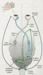
retina--> optic nerve--> lateral geniculate nucleus (thalamus- on other side)--> optic radiations--> occipital lobe--> primary motor nucleus--> pontine nucleus --> facial nucleus--> facial nerve --> blink
tests CNII, visual cortex, cortex and CNVII |
|
|
describe PLR pathway |
Retina--> optic nerve--> pretectal nucleus (brainstem)--> optic chiasm (central decussation)--> oculomotor nerve--> ciliary nerve |
|
|
what are the clinical signs of brain stem disease? |
1. cranial nerve deficits - facial nerve paralysis - central vestibular syndrome (VIII) - loss of gag reflex (IX/X) - tongue paralysis (XII) - abnormal eye positive (III, IV +/- VI) - bilateral masticatory muscle atrophy and loss of jaw tone (V)
2. proprioceptive deficits - bc ascending sensory and descending UMN tracts pass through brain stem - small lesions = unilateral - larger lesions = bilateral 3. hemi or tetraparesis 4. stupor or coma with abnormal pupil size* - due to pressure on CNIII nucleus 5. abnormalities in respiratory and CDV function - abnormal breathing patterns i.e. Cheynes Stokes - neurogenic pulmonary oedema -- mau cause severe dyspnoea, tachypnoea and hypoxaemia unrelated to any direct effect on RR and depth - bradycardia with increased ICP - tachyarrhythmias with brain-heart syndrome - Biots breathing, apneustic breathing - altered HR due to lesion in medulla (vagus nerve) 6. decerebrate injury - occurs with mid-brain injury- rigid limb extension, opisthotonus and coma 7. may also have forebrain involvement - head pressing, aimless circling, unilateral (central blindness), proprioceptive deficits, can have seizures
|
|
|
stuporous and multiple cranial nerve deficits = |
widespread brain stem disease |
|
|
what is the major abnormality associated with brain stem disease? |
stupor or coma with abnormal pupil size |
|
|
would conscious proprioception be affected with cerebellar disease? |
no the cerebellum is anatomically separate from the brain stem so lesions here do not impinge on the ascending and descending tracts in the brain stem
mental status also not affected - anatomically distinct from RAS |
|
|
what are the clinical signs of cerebellar disease? |
- mentally alert - intention tremors - head and eye - hypermetria and dysmetria - ataxia and wide based stance but opposite to side of lesion = paradoxical vestibular syndrome - loss of menace with extensive cerebellar disease - muscle tone remains strong bc long tracts (from brain stem) are intact = normal CP and no UMN signs (unless cerebellar dz combined with CVS*) |
|
|
UMNs originate in the |
Brain stem |
|
|
what are the differentials for forebrain signs? |
intracranial congenital: 1. hydrocephalus 2. lissencephaly 3. cortical dysplasia
acquired: 1. hydrocephalus 2. infectious/inflammatory 3. Neoplastic (mengioma most common) 4. cerebrovascular accident
extracranial 1. hepatic disease (HE) 2. advanced renal disease (less common) 3. electrolyte abnormalities - hypoCa - hyper or hypoNa 4. hypoglycaemia 5. thiamine deficiency 6. toxins - lead - OP (snail baits) |
|
|
acquired hydrocephalus is more common than congenital hydrocephalus. True or false? |
false, congenital hydrocephalus is most common |
|
|
3wk old pomeranian with seizures, behavioural abnormalities and obtundation. What do you suspect? |
congenital hydrocephalus |
|
|
what are the 3 abnormalities associated with congenital hydrocephalus? |
1. open fontanel (normally closes 3-6m) 2. domed skull 3. ventrolateral strabismus (sun setting sign) |
|
|
what are the causes of acquired hydrocephalus? |
due to an obstruction to CSF flow - usually secondary to mass like lesions obstructing flow of CSF in rostrocaudal direction
1. brain tumours 2. infectious - viral, bacterial, rickettsial, fungal, protozoal 3. inflammatory - GME - FIP (pyogranulomatous form) |
|
|
how should you manage acquired hydrocephalus? |
reduce ICP and CSF production 1. mannitol 2. prednisolone +/- acetazolamide (CAI) - pred better success bc anti-inflammatory properties surgical - ventriculoperitoneal shunt treat underlying cause - neoplasia - infectious - inflammatory |
|
|
what are the causes of brain neoplasia |
1. primary brain tumour 2. tumour of supportive structures - meningioma - glioma - choroid plexus tumour 3. invasion from adjacent structure tumours - nose, ear, frontal sinuses 4. metastatic neoplasia - haemangiosarcoma - melanoma - mammary carcinoma - lymphoma |
|
|
treatment options for brain tumours ? |
1. palliative - pred: anti-inflammatory and reduces oedema - low dose frusemide - acetazolamide if worried about hydrocephalus too 2. treat seizures - EAD - phenobarbitone 3. chemotherapy and radiation therapy 4. surgical |
|
|
what is a seizure? |
an involuntary, paroxysmal, synchronous neuronal discharge that always originates in the forebrain |
|
|
describe the phenomenon of seizure severity progression |
kindling = seizure focus expands and then spread throughout brain parenchyma of electrical activity progressively worse over weeks to months |
|
|
what are the 3 clinical phases of a seizure? |
1. aura the period immediately before the seizure when the animal displays a change in behaviour 2. ictus the seizure. can last a few seconds to status epilepticus 3. post ictal period time following seizure, lasts minutes to days, disorientated, drowsy, often blind |
|
|
what do generalised motor seizures indicate? |
that epileptogenic focus is large - involves both cerebral hemispheres |
|
|
describe how to differentiate between syncope and seizures |
- both lose consciousness - often no warning signs with syncope, with seizures there is the aura - syncope: passive development of flaccid limbs seizures: involuntary muscle contractions - syncope occurs during activity, seizures occur while resting - defecation and urination occurs with seizures but not with syncope - syncope lasts seconds, seizures last seconds to minutes - animals with syncope are normal afterwards whereas animals that have seizured will be disorientated after-mins to hours- post ictal stage |
|
|
what are the 2 most common extra cranial causes for seizuring? |
hypoglycaemia hypocalcaemia |
|
|
list toxins that cause seizures |
1. OPs, carbamates 2. Lead 3. rodenticides 4. 1080, strychnine 5. mouldy food - penetrem mycotoxin 6. pyrethrins- cats 7. recreational drugs 8. bufo toxin (cane toad)
most of the toxic causes (except coagulopathy and lead poisoning) need to be diagnosed based on history |
|
|
list differentials for seizuring |
intracranial congenital: 1. hydrocephalus 2. lissencephaly
acquired: 1. hydrocephalus 2. neoplasia - i.e. meningioma 3. inflammatory - GME - necrotising encephalitis 4. infectious - Toxoplasma, Neospora, Cryptococcus, FIP 5. idiopathic epilepsy** 6. CVA 7. head trauma
extracranial 1. hypoglycaemia 2. hypocalcaemia 3. toxins - snail bait, 1080, rodenticides, pyrethrins in cats etc 4. thiamine deficiency 5. hyperlipidaemia 6. polycythaemia 7. hypoxaemia (syncope more common) 8. HE (uncommon cause seizures) 9. renal failure- uraemia (rare) |
|
|
describe diagnostic work up for seizures |
1. history - diet: thiaminases, mouldy food - toxin exposure i.e. snail bait, rodenticides - pyrethrin exposure in cats* - access to recreational drugs 2. general PE - may reveal end stage organ disease 3. neurological exam - with forebrain disease - blindness (loss of menace), PLR may be intact - loss of smell - obtundation - circling towards side of lesion - postural deficits in contralateral limb
rule out extra cranial and simple intracranial causes 1. CBC, biochem, UA - metabolic causes: hypoglycaemia, hypocalcaemia - liver enzymes 2. infectious diseases profile - Toxoplasma - Neospora (dogs) - Cryptococcus - FIP 3. CSF analysis - if suspect inflammatory disease
more specific tests if necessary: 1. SBA - liver failure 2. blood Pb assay 3. coagulation profile - rodenticides - liver failure
** if these tests are abnormal leaves 2 possibilities: 1) idiopathic epilepsy (neuro exam will be normal) 2) brain tumour (need CT/MRI) |
|
|
idiopathic epilepsy is typically seen in dogs and cats <1yo and >7yo true or false? |
false! rarely seen in cats and in dogs is generally > 1yo and < 7yo |
|
|
dogs with idiopathic epilepsy generally have isolated seizures and relatively long (clinically normal) inter-ictal periods. True or false? |
true |
|
|
neurological exam is always abnormal in dogs with idiopathic epilepsy. True or false? |
FALSE neuro exam is always normal in dogs with idiopathic epilepsy |
|
|
when should potassium bromide be added to phenobarbitone for treating seizures? |
if GSD or severe seizures |
|
|
what are the side effects of phenobarbitone? |
- transient drowsiness and ataxia (10-14d) - PU/PD, polyphagia - can still seizure - biochemical hepatopathy (check liver enzymes biannually)
|
|
|
what is the MOA of phenobarbitone? |
increases seizure threshold by potentiating the effect of GABA (preferentially targets RAS) |
|
|
your patient was given phenobarbitone for idiopathic epilepsy 3wks ago but is still seizuring. How do you address this problem,? |
warn owners that phenobarbitone will not eliminate all seizures, but if frequent investigate: - measure serum phenobarbitone - if phenobarbitone in sub therapeutic range increase dose - if phenobarbitone in therapeutic range - add in KBr or revisit diagnosis*
therapeutic range: 70-170umol/L |
|
|
first choice AED if dog has moderate to severe hepatic disease? |
potassium bromide |
|
|
can potassium bromide be used in cats? |
no! causes eosinophilic pneumonitis in up to 50% cases |
|
|
what is refractory canine epilepsy and how can it be managed? |
1) any frequency unacceptable to owner 2) > 1 seizure per 3 months 3) sub therapeutic levels and dose cannot be increased due to side effects 4) unchanged frequency/severity of seizures for 4 months despite therapeutic levels
management: 1. reassess diagnosis - advanced imaging 2. add another drug to KBr + phenobarbitone - levetiracetam - gabapentin - others |
|
|
what are the rules of thumb for seizure management? |
1. except in SE, animals rarely die from seizures 2. most seizures worsen over time in severity an frequency (kindling) if not treated 3. meaningful neurological exam soon after a seizure is not possible 4. except in status epilepticus, there is no correlation between severity of seizures and cause 5. most seizure disorders can be controlled at least partially 6. clients should be prepared for drug and dose adjustments 7. relapses occur despite treatment 8. oral medications take days to weeks to work and transient early sedation and possible seizure continuation during this time is inevitable 9. treatment is life long and should never be suddenly stopped |
|
|
what are the most common causes of status epilepticus? |
1. hypoglycaemia 2. hypocalcaemia 3. toxins 4. breakthrough epilepsy 5. rapidly progressive inflammatory or neoplastic brain disease
|
|
|
describe treatment of status epilepticus |
1. stop the seizure - get IV access - diazepam (drug of choice) or midazolam 2. ABC 3. investigate aetiology - hx- toxins - bloods- hypoCa, hypoglycaemia 4. refractory seizuring - propofol IV or CRI (not cats) - phenobarbitone, pentobarbitol or levetiracetam IV - maintain on isoflurane, tapering doses by 25% every 2-4h 5. manage complications - mannitol - cool body core- hyperthermia |
|
|
what are the levels of consciousness? |
alert obtunded- decreased or inappropriate functional activity - mentally dull and poorly responsive to stimuli stupor - unresponsive to environment but roused by noxious/painful stimuli coma - unresponsive to any stimuli - unconscious |
|
|
what are the causes of stupor and coma? |
1. diffuse bilateral cerebral disease 2. metabolic or toxic encephalopathies 3. compression of rostral brainstem (midbrain and pons) - trauma, neoplasia, iatrogenic i.e. CSF tap - if there is pressure when do it, the release of pressure causes subtentorial herniation 4. destructive lesions of rostral cerebellum - trauma, inflammatory, neoplastic, vascular |
|
|
what are the causes of brain stem injury? |
increased ICP--> subtentorial herniation - inflammatory brain disease - neoplasia - hydrocephalus - iatrogenic- CSF tap blunt trauma - kicks, falls, MVA - Bites from larger animal - gun shot |
|
|
what is Cheynes stokes? |
waxing and waning depth of respiration with periods of hyperpnoea and apnoea- due to brainstem injury caused by bilateral cerebral disease
|
|
|
what is apneustic breathing? |
short breathing cycles followed be periods of apnoea- indicates pontine injury |
|
|
what is Biots breathing? |
irregular periods of apnoea with alternating periods of 3-4 breaths of identical depth - indicates increased ICP |
|
|
what are the causes of non-cardiogenic oedema and what do you see on radiographs? |
causes: (hypoxaemic events) - neurogenic pulmonary oedema - near drowning - seizures - UAO - near electrocution - also important complication of laryngeal paralysis
will see alveolar pattern in caudal lung lobes |
|
|
how should head trauma be managed? |
1. mannitol - reduce ICP 2. oxygenation +/- ventilation - reduces ICP and helps with neurogenic pulmonary oedema 3. careful head and neck elevation - reduce ICP 4. judicious fluid therapy - want to keep SBP > 60mmHg 5. seizure control with barbituate or proposal CRI |
|
|
what eye appearance would you expect in the following cranial nerve injuries: a) III (oculomotor) b) IV (trochlear) c) VI (abducens) |
a) oculomotor: ventrolateral strabmismus = 'sun setting sign' b) trochlear: dorsolateral rotation c) abducens: medial strabismus |
|
|
is trigeminal neuropathy a LMN or UMN disease? |
LMN |
|
|
how can you differentiate trigeminal neuropathy from masticatory myositis? |
in trigeminal neuropathy: 1. mandibular paralysis- dropped jaw/reduced tone (whereas in masticatory myositis you get trismus) 2. difficulty pretending food 3. some animals have loss of facial sensation and Horner's syndrome 4. usually recover |
|
|
what are the 2 most common causes of facial nerve paralysis? |
otitis media idiopathic |
|
|
how can you differentiate between central vestibular syndrome and peripheral vestibular syndrome? |
in both there will be ataxia, circling towards side of lesion, head tilt and nystagmus but with central vestibular syndrome you see:
- nystagmus changes direction - vertical nystagmus - CP and postural deficits - Horners syndrome and cranial nerve VII and VI signs are RARE |
|
|
do isolated cerebellar lesions cause circling? |
no |
|
|
vertical nystagmus |
central vestibular syndrome |
|
|
what are the causes of peripheral vestibular syndrome and what is the most common cause? |
1. idiopathic (geriatric) = most common - acute and severe onset - facial nerve paralysis are NOT features** - gets better in a few days, no residual signs, unlikely to occur 2. otitis interna 3. ototoxic drugs 4. trauma to lateral skull 5. tumours of middle ear |
|
|
what are the causes of central vestibular syndrome? |
1. brain neoplasia 2. inflammatory- GME 3. infectious meningoencephalitis - canine distemper - Cryptococcus - Toxoplasma, Neospora 4. Metronidazole toxicity 5. head trauma 6. CVA
**hence will see forebrain signs too |
|
|
describe the pathway for innervation of the eye |
sympathetic = sympathetic nerve 1st order neuron in hypothalamus--> cervical spinal cord in tectotegmental tract--> 2nd order neuron in thoracic cell bodies (C8-T2)--> leave T1-3 spinal nerve roots --> vagosympathetic trunk in cranial thoracic cavity and travel cranially up neck--> 3rd order neuron in cervical ganglia ventromedial to tympanic bulla--> enter skull--> ciliary nerve
parasympathetic: oculomotor nerve (CNIII) cell body in midbrain |
|
|
possible causes of Horner's syndrome? |
1st order neuron (hypothalamus) - disease in brain stem or hypothalamus - cervical spinal injury i..e FCE, IVDD 2nd order neuron (thoracic cell bodies) - BNRA - thoracic masses - neck injury i.e. choker chains, needle sticks - cervical abscess - idiopathic* (50% cases) i.e. golden retrievers - mediastinal lesions i.e. thymoma, lymphoma - trauma 3rd order neuron - otitis media - trauma or injury i.e. head trauma, bite wound - PVS - retrobulbar neoplasia or infection |
|
|
prognosis for idiopathic Horner's syndrome? |
usually recover in 3-4 weeks (months?) |
|
|
describe how to localise Horner's syndrome |
1. neurological exam - function of brain stem (CN deficits, CP deficits) - function of cervical spinal cord (reflexes, pain, CP) - function of forelimbs i.e. BNRA = LMN 2. denervation hypersensitivity testing- topical 10% phenylephrine examine every 5-10mins until 75% dilation - < 20 mins = 3rd order neuron - 20-45mins = 2nd order neuron - > 45mins = 1st order neuron 3. investigate cause - diagnostic imaging |
|
|
describe the levels of nerve injury |
1. neuropraxia - short term transient loss of nerve function due to compression - complete recovery in 6-12m 2. axonomesis - disruption of axons with intact supporting structures - nerve sheaths can regrow but takes months to years 3. neurotmesis - irreversible injury - complete severance of nerve - will never recover |
|
|
cerebellar disease results in alteration in force of limb movements. true or false? |
false! cerebellar disease results in abnormalities in rate, range and direction of limb movements NOT force cerebellum does not initiate movement it just controls it once it has been initiated |
|
|
roles of the cerebellum? |
controls movement once it has been initiated and influences posture coordination of eye position, movement, muscle tone and unconscious proprioception |
|
|
what are the causes of cerebellar dysfunction? |
congenital: 1. cerebellar abiotrophy - kelpies OR - < 6m old 2. cerebellar hypoplasia 3. neuroaxonal dystrophy in rottweilers
acquired: 1. inflammatory and infectious causes - GME 2. infarct - haemorrhagic or ischaemic 3. neoplasia |
|
|
what are the differentials for neck pain? |
1. IVDD 2. inflammatory CNS disease - GME - SRMA 3. trauma - spinal fracture/luxation 4. atlantoaxial instability - congenital in toy breeds - trauma 5. Wobblers 6. vertebral or spinal neoplasia 7. discospondylitis 8. polyarthritis 9. polymyositis 10. jaw disease/injury - fracture - TMJ disease - dental dz - masticatory myositis |
|
|
describe diagnostic investigation for neck pain |
1. CBC, biochem, UA 2. Rads/CT 3. CSF - not if ICP, stupor or obtundation (give mannitol) 4. myelogram? 5. further investigation if necessary
|
|
|
hallmark for diagnosis of GME? |
mononuclear pleocytosis granulomatous type inflammation - macrophages, lymphocytes and plasma cells and giant cells in brain and spinal cord |
|
|
what are the clinical signs of GME ? |
- multifocal neurological signs or if focal can just have specific signs i.e. forebrain signs, vestibular signs - neck pain - ADR for weeks to a couple months - blindness - optic neuritis (ocular form) - 2-6yo small breeds |
|
|
the breed that most commonly gets SRMA? |
beagles |
|
|
contraindications to CSF tap? |
1. suspect increased ICP - severe obtundation, stupor, anisocoria, papilloedema, low coma score - can cause tentorial herniation and brainstem compression = death 2. coagulopathy - may lead to haemorrhage in brain or spinal cord 3. cervical instability 4. large SOL - risk causing tentorial herniation |
|
|
treatment of choice for GME? |
pred + cytarabine - pred lifelong, taper to lowest dose possible - cytarabine 4 doses over 2 days then repeat every 3 wks
or prednisolone + cyclosporine A |
|
|
signalment for SRMA? |
- beagles OR - large breed young dogs 6-18 months |
|
|
clinical signs of SRMA ? |
- multifocal CNS signs - neck pain, rigidity - fever, anorexia - inappetance and reluctance to move |
|
|
treatment of SRMA ? |
Pred usually better in 24-48h but keep on for 6 months |
|
|
differentials for meningitis in cats |
1. Toxoplasma 2. Cryptococcus 3. FIP- dry form (pyogranulomatous CNS disease) 4. bacterial |
|
|
what antibiotics should be used to treat bacterial meningitis? |
antibiotics that cross BBB: 3rd generation cephalosporin or fluroquinolone
aminoglycosides NOT recommended |
|
|
animals with widespread LMN disease often have cranial nerve signs. True or false? |
true - cranial nerves are lower motor neurone |
|
|
which muscles does the sciatic nerve innervate? |
biceps femoris, SM, ST, cranial tibial, gastrocnemius |
|
|
what are the major systems that could be involved in an animal with weakness? |
- neuromuscular - cardiovascular, respiratory - metabolic, endocrine |
|
|
what are the causes of UMN tetra paresis? |
(weakness in all four limbs but increased muscle tone and reflexes)
acute: 1. cervical IVDD 2. cervical vertebral fracture 3. FCE
subacute-chronic: 1. CNS inflammation - GME - SRMA 2. spinal neoplasia 3. discospondylitis |
|
|
what are the causes of LMN tetra paresis? |
1. tick paralysis 2. snake envenomation 3. terodotoxin (puffer fish) 4. botulinum toxin 5. OPs and carbamates - block NMJ 6. chemotherapy drugs - vincristine - cisplatin 7. heavy metals 8. diabetes mellitus (cats) 9. hyperinsulinism 10. hypothyroidism 11. electrolyte disturbances 12. ischaemic neuropathy (cats) - rapid onset - ATE 13. inflammatory - polyradiculoneuritis- inflammation of spinal nerve roots - polyneuritis - myasthenia gravis - polymyositis - Toxoplasma, Neospora 14. neoplasia - lymphoma - paraneoplastic syndrome
|
|
|
prednisolone is the mainstay of treatment for idiopathic polyradiculoneuritis. True or false? |
false corticosteroids are NOT indicated - promotes infection in immobile patients esp UTI and pneumonia |
|
|
describe diagnosis and treatment of myasthenia gravis |
diagnosis: 1. clinical signs - episodic weakness esp wit exercise that resolves with rest - +/- megoesophagus 2. response to edrophonium chloride injection 3. seroloy - anti-ACh receptor assay - confirms diagnosis
Treatment: 1. pyridostygmine - anticholinesterase 2. +/- with pred if doesnt work on oown 3. supportive care - feeding, IVF |
|
|
what is a reverse sneeze? |
like a repeated snorting noise = paroxysmal inspiratory stertorous noise that occurs with caudal nasal and nasopharyngeal disease |
|
|
dull lung sounds on auscultation with resonance on percussion suggests? |
pneumothorax |
|
|
you auscultate increased breath sounds dorsally and decreased sounds ventrally. what does this suggest? |
pleural effusion |
|
|
what respiratory pattern will you see with lesions/disease localised to extra thoracic airway i..e larynx, nasopharynx? |
inspiratory, obstructive |
|
|
what are the indications to investigate respiratory signs? |
1. chronicity > 2-3wks - or fails to respond to empirical therapy - or recurring 2. dyspnoea 3. blood 4. malaise 5. exercise intolerance 6. if suspect FB 7.mass or distortion of nose * unless you know is FHV |
|
|
what are the contraindications to using anti-tussives? |
1. productive cough/respiratory disease 2. poor mucociliary clearance 3. FB 4. weakness |
|
|
what are the centrally acting and peripherally acting anti-tussives ? |
centrally acting: 1. opioids - block cough centre in medulla
peripherally acting: 1. bronchodilators i.e. terbutaline (beta 2 agonist) - increases airway diameter 2. methylxanthines i.e. theophylline promote cilia beating= enhanced mucociliary claeance 3. anticholinergics - use in emergency setting only |
|
|
list indications for multi dose inhalers i.e. with fluticasone, salbuterol |
- BAS - feline asthma - laryngeal paralysis - canine chronic bronchitis - lymphocytic plasmacytic rhinitis |
|
|
what are the non-nasal diseases that may present with the following nasal signs: a) epistaxis b) nasal discharge c) stertor |
a) epistaxis 1. coagulation defect - IMT, DIC, rodenticide, vWD, vasculitis 2. small vessel trauma - polycythaemia, hyperviscosity, hypertension 3. hypereosinophilic syndrome
b) nasal discharge 1. bronchopulmonary disease 2. ciliary dyskinesia (rare) 3. regurgitation/reflux/vomiting esp brachycephalics can end up in nasopharynx
c) stertor 1) Rathkes pouch anomoly - young animals 2) otopharyngeal or retropharyngeal mass |
|
|
describe diagnostic tests for nasal disease |
1. examine under GA - palpate bony limits including hard palate - dental exam - probe gingival sulcus and flush with 3ml to see if comes out nose (oronasal fistula) - dorsal recumbency and perform nasopharyngeal exam - make sure look behind soft palate - otoscope rostral nasal cavity via nares 2. rads - can assess whether invasive or non-invasive and prognosticate - less sensitive than CT and extent of disease can't be accurately determined - = can't plan treatment for invasive disease 3. CT - 3 patterns: non-invasive, neoplasia and fungal disease - can use for surgical planning 4. LCAT- Cryptococcus 5. rhinoscopy +/- biopsy - cotton tip cytology, traumatic nasal flush or pinch biopsies - pinch biopsies can be done with rhinoscope (fungal disease) or blind (cancer)
|
|
|
what views are used for nasal radiography and what lesions/abnormalities may be seen? |
views: - lateral - skyline - open mouth with angled beam +/- dental films
- invasive vs non invasive disease i.e. soft tissue densities and lysis of turbinates = invasive (neoplasia or fungal) - radiodense FBs - alveolar bone loss |
|
|
what are the 3 types of patterns seen on CT with nasal disease ? |
1. non invasive 2. neoplasia - turbinate destruction, bony lysis extending beyond turbinates and mass effect 3. fungal - cavitatory bony loss, lysis but not as proliferative as neoplasia - more cavitatory |
|
|
nasal neoplasia can be accurately diagnosed using rhinoscope guided pinch biopsy. True or false? |
false this biopsy is too small to detect cancer and needs to be done without biopsy to accurately diagnose cancer rhinoscopy guided pinch biopsy is best for diagnosing fungal nasal disease |
|
|
describe diagnosis and treatment of chronic rhino sinusitis (snufflers) in cats |
diagnosis: is by exclusion of other causes of turbinate destruction 1. clinical signs - chronic bilateral mucopurulent discharge - otherwise well 2. CT - turbinate loss 3. rhinoscopy + saline flush + biopsy - saline flush is therapeutic - histopath- turbinate necrosis
Treatment: 1. appropriate antibiotics for 6 months - bone penetrating ABs - clindamycin or doxycycline 2. supportive - steam cleaning face - piroxicam to control epithelial inflammation |
|
|
extensive turbinate destruction, sparing bony limits of the nasal cavity and neutrophilic rhinitis without evidence of other aetiological agents is most consistent with? |
chronic rhinosinusitis (snufflers) |
|
|
most common type of nasal neoplasia ? |
adenocarcinoma (nasal neoplasias do have potential to met but it is the invasiveness that causes morbidity and mortality) |
|
|
describe clinical signs diagnosis and treatment of nasal neoplasia |
clinical signs: - reverse sneezing, sneezing - epistaxis, stertor (obstruction) - distortion, discharge (esp with adenocarcinoma because mucus secreting cells) - may extend to orbit, mouth, brain --> clinical signs such as epiphora, ocular displacement, neurological signs
diagnosis: 1. CT/rads - CT needed for surgical planning and prognosticating 2. large (non-guided) pinch biopsy - definitive diagnosis - histopath and grading
Treatment: 1. radiation = tx of choice - even for cats with nasal lymphoma 2. chemotherapy - doxorubicin + carboplatin + piroxicam 3. palliative - piroxicam 4. vincristine if TVT*
|
|
|
list 3 ways to differentiate between fungal nasal disease and nasal neoplasia |
1. CT pattern: with neoplasia is more proliferative whereas fungal more cavitatory 2. fungal disease more likely to cause serosanguineous or mucoid discharge whereas neoplasia more likely to cause epistaxis 3. fungal disease causes nasal depigmentation |
|
|
surgery is the treatment of choice for nasal neoplasia. True or false ? |
false! Surgery is NOT indicated radiation is the tx of choice |
|
|
sinonasal aspergillosis tends to occur in doliocephalic dogs and brachycephalic cats. True or false? |
true |
|
|
most common aetiologic agent of sinonasal aspergillosis? |
Aspergillus fumigatus |
|
|
describe diagnosis, treatment and prognosis for sinonasal aspergillus |
diagnosis:
1. clinical signs
- nasal depigmentation and pain and malaise 2. CT/rads - cavitatory, invasive - classic CT appearance: fluffy fungal bulls in frontal sinus, no turbinates further into nasal cavity 3. rhinoscopy + biopsy
Treatment: 1. rhinoscopic debridement of fungal plaques 2. frontal sinus trephination - involved in 80% dogs 3. topical antifungals - clotrimazole, enilconazole 4. reassess +/- retreat every 3wks using rhinoscopy
prognosis: - good- excellent for dogs with thorough tx - if complete cure then seldom recurs - poor for cats with orbital involvement |
|
|
describe clinical signs, diagnosis and treatment of cryptococcus in cats |
clinical signs: - stertor - sneezing, reverse sneezing - discharge - facial deformity - CNS signs if crosses cribiform plate diagnosis: 1. UA exam under GA 2. rads/CT 3. traumatic flush - dislodge granulomas and push into swab 4. nasal swab and cytology - diff quik - see yeasts (non-staining capsule) 5. LCAT - latex cryptococal antigen agglutination test - titres should always be obtained for monitoring response to treatment 6. identify serovar
Treatment: 1. local non-invasive debridement 2. itraconazole or fluconazole - 6 months, or til negative LCAT - + amphotericin B if nervous signs - fluconasole has less side effects 3. monitor titres via LCAT - do at 3-4m then every 6 weeks - treat until titre is zero |
|
|
which is the more virulent Cryptococcus serovar? |
Cryptococcus gatti |
|
|
describe clinical signs, diagnosis and treatment of lymphocytic, plasmacytic rhinitis |
clinical signs: - chronic mucopurulent discharge - systemically well - non-progressive signs - partial response to ABs
diagnosis: 1. CT/rads - minimally destructive, normal turbinates or mild loss - soft tissue density due to mucus and congestion 2. rhinoscopy - hyperaemia, congestion, and inflammation of mucosa - mild turbinate atrophy - no cavitation - rule out other causes 3. biopsy - generalised lymphoplasmacytic inflammation +/- neutrophils
Treatment - 6 months to life long 1. MDI fluticasone 2. immunomodulatory ABs- doxycycline 3. piroxicam - epithelial anti-inflammatory 4. itraconazole |
|
|
what should you always do when you suspect laryngeal disease? |
take chest rads |
|
|
what is a really good way to reduce laryngeal oedema/inflammation and subsequent decompensation? |
inhaled fluticasone |
|
|
what does it mean if a patient has inspiratory and expiatory obstructive dyspnoea ? |
it means they have a fixed obstruction ie. oedema, inflammation or a mass |
|
|
what are the clinical signs associated with laryngeal disease ? |
1. stridor - if only during inspiration = dynamic i.e. everted laryngeal saccules - if during inspiration and expiration = fixed i..e oedema, mass 2. forced expirations, dysphonia 3. respiratory distress 4. often present with heat stress 5. secondary laryngeal oedema and inflammation causes them to decompensate - resp distress and non-cardiogenic pulmonary oedema |
|
|
describe the diagnostic steps in investigating laryngeal disease |
1. PE - stridor hallmark -auscultation: rhonchi loudest over larynx 2. UA exam under light IV anaesthesia 3. radiographs - * always take if suspect laryngeal disease* - non-cardiogenic pulmonary oedema with laryngeal paralysis |
|
|
treatment of choice for laryngeal paralysis? |
arytenoid lateralisation |
|
|
what are the causes of laryngeal paralysis? |
1. idiopathic - large breed dogs 2. congenital 3. anterior thoracic masses 4. polyneuropathy, polymyopathy |
|
|
what are the grades of laryngeal collapse? |
1. stage 1: everted laryngeal saccades (BAS) 2. stage 2: malacia and collapse of corniculate proceed and aryepiglottic folds into lumen 3. stage 3: progression of stage 2 to complete collapse |
|
|
what are the clinical signs of tracheal and bronchial disease? |
- honking cough triggered by excitement - obstructive dyspnoea, inspiratory if extra thoracic (cervical trachea), expiratory if intrathoracic (thoracic trachea and bronchi) - audible wheezes - auscultation: rhonchi +/- crackles if intrathoracic - percussion and palpation normal - tracheal pinch elicits cough |
|
|
what are some sequelae to bronchial disease that may be seen on chest rads? |
- bronchiectasis - dependent lobar pneumonia (caudal part of left cranial and right middle) - cor pulmonale |
|
|
describe the indications to do a transtracheal aspirate and an endotracheal tube wash |
transtracheal aspirate: - large dogs with productive disease - of whom risk of decompensation with GA can be avoided - dont need GA - percutaneous trochar - NOT indicated in cats - cytology, culture, Mycoplasma PCR
endotracheal tube wash - best for cats and small dogs - GA required - sterile ET placed with as minimal pharyngeal contamination as possible - presence of alveolar macrophages confirms BALF (bronchoalveolar lining fluid) |
|
|
what is the pathognomonic sign of chronic bronchitis? |
Kirschmanns spirals- inspisated mucus causing little casts of bronchioles and indicate excess mucus secretion |
|
|
many dogs with dynamic airway collapse develop elevated ALP, vascular hepatopathy and mild bile acid elevations without significant liver dysfunction. True or false? |
true unknown why take care not to confuse with concurrent hepatopathy or hyperA |
|
|
6yo obese chihuahua with protracted history of intermittent honking cough, most likely diagnosis? |
tracheal collapse / tracheobronchial collapse |
|
|
describe diagnosis and treatment of tracheobronchial collapse |
diagnosis: 1. clinical signs/hx - obese toy breeds - long history of paroxysmal honking cough - inspiratory or expiratory obstructive dyspnoea - often have concurrent mitral murmurs and important to determine if cough is respiratory or cardiac - generally if tracheal collapse is primary cause of signs they will have slow HR and pronounced arrhythmia 2. chest rads - inspiration and expiration - rule out cardiac disease 3. fluroscopy 4. tracheobronchoscopy +/- BAL - loose dorsal ligament folding into lumen and flatted airway cross section - graded 1-4
treatment: 1. eliminate exacerbating factors - weight loss! - chest harness, avoid exercise in hot weather - hair clipped short to reduce heat stress 2. medical - codeine (anti-tussive) +/- bronchodilators - theophylline, aminophylline - improves mucociliary clearance and decreases diaphragmatic fatigue - fluticasone inhaled 3. surgical - only if dyspnoea* - stent
|
|
|
most common clinical sign associated with tracheal rupture? |
subcutaneous emphysema (trachea is in mediastinum which communicates with SC at thoracic inlet) |
|
|
define bronchiectasis |
= distortion and dilation of bronchi caused by failure of elastic and muscular components of bronchial wall- bronchi become torturous
chronic inflammation and infection are most common cause i.e. chronic bronchitis, eosinophilic bronchopneumopathy , ciliary dyskinesia
frequently accompanied by squamous metaplasia and secondary loss of mucociliary clearance, secondary dependent lobar pneumonia |
|
|
what are the 2 most common conditions of the trachea and bronchi? |
laryngeal paralysis tracheobronchial collapse |
|
|
paroxysmal expiratory dyspnoea in a cat, you should suspect? |
feline asthma |
|
|
what are the clinical signs of small airway/bronchiole disease? |
- honking cough - obstructive pattern expiratory dyspnoea - auscultation: crackles and wheezes (high pitch) - normal percussion - cough elicited on tracheal pinch - obstructive pattern and wheezing becomes more obvious with exercise challenge |
|
|
describe diagnostic tests used for small airway disease |
1. radiographs - bronchial or bronchointersitial pattern - sometimes see sequelae: bronchiectasis, dependent lobe consolidation or pneumonia secondary to poor mucociliary clearance (alveolar pattern in dependent lobes) 2. tracheobronchoscopy + BAL - minimal changes unless concurrent large airway disease - will see mucus and discharges from small airways - rule out other causes of coughing - guided BAL - when sampling for culture take samples from right middle and caudal part of cranial lung lobe (dependent lobes tend to accumulate secretions) 3. rule out parasites - ascarids: faecal samples - lungworm: Baermann flotation in faeces - HW: snap test |
|
|
what is the most important small airway disease in dogs? |
canine chronic bronchitis |
|
|
what are the possible sequelae to canine chronic bronchitis? |
1. bronchiectasis 2. pneumonia (failed mucociliary clearance) 3. cor pulmonale 4. COPD - irreversible 5. hepatopathy |
|
|
what are the clinical signs of canine chronic bronchitis? |
- coughing < 2 months - coughing may be only sign in mild disease - waxes and wanes but is slowly progressive over yrs, may be exacerbated by cold weather - productive cough in morning but then dry for rest of day - in advanced disease: dyspnoea, exercise intolerance, auscultation abnormalities |
|
|
squamous metaplasia of the respiratory epithelium is reversible. True or false? |
false it is irreversible and causes poor mucociliary clearance |
|
|
diagnostic criteria for canine chronic bronchitis? |
- cough > 2 months - excessive airway mucus- Kirschmanns spirals - exclusion of lower airway diseases |
|
|
mainstay of therapy for canine chronic bronchitis? |
MDI fluticasone |
|
|
describe diagnosis and treatment of canine chronic bronchitis |
diagnosis: (diagnosis of exclusion) 1. PE - usually BAR - middle aged, often obese - hx of cough - pronounced sinus arrhythmia common (high vagal tone from primary disease) - auscultation: can be normal, may hear wheezes or crackles, expiratory snaps in severe cases (developing COPD) - cyanosis = really severe 2. chest rads - bronchial or bronchointersitial pattern - normal in early disease - may see sequelae 3. bronchoscopy + BAL - mucus, oedema, inflammation, hypertrophy - BAL: Kirschmanns spirals pathognomonic but dont need to be see to make diagnosis, mixed inflammation usually non degenerate neutrophils - culture to rule out infection
Treatment: 1. environmental management - weight loss - reduce triggers i.e. passive smoking, dust - walking harness 2. mucus removal - nebuliser - coupage 3. corticosteroids - inhaled fluticasone mainstay* - systemic steroids are last resort 4. anti-tiussive - only if dry cough predominant sign* 5. doxycycline if haven't ruled out Mycoplasma 6. bronchodilator - xanthine derivative i.e. amydophilone - may allow to use less glucocorticoid |
|
|
what are the clinical signs of feline asthma? |
- coughing - audible wheezing (bronchoconstriction) - obstructive pattern expiratory dyspnoea, commonly present in respiratory distress and may be barrel chested due to air trapping - paroxysmal or continuous |
|
|
describe diagnosis of feline asthma |
1. PE - BAR usually but may present in respiratory distress - coughing and audible wheezes - obstructive pattern expiratory dyspnoea - air trapping when bronchoconstricted makes them barrel chested - may be bit hyper resonant on percussion due to air trapping 2. chest rads - bronchial pattern (railways and donuts) - hyperinflation and flattening of diaphragm in some cats 3. CBC - peripheral eosinophilia in >20% 4. +/- bronchoscopy and BAL - may avoid because dangerous, if going to do pre-med with terbutaline - may just do doxycycline trial for Mycoplasma 5. Baermann faecal floatation
|
|
|
describe treatment of feline asthma for the following presentations: a) emergency b) severe stable c) moderate daily signs d) mild daily signs e) refractory cases |
a) emergency - will have expiratory obstructive dyspnoea and tachypnoea 1. terbutaline IM or salbuterol inhaled 2. oxygen (40% FiO2) and cage rest 3. +/- dexamethasone IV 4. +/- atropine (abolishing vagal tone) b) severe stable - have dyspnoea at rest 1. MDI fluticasone 2. terbutaline or salbuterol 3. pred PO - likely to have compliance issues with inhaled fluticasone 4. reduce environmental triggers c) moderate daily signs 1. MDI fluticasone 2. salbuterol or terbutaline if flares up 3. reduce environmental triggers d) mild daily signs 1. fluticasone 2. salbuterol as needed 3. reduce environmental triggers e) refractory cases 1. cyclosporine 2. cyproheptadine 3. reduce environmental triggers |
|
|
what are the clinical signs of pulmonary parenchymal disease? |
- restrictive pattern inspiratory dyspnoea (small expiratory phase too but subtle) - increased breath sounds - crackles on auscultation - soft productive cough - exercise induced cyanosis |
|
|
list the different pulmonary patterns/distributions on radiographs |
- cranioventral - pneumonia, aspiration pneumonia - hilar- cardiogenic pulmonary oedema - lobar- focal disease i.e. FB, abscess, if in dependent lobes = failed mucociliary clearance - caudodorsal- non cardiogenic pulmonary oedema - mass- tumour |
|
|
you suspect pneumonia but there are no bacteria, what is the likely diagnosis and what will you use to treat? |
intersitial lung disease i.e. idiopathic pulmonary fibrosis in WHWT, eosinophilic bronchopneumopathy tx: immunosuppressive steroids |
|
|
ground glass appearance of lungs on CT? |
intersitial pneumonia |
|
|
most common causes of aspiration pneumonia ? |
- megoesophagus - laryngeal paralysis - following anaesthesia |
|
|
describe treatment of bacterial pneumonia |
1. antibiotics- empirical and then based on C+S - if stable: PO amoxyclav, TMS or cephalexin - if unstable: IV cephazolin or ampicillin +/- enrofloxacin SC 2. nebulisation + hydration 3. coupage 4. acetylcysteine (mucolytic) 5. high fat diet 6. lobectomy if one lobe consolidated 7. repeat rads at 2 and 6-8wks |
|
|
list predisposing conditions to pulmonary thromboembolism |
- neoplasia - SIRS, DIC - PLE, PLN - pancreatitis - hyperadrenocorticism - PLE, PLN - cardiac disease, heart worm - IMHA
|
|
|
what are the general clinical signs of pleural, mediastinal and chest wall disease? |
- restrictive pattern inspiratory dyspnoea - +/- cough - paradoxical chest movements - precaval syndrome |
|
|
why are pleural diseases typically bilateral? |
because mediastinum in dogs and cats is typically incomplete |
|
|
clinical signs specific to mediastinal disease |
- dysphagia - regurgitation - abnormal location of cardiac impulse - vocalisation changes (CN X) - Horners syndrome (sympathetic N) - loss of normal compliance of thorax in cats |
|
|
where should you perform thoracocentesis for pleural effusion? |
4th and 6th ICS for fluid direct ventrally and for air direct dorsally |
|
|
describe the tests that can be run on pleural fluid |
1.protein - 25-30g/L = modified transudate i.e. heart failure, lymphatic obstruction, large PTE - > 35g/L = exudate i.e. pyothorax, pleuritis - < 25g/L = low protein transudate associated with reduced oncotic pressure i.e. PLE, GN, IVF overload in cats 2. cytology 3. culture - do on exudates 4. IHC - working out if B-cells or T-cells 5. Coronavirus IFA test - accurate test for FIP - antigen in macrophages 6. TAG and cholesterol - compare to serum - chylothorax and pseudochyle 7. flow cytometry - phenotypical analysis |
|
|
list diagnostic steps for pleural disease |
1. ultrasound 2. thoracocentesis 3. rads/CT 4. biopsy of masses |
|
|
sterile, poorly cellular, high protein pleural effusion in the cat? |
FIP |
|
|
what are the causes of non-septic pleural exudate and how will you investigate further? |
causes: 1. FIP 2. Bartonella 3. neoplasia 4. chronic diaphragmatic hernia 5. lung lobe torsion 6. resolving septic exudate
next diagnostics: 1. systemic evaluation 2. +/- serology 3. thoracic rads 4. US |
|
|
what are the causes of chyle pleural effusion and how will you investigate further? |
*anything that causes high venous pressures can cause chylothorax 1. Heart disease- cats 2. neoplasia 3. congenital 4. traumatic 5. lung lobe torsion 6. cranial mediastinal mass 7. idiopathic 8. diaphragmatic hernia
diagnostics: 1. systemic evaluation 2. thoracic rads 3. echo +/- thoracic US 4. vascular studies 5. CT |
|
|
describe treatment and prognosis for idiopathic chylothorax |
1. low fat diet 2. periodic thoracocentesis 3. surgery (only moderate success rates) 4. chemical pleurodesis 5. rutin (lily root) 6. octreotide
prognosis: poor |
|
|
absence of dyspnoea rules out haemothorax. True or false? |
false, there needs to be 30ml/kg fluid in the pleural space before dogs show signs of respiratory difficulty |
|
|
if fluid is pH < 6.9 and exudative it is likely to be? |
pyothorax and immediate antibiotic therapy is indicated |
|
|
mainstay of treatment of pyothorax? |
bilateral indwelling chest drains
- perform lavage 2-4x daily for 2-5 days - once < 2-3ml/kg/day is drained from each drain and it becomes modified transudate the drains can be removed |
|
|
antibiotic of choice for pyothorax in cats ? |
tiracillin + clavulonic acid (Timentin®) |
|
|
what are the causes of pyothorax in a) cats b) dogs |
a) cats: 1. bite wounds 2. dental disease 3. perforation of mediastinal structures 4. migrating FB 5. extension of bacterial pneumonia
b) dogs 1. migrating FB i.e. grass seed 2. extension of bacterial pneumonia |
|
|
what are the causes of pneumothorax? |
1. blunt trauma- most common 2. penetrating chest injuries 3. ruptured lung lesions 4. chest wall or oesophageal penetration 5. pneumomediastinum with mediastinal breach |
|
|
describe clinical signs, diagnosis and treatment of pneumothorax |
clinical signs: - restrictive pattern inspiratory dyspnoea - reduced breath sounds - resonant percussion - may be barrel chested and dependent on abdominal effort for tidal volume in tension pneumothorax - +/- cyanosis from under ventilation or V/Q mismatch and circulatory shock as venous return is impeded by increased intrathoracic pressure
diagnosis 1. thoracic rads - lobar retraction - collapsed lungs with no bronchial markings peripherally - loss of cardiosternal contact - may see underlying cause 2. thoracocentesis - dorsal for air
treatment: 1. thoracocentesis - slowly otherwise can cause re-expansion pulmonary oedema - often after MVA only single drainage required 2. oxygen 3. analgesia 4. seal breach in chest wall if present 5. thoracotamy and lobectomy may be required |
|
|
pneumothorax can cause pneumomediastinum. True or false? |
false pneumothorax does not cause pneumomediastinum but pneumomediastinum may cause pneumothorax |
|
|
treatment for thymoma? |
pred and L-asparginase therapy first (reduce size of mass) then surgical incision |