![]()
![]()
![]()
Use LEFT and RIGHT arrow keys to navigate between flashcards;
Use UP and DOWN arrow keys to flip the card;
H to show hint;
A reads text to speech;
112 Cards in this Set
- Front
- Back
|
what organs does the renal system consist of ? |
-2 kidneys -2 ureters - urinary bladder -urethra |
|
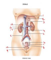
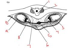
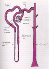
label the structures 1-14 |
1. diaphragm 2.oesophagus 3. L adrenal gland 4. abdominal aorta 5. IVC 6. Rectum 7. ovary 8. uterus 9. urethra 10. urinary bladder 11. ureter 12- renal artery 13. renal vein 14. R kidney |
|
|
where are the kidneys located in relation to the peritoneum |
-in the retroperitoneal space |
|
|
which border faces the vertebral column |
the concave medial border |
|
|
which level vertebra is the upper and lower poles of the liver located |
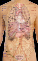
T12 and L3 |
|
|
which ribs protect the kidneys |
11 and 12 |
|
|
which kidney is higher than the other |
left (light so it rises like air )
|
|
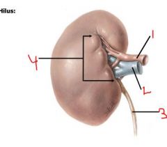
label structures 1-4 |
1. renal artery 2. renal vein 3. ureter 4. hilus |
|
|
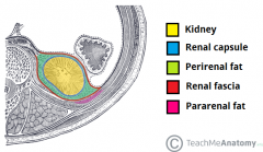
what are the layers which surround the idneys from deepest to most superficial |
- renal capsule -adipose capsule -renal fascia |
|
|
what is the function of the layers of the kidney |
-maintains kidney shape - trauma barrier |
|
|
what is the deep layer of the kindey continuous with |
outer layer of the ureter |
|
|
what type of tissue is the renal capsule made from ? |
dense irregular connective tissue |
|
|
what type of tissue does the middle layer of the kidney consist of? |
adipose tissue adipose capsule |
|
|
what tissue does the superficial layer of the kidneys consist of ? |
dense irregular CT (renal fascia) |
|
|
which layaer anchors kidney to abdominal wall and surrounding structures |
renal fascia |
|
|
which layer maintains position of the kidneys |
adipose capsule |
|
|
which layer maintains shape of kidneys |
renal capsule |
|
|
which layers act as trauma barriers of the kidneys |
adipose and renal capsule |
|
label the following structures on ( lying on stomach) |
1. body of L2 2. psoas muscle 3. LK 4. ureter 5. Abd. Aorta 6. IvC 7. retroperitoneal space 8. adipose capsule 9.renal fascia 10. renal capsuke |
|
|
What is nephroptosis
Ptosis (falling) |
-a condition also known as floating kidney where a kidney descends more than 2 vertebral bodies (5cm) during positional change e.g supine to standing |
|
|
Who is predisposed to nephroptosis and why ? |
women- - kidneys enlarged during menstrual cycle making them heavier and so likely to drop - intra-abdominal pressure birth giving. (partuition) -abdominal wall is weakened during pregnacny -corsets, tight skirts, belts women to men ratios is 10:1
Skinny people/ extreme weight loss- decreased muscle tone to abdominal wall. -fatty renal capsule keeps position of kidneya |
|
|
What serious consequences might arise from nephroptosis? |
Pyelonephritis Renal calculi Hematuria Hypertension Renal ischemia Flank pain |
|
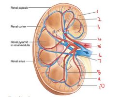
what are the two regions inside the kidneys ? Which is outter which is inner ? |
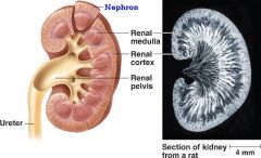
Renal cortex- outer/superficial (smooth texture -Renal Medulla (inner, striated) |
|
|
what do , the renal cortex and renal medulla form together ? |
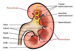
-renal parenchyma. (functional unit of the kidney) -(renal pelvis also a functional unit) |
|
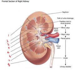
label diagrams |
1. renal cortex 2. renal medulla 3. renal column 4. renal pyramid 5. renal sinus 6. renal papillae 7. fat 8- renal capsule |
|
what does a renal lobe consist of ? |
-1 renal pyramid -overlaying area of renal cortex -½ of each adjacent renal column |
|
|
how much cardiac output do the kidneys receive and via what ? |
20-25% via left and right renal arteries |
|
|
divisions of renal blood vessels
RSIAIAGE |
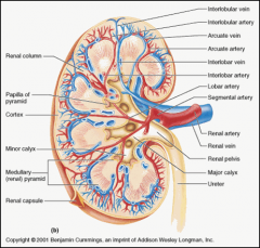
-Renal artery --> segmental arteries in the hilus and sinus -Segmental arteries --> interlobar (arteries between renal lobes) -Interlobar arteries --> arcuate arteries (arching over top of renal pyramids) -Arcuate arteries --> interlobular arteries ( in the renal cortex) -Interlobular arteries --> afferent arterioles (in the renal cortex ) Afferent arterioles --> glomerular capillaries (forming part of nephron) -Glomerular capillaries efferent arterioles -efferent arterioles -> peritubular capillaries |
|
|
what can efferent arterioles be thought of as ? |
portal vessels
|
|
|
what are portal vessels |
Portal vessels carry blood from one capillary bed to another. |
|
|
what are glomerular capillaries a part of |
nephron |
|
|
where are interlobular arteries |
in the renal cortex |
|
|
where are segmental arteries ? |
in the hilus and sinus of the kidney |
|
|
what's the name given to the second type of capillary given off by the efferent arterioles that surround the part of the nephron which extends into the medulla ? |
vasa recta (capillaries near loop of henle) |
|
|
where are interlobar arteries located |
between renal lobes |
|
|
renal venous drainage |
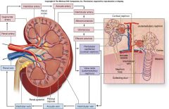
-Peritubular capillaries -> peritubular venules Peritubular venules -> interlobular veins (in the renal cortex) -Interlobular veins -> arcuate veins (arching over the top of renal pyramids) Arcuate veins -> interlobar veins (running between renal lobes) Interlobar veins -> Segmental veins (in hilus and sinus) Segmental veins Renal vein (to inferior vena cava) |
|
|
where do the vasa recta drain ? |
in the interlobular and arcuate veins |
|
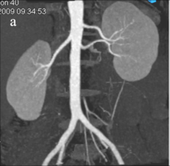
label diagram |
1. interlobular arteries 2. arcuate arteries 3. interlobar arteries 4.segmental arteries 5. renal arteries 6. renal vein 7. segmental vein 8. interlobar vein 9. arcuate vein 10. interlobular vein |
|
|
what are the 2 main parts which nephrons concist of ? |
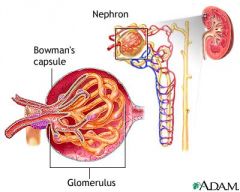
1. renal corpuscle 2. renal tubule |
|
|
2 main parts of the renal corpsucle |
1. the glomerulus 2. the Bowman’s capsule |
|
|
what's the glomerulus and what supplies it ? |
ball of capillaries supplied by afferent arterioles |
|
|
where in the kidneys is urine specifically produced |
glomerulus |
|
|
what drains the glomerulus |
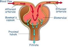
efferent arterioles |
|
|
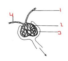
1. efferent arterioles 2. glomerulus 3. bowmans capsule 2&3: renal corpsicle |
|
|
what marks the end of the blind end of the nephron |
bowmans capsule |
|
|
which layer is the B's is continuos with renal tubule outer layer |
outer parietal layer |
|
|
where do filtrates collect in the B's C |
the capsular space |
|
|
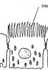
what cells is the inner layer of B's composed of |
podocytes |
|
|
whats the function of podocytes |
envelop glomerular capilarries |
|
|
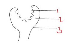
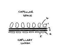
1. inner layer 2. capsular space 3. outer parietal layer |
|
|
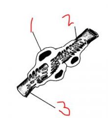
1. podocytes 2. pedicel- they interdigitate 3. glomerular capillary |
|
|
1. Pedicel 2.Slit membrane 3. Basal lamina 4. Endothelial cell 5. Fenestration of endothelial cell 6.Filtration slit |
|
|
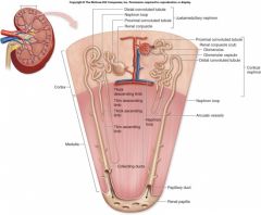
what are the tubular segments of the renal tubule |
1. Proximal Convoluted Tubule 2. The Loop of Henle 3. Distal Tubule 4. Collecting Duct |
|
|
where does the nephron drain into ? |
renal sinus |
|
|
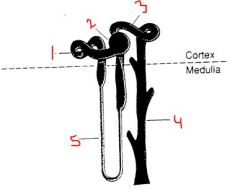
1.PCT 2. 3. Distal convoluted 4 collecting duct 5. loop of henle |
|
|
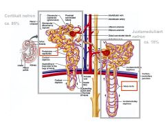
what are the 2 tyes of nephrons |
- cortical nephrons - juxtamedullary |
|
|
where do cortical (85%) nephrons lie |
in the outer 2/3 of the cortex |
|
|
which nephrons have a short loop of henle |
corticle nephrons |
|
|
where do the juxtamedullary nephrons lie
(juxta- next to) |
in the inner 1/3 of the cortex |
|
|
where do the loops of henle of JMN pass ? |
deep in the medulla |
|
|
which part of the proximal tube is convoluted |
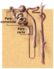
pars convoluta (early part) |
|
|
which part of the proximal tube is straight |
pars recta |
|
|
what epithelium lines the proximal tube ? |
Cuboidal/columnar epithelium |
|
|
how long is the PCT |
15 mm |
|
|
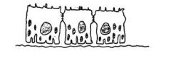
describe the cells in the pars convoluta |

-busier -more microvilli - more mitochondria - more invaginations |
|

- |
1. basal lamina 2. microvilli 3. nucleus 4. mitochondrion 5. tight junction (Zona occludens) 6. large intercellular space |
|
|
describe the cells in the pars recta |
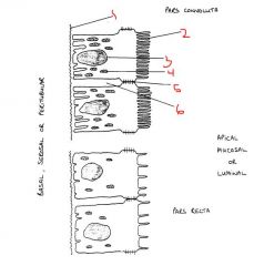
-less busy -less mitochondria
|
|
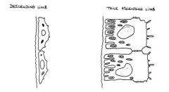
what cells make up the thick segments of the loop of henle |
simple cuboidal ep |
|
|
which segment of the loop has high metabolic activity and lots of mitochondria ? |
thick ascending limb filer ions and things that we need so they do more work |
|
|
what cells line the thin segments |
simple squamous |
|
|
where does the final part of the ascending loop of henle return to ? |
Efferent or afferent arterioles of the same nephron |
|
|
which segments of the loop have ow metabolic activty? |
thin segments ( highly permeable to water) |
|
|
macula densa |
(thick ascending limb columnar cells) |
|
|
juxtaglomerular cells |
(afferent arteriole smooth muscle cells) |
|
|
what lines early and late parts of the dct? |
cuboidal epithelium |
|
|
the late parts of the DCT contain cuboidal ep composed of what cell types ? |
-principal cells - intercalated cells |
|
|
What type of receptors do the principle cells contain ? |
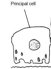
-anti-diuretic hormone receptors -aldosterone receptors
(bald really accessible) |
|
|
whats the function of the intercalated cells |
carbonic anhydrase activity (pH)
|
|
|
how many tubules do collecting ducts receive fluid from |
6 distal tubules (aprox) |
|
|
in the medulla collecting ducts pair to form what |
ducts of bellini |
|
|
function of ureters |
transfer urine from kidneys to bladder |
|
|
what happens as the bladder fills up? |
the ureters compress preventing urine backflow |
|
|
why is the lumen of the ureter susceptible to obstruction/ injury ? |
it has a v. narrow lumen |
|
|
layers of the ureter |
-adventitia -muscularis -mucose |
|
|
what tissue does the adventitia of the ureters maed of and what is its function |
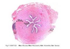
-connective tissue -binds ureter to surrounding tissues |
|
|
whats the muscularis layer of the Ureter made from? whats its function ? |
-smooth muscle - contracts in response to expansion caused by urine enrty |
|
|
describe the mucosal layer of the ureters |
inner layer transitional epithelium continuous with renal pelvis and bladder |
|
|
function of the bladder ? |
Retains urine until micturition is convenient |
|
|
where is the bladder |
• Muscular sac on the floor of the pelvic cavity • Inferior to peritoneum • Posterior to pubic symphysis |
|
|
layers of the bladder |
1. adventitia: fibrous outer layer (except superior surface which is covered by parietal peritoneum) 2.muscularis: (detrusor muscle) three layers of smooth muscle mucosa 3. transitional epithelium covered in wrinkles called rugae |
|
|
what do the openings of the two ureters mark ? |
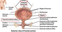
a smooth triangular area called the trigone – a common site of infection. |
|
|
why is the trigone a common site for infection ? |
near urethra. bacteria goes up urethra |
|
|
what happens to rugae as bladder expands |
rugae flatten |
|
|
which way does the bladder expand |
superiorly |
|
|
what happens to bladder walls as it expands |
they become very thin |
|
|
function of urethra |
Transfers urine from bladder to exterior |
|
|
where is the urethral bound in females |
Bound to anterior wall of vagina by connective tissue
|
|
|
where do externa urethral orifices lie in women |
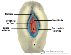
o The external urethral orifice lies between the vaginal orifice and the clitoris |
|
|
how long is the urethra in females |
3-4 cm |
|
|
how long is the uretra in males |
18 cm |
|
|
what are the 3 urethral regions in the male |
-prostatic -membraneous -penile |
|
|
describe the prostatic urethra |
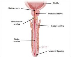
o 2.5cm long o Begins at bladder and passes through prostate gland |
|
|
membranous uretra |
o Short, thin-walled portion (0.5cm) o Where urethra passes through the muscular floor of the pelvic cavity |
|
|
Penile urethra |
o 15cm long o Passes through the penis to the external urethral orifice |
|
|
The detrusor muscle is thickened near the urethral opening in the bladder to form what ? |
internal URETHRAL SPHINCTER |
|
|
describe internal urethral sphincter |
o Compresses urethra and retains urine o Smooth muscle o Involuntary control |
|
|
decribe the external urethral sphincter |
-situated where the urethra passes through the pelvic floor. o Skeletal muscle o Voluntary control |
|
|
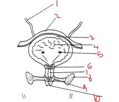
1. ureter 2. parietal peritoneum 3.detrussor muscle 4. rugae 5.uteric orifices/openings 6. internal urethral sphincter 7. urethra ? 8. inferior pubic ramus 9.external urethral sphincter 10. external urethral orifice |
|
|
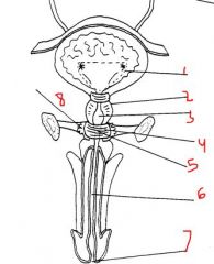
1. urethral orifices 2. prostate 3. prstatic urethra 4. urethral sphincter 5.bulbourethral gland 6. penile urethra 7.external urethral orifife 8. ? |
|
|
cystitis |
Lower urinary tract infection. occurs commonly in women believed to be caused by sexual intercourse. pain during urination |
|
|
pyelitis |
inflammation of renal pelvis can be caused by kidney stones result of inflammed ureters and bladder as they fail to excrete urine thus it remains in pelvis and leads to inflmmation there. also caused by bacteria infections or STDs such as gonorrhea |
|
|
pyelonephritis |
kidney infection can be a result of STD, kidney stones lower UTI infection and is a progression of ppyelitis
-pain during urinatioon fever vomotting |