![]()
![]()
![]()
Use LEFT and RIGHT arrow keys to navigate between flashcards;
Use UP and DOWN arrow keys to flip the card;
H to show hint;
A reads text to speech;
60 Cards in this Set
- Front
- Back
|
Central nervous system |
Comprises the brain and spinal cord. The spinal cord connects the brain and the peripheral nervous system. |
|
|
Peripheral nervous system |
Includes all parts of the nervous system that lie outside the brain and the spinal cord. |
|
|
Cerebral cortex |
The convoluted outer layer or covering of the two cerebral hemispheres. - Contains about three quarters of all the brains neurons |
|
|
Roles of cerebral cortex |
Information processing activities: Perception, language, learning, memory, thinking, problem-solving, Planning Control of voluntary body movements |
|
|
Sensory cortex areas |
Receive and process information from our different senses |
|
|
Motor cortex areas |
Receive, process, and send information about voluntary body movements |
|
|
Association cortex areas |
The entire network of nerves located outside the Central Nervous System. It extends from the top of the head, throughout the body to the tips of the fingers and toes and to all parts of the skin. The association areas make up the rest of the brain outside of the lobes and cortices. - The association areas receive information from the other lobes and incorporate/merge that information with the information already in that lobe.
|
|
|
Cerebral hemispheres |
Two almost symmetrical brain structures that appear to be separated by a deep groove running from the front to the back of the brain (longitudinal fissure). |
|
|
Corpus callosum |
A strand, or bridge of nerve tissue that connects the left and right cerebral hemispheres and serves as the main communication pathway between them. |
|
|
Four lobes |
Frontal, Parietal, Occipital and Temporal |
|
|
Frontal Lobe |
The largest of the four lobes and occupies the upper forward half of each cerebral hemisphere, right behind your forehead. |
|
|
Association areas in the frontal lobe |
Receive and combine information from elsewhere in the lobe. They also allow us to perform complex mental functions like planning, estimation, comprehension. |
|
|
Roles of the frontal lobe |
Body movements Language Planning Judgement Problem solving Personality Regulating emotion |
|
|
Primary motor cortex |
Involved in controlling voluntary bodily movements through its control of skeletal muscles. |
|
|
Broca's Aphasia |
An area in the frontal lobe responsible for the production of articulate speech, coordinating movements of the muscles required for speech and supplying this information to the appropriate motor cortex areas. |
|
|
Aphasia |
A language disorder apparent in speech (comprehension or production), writing or reading, produced by injury to brain areas specialised for these functions. |
|
|
Damage to Broca's Aphasia |
Damage to Broca’s area often produces speech that is very deliberate, consisting of a few words with very simple grammatical structure, but damage rarely results in the total loss of speech. |
|
|
Parietal Lobe |
Receives and processes sensory information from the body and skin senses and other sensory areas in the brain. It also sends information to other areas of the brain. |
|
|
Primary Somatosensory Cortex |
Receives and processes sensory information from the skin and body, enabling us to perceive bodily sensations. Homunculous man |
|
|
Association areas of the parietal lobe |
Receive and combine information from within the lobe and other structures and areas of the brain |
|
|
Roles of the Parietal Lobe |
Spatial navigation Recognise facial emotion Receives sensations such as touch, pressure, temperature and pain |
|
|
Occipital Lobe |
Primarily involved in vision |
|
|
Damage to the Occipital Lobe |
Can produce blindness, even if the eyes and their neural connections to the brain are normal. |
|
|
Primary Visual Cortex |
Located at the base of each occipital lobe and this is where visual information from the two eyes is received and processed |
|
|
Association Areas in the Occipital Lobe |
Associated areas in the occipital lobe work with other lobes in the brain to integrate visual information. This allows visual information to be organised and interpreted in a meaningful way. |
|
|
Temporal Lobe |
Primarily involved with auditory perception, but also plays an important role in memory, in aspects of visual perception such as our ability to recognise faces and identify objects, and in our emotional responses to sensory information and memories |
|
|
Primary Auditory Cortex |
Receives and processes sounds from both ears, receiving and processing different features of sound and therefore playing a vital role in the identification of sounds. |
|
|
Association areas of the Temporal Lobe |
Different association areas in the temporal lobe play roles in memory and perception - Receiving, processing and storing memories of facts - How to do things - Personal experiences like family holidays and birthdays. |
|
|
Damage to the Temporal lobe |
Can leave a person with the ability to describe someone’s facial features, to identify their sex, and to judge their approximate age, but without the ability to recognise the person as someone that they know, even if it is their mother. |
|
|
Role of the Temporal Love |
- Receive, process and store memories and facts - Auditory perception - Processing of sounds |
|
|
Wernicke's Area |
Involved with the comprehension of speech |
|
|
Wernicke's Aphasia |
A type of aphasia in which a person has considerable difficulty comprehending speech and speaking in a meaningful way. |
|
|
Hippocampus |
Found in the medial temporal lobe in both hemispheres - responsible for the formation of long-term memories |
|
|
Amygdala |
Found in the medial temporal lobe in both hemispheres - responsible for linking emotions to memory |
|
|
Hemispheric specialisation |
The idea that on hemisphere has greater control over a particular function |
|
|
Left hemisphere specialises in... |
- Verbal Tasks (involve the recognition of words) eg. Reading, writing, understanding of speech - Analytical tasks (involve breaking down a task into its key parts and then approaching it in a step by step way) eg, following instructions on how to bake a cake. - Left hemisphere receives and processes sensory information from the right side of the body. - Controls voluntary movement on the right side of the body |
|
|
Right hemisphere specialises in... |
- Non verbal tasks (tasks that are not dependent on language skills) eg. Completing a jigsaw puzzle. - Spatial and visual thinking eg. Reading a map, visualising a place in your mind, recognising faces. - Creative tasks - More involved in recognising emotions from facial cues (signals), such as raised eyebrow and smiling. - Right hemisphere receives and processes information from the left side of the body and controls voluntary movements on the left side of the body. |
|
|
Spatial neglect |
An attentional disorder in which individuals fail to notice anything either on their left or right side. They tend to behave as if one side of their world does not exist. |
|
|
Split brain surgery |
Involves surgically cutting the corpus callosum thereby disconnecting one hemisphere of the brain from the other. |
|
|
Spinal cord |
- connects the central nervous system with the peripheral nervous system - long column of nerve tissue that extends from the base of the brain and is encased in the spinal column which runs from the skull to the lower back |
|
|
Functions of the spinal cord |
- To pass sensory information from the Peripheral Nervous System to the brain. - To pass motor information from the brain to the Peripheral Nervous System so that the appropriate actions can be taken. |
|
|
Peripheral nervous system (PNS) |
The entire network of nerves located outside the Central Nervous System. It extends from the top of the head, throughout the body to the tips of the fingers and toes and to all parts of the skin. |
|
|
Functions of PNS |
1. To carry information from the sensory organs to the CNS 2. To convey information from the CNS to the muscles, organs and glands. |
|
|
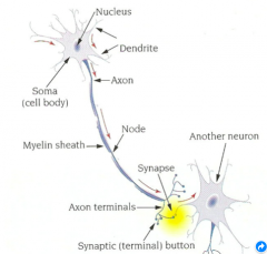
Neuron |
|
|
|
Dendrite |
Receives the message first |
|
|
Soma (cell body) |
Decides if the message is important and should be carried further |
|
|
Axon |
Carries the message to the next neuron through nodes |
|
|
Node |
Attaches two axons |
|
|
Myelin sheath |
fatty substance covering most of the axon - allow the message to bounce along and be transmitted faster. |
|
|
Synapse |
The gap between the pre-synaptic neuron and the post-synaptic neuron |
|
|
Types of neurons |
Sensory, motor and interneurons |
|
|
Sensory neurons (afferent) |
Specialised cells that receive information from both the external environment and from within the body and transmit the information to the CNS. |
|
|
Motor neurons (efferent) |
Transmits messages from the CNS to the muscles, glands and organs. They enable muscles to move, cause glands to secrete chemicals and activate internal organs such as the heart, lungs and intestines |
|
|
Interneurons |
Provide neural links between sensory and motor neurons and have a specialised role of carrying and integrating messages between sensory and motor neurons. Interneurons exist only within the CNS. |
|
|
Somatic Nervous System |
A network of sensory (afferent) nerves that carry information received by sensory receptors in the body to the CNS, and motor (efferent) nerves that carry information from the CNS to control voluntary movements of skeletal muscles. The Sensory function of the SNS is activated when you sense or feel something, on your skin, for example, and the SNS sends signals from that point to your brain via the spinal cord, resulting in you experiencing the sensation. The motor function of the SNS is demonstrated whenever you voluntarily move a body part. |
|
|
Autonomic Nervous System |
The Autonomic Nervous System is a network of nerves that connects the CNS to the body’s internal organs and glands providing feedback to the brain about their activities. The ANS is called autonomous because many of the organs, glands and processes under its control are self-regulating and not usually under voluntary control. Can be broken down into sympathetic and parasympathetic. |
|
|
Parasympathetic Nervous System |
Responsible for decreasing the activity of most visceral muscles, organs and glands, and keeping the body functioning in a normal state. (counterbalances the activities of the SNS |
|
|
Functions of the Parasympathetic Nervous System |
It has two main functions: 1. It keeps the systems of the body functioning efficiently and in times of minimal stress and in the absence of threats, helps it to maintain the internal body environment in a steady, balanced, state of normal functioning (homeostasis). 2. It also restores the body to a state of calm, once the need for activity of the SNS has passed. |
|
|
Sympathetic Nervous System |
Responsible for increasing the activity of most visceral muscles, organs and glands in times of vigorous activity, stress or threat. |
|
|
Result of SNS Activation |
- heart rate and blood pressure increase, - breathing rate increases so more oxygen can be taken in, - sugar and fat are released from storage to provide instant energy to the skeletal muscles, - the pupils dilate to allow more light to enter the eye and enhance vision, - and sweat glands increase their production of sweat which cools the body. |