Reading...
![]()
Play button
![]()
Play button
![]()
Use LEFT and RIGHT arrow keys to navigate between flashcards;
Use UP and DOWN arrow keys to flip the card;
H to show hint;
A reads text to speech;
39 Cards in this Set
- Front
- Back
|
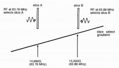
How does the MR computer know where the signal is coming from
|
|
|
|
What is the magnetic field strength at the isocentre
|
same as Bo
|
|
|
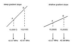
What determines the slope of the gradient magnetic field
|
the amplitude of the magnetic field gradient
|
|
|
What does the slope of the gradient magnetic field determin
|
it will determine the rate of change along the gradient axis
|
|
|
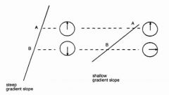
What is the difference between as gradient with a steep slop and a shallow slope
|
the steep slope have a greater change in gradient between to points
|
|
|
What causes a greater alteration in the precession of 2 protons; shhallow or steep slope
|
steep
|
|
|
Sleep stop Vs shallow slope
|
|
|

NOTE THE GRADIENTS ARE CHANGED DEPENDENDING ON THE IMAGE BEING OBTAINED
|
|
|
|
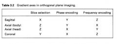
What is the are the axis of the gradients of the head
|
|
|
|
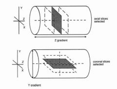
How is a slice selection gradient aligned
|
the same direction as the main magnetic field
|
|
|
How is a particular slice chosen
|

since there is slightly different precession along the gradient just give a RF pulse that will only react to that pulse (according to the lamor equation)
|
|
|
How do you know which direction is going to be the slice selection gradient
|

by the imaging plane being imaged
|
|
|
In axial imaging what plane is the slice selecting gradient
|

z-plane
|
|
|
In coronal imaging what axis is the slice selection gradient
|

Y axis
|
|
|
What happens if you want to image oblique images
|
a combination of 2 magnetic field gradients will have to be used for the slice selection (not shown but use your imagination)
|
|
|
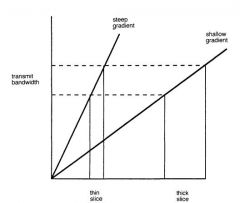
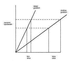
How is the size of the slice selected
|

depends on the steepness of the magnetic field gradient being used. But steeper is thinner
|
|
|
How is the frequency bandwidth that is recieved in thin slices compared to thick slices.
|

The same
|
|
|
Error previous page. This slide is misleading becauses either the size of the bandwith being recieved or the steepness of the gradient can be adjusted to change the slice thickness.
|

|
|
|
How is the bandwidth or steepness of the magnetic gradient changed.
|
It is automaticaly done by selecting the slice size in the MRI scanner
|
|
|
What is the bandwidth in these images the RF pulse or the bandwitdth that is recieved by the machine when the signal occurs
|

The RF pulse bandwidth
|
|
|
What determines the size of the gap between the gradient
|

The gap between the slices is determined by the gradient slope and by
the thickness of the slice. (NOT SHOWN IN THIS IMAGE AND THIS EXPLANATION DOES NOT FULLY EXPLAIN WHY) |
|
|
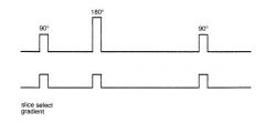
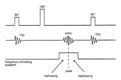
Why is the slice selection gradient turned on during RF pulse administration.
|

|
|

The slice selection gradient is turned on because the RF pulse will be tuned to that precessional frequency and needs to know where to go.
|
k
|
|
|
Which axis is typically the frequency encoding direction
|
long axis
|
|
|
What is the direction of the frequency encoding (long axis) of the body
|
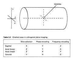
X
|
|
|
What is meant by the long axis
|

the longer of the two remaining dimensions once the slice selection gradient is chosen (IE look at the frequency encoding direction of the head (nose to occiput)
|
|
|
When is the frequency encoding (readout gradient) turned on
|

When the signal is going to be recieved.
|
|
|
how does the turning on the frequency encoding gradient effect the rephasing of the protons
|
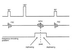
I am assuming it will make them rephase quicker then dephase
|
|
|
What determines the field of view (FOV) when doing a scan
|

|
|
|
What happens to the protons as the readout gradient occurs
|

continued rephasing
|
|
|
What happens to the protons as the readout gradient occurs
|
continued rephasing
|
|
|
What does the steepness of the frequency encoding gradient determine
|
he steepness of the slope of the frequency encoding
gradient determines the size of the anatomy covered along the frequency encoding axis during the scan. This is called the field of view (FOV). The steeper the less anatomy that will be covered. |
|
|
What happens to the nuclei at the isocentre of the phase encoding gradient
|
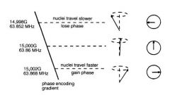
No change
|
|
|

|
|
|
How are these frequencies mapped
|
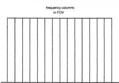
Each frequency is allocated a
frequency column that is mapped onto the FOV along the frequency axis |
|
|
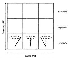
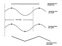
What does 1 cycle look like
|
|
|
|
What must occur to the bottom for nyquist thereom to be followed
|
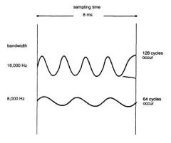
increased sampling time. (nyquist only says 2 samples per cycle not at least 2 samples)
|
|
|
What does reducing the frequency bandwidth do to the sampling time
|
Reducing the receive bandwidth affects the minimum TE because the
echo is usually centred on the middle of the frequency encoding gradien |
|
|
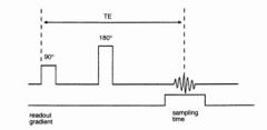
What does increasing the recieve bandwidth do
|
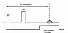
increases TE time
|