Reading...
![]()
Play button
![]()
Play button
![]()
Use LEFT and RIGHT arrow keys to navigate between flashcards;
Use UP and DOWN arrow keys to flip the card;
H to show hint;
A reads text to speech;
57 Cards in this Set
- Front
- Back
|
Are coils sensitive to transverse magnetization
|
Coils are only sensitive to variations of transverse magnetization vector
|
|
|
What signal occurs after a 90 degree pulse
|
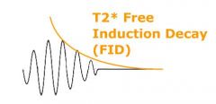
After a 90° RF pulse, the Free Induction Decay (FID) signal is oscillating at resonance frequency and signal enveloppe is a decay curve described as an exponential curve, depending on tissue-specific spin-spin relaxation and static field inhomogeneities
|
|
|
What is another name for FID
|
This decay is characterized by time constant T2*
|
|
|
Is T2* longer than T2
|
T2* is always shorter than T2.
|
|
|
What is the function of the 180 pulse in spin echo sequences
|
The 180° RF pulse reverses dephasing due to static field inhomogeneities (T2* effects) but not random spin-spin relaxation (T2 effects, tissue-specific)
|
|
|
What are the 2 pulses involved in a spin echo sequence
|
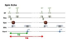
Spin Echo sequence requires an excitation pulse (90° RF pulse) and a 180° rephasing pulse
|
|
|
What is the time between the the 90 pulse and the 180 pulse
|
the TE/2
|
|
|
What does a spine echo sequence look like
|
|
|
|
When is the MR signal acquired
|
at TE when the signal is the strongest
|
|
|
How many times must the pulse sequence be repeated
|
The 90° - 180° RF pulses sequence must be repeated as many times as the number of lines in the data matri
|
|
|
What is the time between each pulse sequence called
|
The time between each 90° RF pulse (excitation pulse) is called Repetition Time (TR).
|
|
|
Are transverse relaxation and longitudinal relaxation simultaneous
|
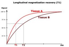
tranverse relaxation (transverse magnetization decay, producing MR signal) and longitudinal relaxation (longitudinal magnetization recovery) are simultaneous
|
|
|
What happens during a long TR
|
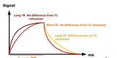
The longer the TR is, the more longitudinal magnetization will recover.
|
|
|
What happens if there is a long TR
|
TR modifies T1-weighting : the longer is the TR, the more T1-weigthed the image is
|
|
|
What happens if there is a long TE
|
TE modifies T2-weighting : the shorter is the TE, the less T2-weigthed the image is
|
|
|
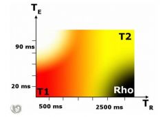
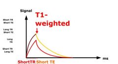
What if there is a short TR and a short TE
|
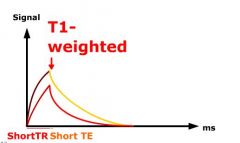
A short TR court and a short TE court give a T1-weighted image
|
|
|
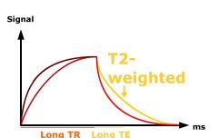
What if there is a long TR and a long TE
|
A long TR long and a long TE long give a T2-weighted image
|
|
|
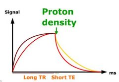
What if there is a long TR and a short TE
|
PD
|
|
|
What happens when the 90 pulse occurs
|
After a 90° RF pulse, net magnetization tips down so that longitudinal magnetization has disappeared and transverse magnetization has appeared.
|
|
|
What 3 things occur after the 90 degree RF pulse
|
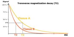
transverse magnetization decays
longitudinal magnetization recovers protons re-radiate the absorbed energy |
|
|
How do coils in the transverse plane receive a signal
|
Coils can receive the signal in the transverse plane due to variations of transverse magnetization vector
|
|
|
What is T2* in the abscence of a magnetic gradient
|
In absence of any magnetic gradient, this signal is called Free Induction Decay (FID).
|
|
|
What does T2* take into account when decaying
|
tissue specific spin-spin relaxation (random interactions between spins) responsible for pure T2decay
static inhomogeneities in magnetic fields which accelerate spins dephasing |
|
|
FID and T2*
|
|
|
|
What is the function of the 180 degree pulse
|
rephase spins and reverse static field inhomogeneities
|
|
|
What happens after the 90 degree pulse
|
After a 90° RF pulse, spins dephase and transverse magnetization decreases. If we apply a 180° RF pulse, spins rephase and transverse magnetization reappears
|
|
|
What is the time after the 90 pulse called
|
After the 90° RF pulse spins dephase (during a time defined as TE/2)
|
|
|
What are the 2 parameters of spin echo sequences
|
echo time
repitition time |
|
|
What is repetition time
|
Repetition Time is the time between 2 excitations pulses (time between two 90° RF pulses)
|
|
|
What is echo time
|
Echo Time (TE) is the time between the 90° RF pulse and MR signal sampling, corresponding to maximum of echo. The 180° RF pulse is applied at time TE/2.
|
|
|
What is the definition of T1
|
After time T1, longitudinal magnetization has returned to 63 % of its final value. T1 defines the recovery rate of longitudinal magnetization
|
|
|
What does a T1 curve look like
|
|
|
|
What is T1 time referring to
|

the time at which the curve has recovered 67% look at the lines. If there is a short T1 (fat) that line will be at a short time
|
|
|
What does a T2 curve look like
|
|
|
|
How does TR effect 2 different tissues
|
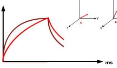
Let's consider 2 tissues A and B with different T1s. If TR is very long, even if tissue A has a longuer T1 than tissue B, the longitudinal magnetization of both tissues will recover completely before the next excitation.
|
|
|
What is the relative magnitude (amplitude)of the transverse magnetization of the hypotheical Tissue A and tissue B after a long TR
|
they will have the same amplitude
|
|

Long TR and notice both of these lines are the same height (magnitude)
|
Long TR and notice both of these lines are the same height (magnitude)
|
|

Shorter TR and tissue B (dark red) transverse amplitude is not as high
|
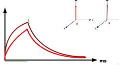
Shorter TR and tissue B (dark red) transverse amplitude is not as high
|
|
|
What happens if TR is set long
|
|
|
|
What is the result of a short TR and TE
|
|
|
|
Long TR and short TE
|
|
|
|
Long TR and TE
|
|
|
|
Do all images really have a combination of T1 T2 and PD
|
yes
|
|
|
What is the appearance of a tissue with a long T1 and T2
|
A tissue with a long T1 and T2 (like water) is dark in the T1-weighted image and bright in the T2-weighted image.
|
|
|
Are long T2 tissues bright
|
yes
|
|
|
Are long T1 tissues dark
|
yes
|
|
|
What is the appearance of fat in a T2W image
|
A tissue with a short T1 and a long T2 (like fat) is bright in the T1-weighted image and gray in the T2-weighted image.
|
|
|
What is the appearance of gadolinium
|
Gadolinium contrast agents reduce T1 and T2 times, resulting in an enhanced signal in the T1-weighted image and a reduced signal in the T2-weighted imag
|
|
|
In clinical practice what is considered a short TE
|
A short TE is usually lower than 30 ms
|
|
|
In clinical practice what is considered a long TR
|
A long TR = 3 times the short TR, usually greater than 1500 ms
|
|
|
In clinical practice what is considered a short TR
|
A short TR = value approximately equal to the average T1 value, usually lower than 500 ms
|
|
|
Is TE always shorter than TR
|
yes, TE is always shorter than TR
|
|
|
What is considered a long TE in clinical practice
|
A long TE = 3 times the short TE, usually greater than 90 ms
|
|
|
What is the key to a good MR sequence
|
a good MRI sequence gives high tissue contrast but lasts the shortest time possible
|
|
|
What is the incline portion of this graph
|
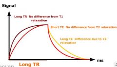
before the 180 pulse
|
|
|
What is the decline portion of this graph
|

after the 180 pulse
|
|
|
Can you see how the differenc that is detected is because of T1 effect (short TR and TE)
|
|