Reading...
![]()
Play button
![]()
Play button
![]()
Use LEFT and RIGHT arrow keys to navigate between flashcards;
Use UP and DOWN arrow keys to flip the card;
H to show hint;
A reads text to speech;
41 Cards in this Set
- Front
- Back
|
bony pelvis, true and false pelvis, pelvic cavity and perineum
|
|
|
|
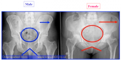
differences between the male and female pelvis
|
-
|
|
|
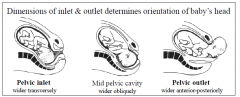
orientation of the foetal head during descent through the pelvic inlet and outlet
|
|
|
|
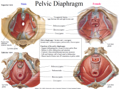
pelvic diaphragm and its function
|
Pelvic diaphragm = levator ani + coccygeus Levator ani = pubococcygeus, puborectalis, iliococcygeus
Functions of the pelvic diaphragm: - Support abdominopelvic viscera & raises pelvic floor - Relaxes to allow defaecation and urination - Contracts when intraabdominal pressure increases - Lifts and shortens wall of anal canal at defaecation - Directs head of foetus into AP orientation at pelvic outlet |
|
|
contents of the pelvic cavity in males and females
|
Pelvic cavity contains coils of intestines, bladder, rectum and in:
Males, the prostate, seminal vesicles, ductus (vas) deferens Females, ovaries, uterine tubes, uterus, upper vagina |
|
|
location and function of the perineal body and the structures that provide support for pelvic viscera
|
-
|
|
|
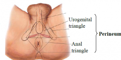
anatomy of the perineum, its boundaries, divisions into triangles and the contents of each triangle
|
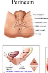
The 2 triangles of the perineum
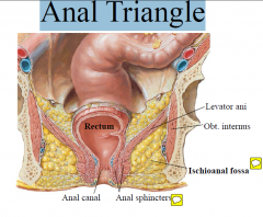
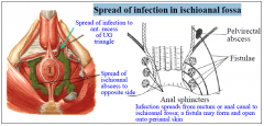
1. Urogenital triangle: external genitalia (in male and female), perineal membrane, anterior horns of ischioanal fossa 2. Anal triangle: anal canal, anal sphincters & ischioanal fossa |
|
|
locations of the superficial and deep perineal pouches
|
-
|
|
|
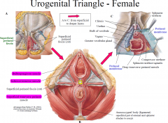
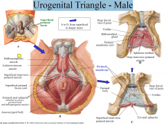
structures in the male and female urogenital triangle
|
-
|
|
|
neurovascular supply to the pelvis and perineum
|
-
|
|
|
location of the pelvic pain line and the general principles of pain referral from viscera
|
-
|
|
|
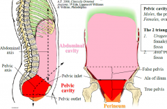
Bony Pelvis
|
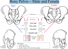
- hip bones, sacrum and cocyx
1. True pelvis – area between pelvic inlet & outlet 2. False pelvis – area above pelvic inlet to top of iliac crest (part of abdominal cavity) |
|
|
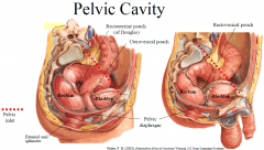
Pelvic cavity
|
extends from pelvic inlet to pelvic diaphragm
|
|
|
Perineum
|
is the region below pelvic diaphragm to skin of lower trunk
|
|
|
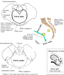
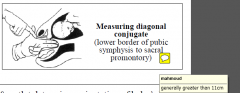
Pelvic Inlet and Outlet
|
|
|
|
Measuring diagonal conjugate
|
|
|
|
Bony Pelvis – Male and Female
|
|
|
|
Boney Pelvis- Male and female X-rays
|
|
|
|
Pelvic Walls and Floor
|
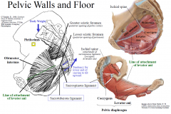
Sacrotuberous & sacrospinous ligaments tie lower end of sacrum to ischium. They resist the tendency of the lower end of the sacrum to tilt upwards when body weight passes to the sacrum, and hence stabilises the SI joint
|
|
|
Pelvic Diaphragm
|
|
|
|
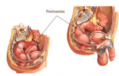
Peritoneal Pouches
|
-
Peritoneum extends into pelvic cavity and reflects on pelvic viscera to form dependent pouches - Pouches in female: uterovesical and rectouterine (Pouch of Douglas) - Pouch in male: rectovesical - When supine or erect, infectious material and fluid gravitates into pouches - On digital rectal examination (or transvaginal in female) there can be tenderness to palpation or bogginess |
|
|
Perineum the 2 triangles
- borders - plane |
|
|
|
Anal Triangle
|
|
|
|
Spread of infection in ischioanal fossa
|
|
|
|
Urogenital Triangle - Female
|
|
|
|
Urogenital Triangle - Male
|
|
|
|
Superficial & Deep Perineal Pouches
|
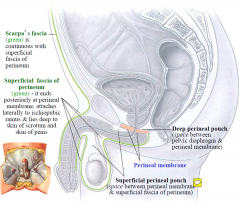
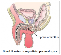
Urogenital triangle divided into superficial and deep perineal pouches for descriptive purposes. Boundaries are: pelvic diaphragm to perineal membrane superficial fascia of perineum
|
|
|
Blood or fluid in superficial perineal space
|
|
|
|
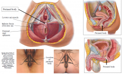
Perineal Body
|
-
Comprised of connective tissue (collagen, elastin), smooth muscle and skeletal muscle - At center point of perineum between urogenital and anal triangles - Critical stabilising structure in the perineum - Receives insertion of levator ani, external anal sphincter, bulbospongiosus and transverse perinei muscles |
|
|
Prolapse of Viscera
|
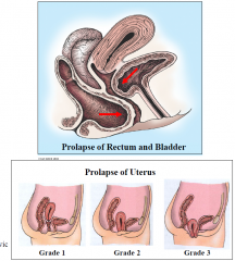
Failure of support leads to prolapse.
Weak muscles or problems with integrity of perineal body result in the ligaments taking more tension Kegel exercises to strengthen pelvic floor in females may help prevent prolapse |
|
|
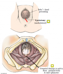
Damage to PF, perineal body & anal sphincters during birth
|
|
|
|
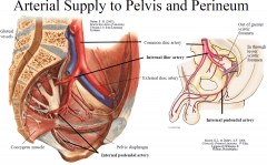
Arterial Supply to Pelvis and Perineum
|
Internal iliac artery supplies pelvic viscera, perineum, gluteal area and has branches to bladder, uterus, vagina, prostate and vas deferens. Internal pudendal artery is a branch of the internal iliac artery and provides arterial supply to the perineum. Also supplies erectile tissue.
Exception is the testicular artery which supplies testes & epididymis and originates from abdominal aorta. |
|
|
Arterial Supply to pelvis
|
|
|
|
Venous Drainage of plevis and perineum
|
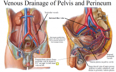
Valveless venous plexus drains pelvic viscera; forms a communicating network (trauma to pelvis oozing of blood which is difficult to stem). These drain to internal iliac veins with some drainage to internal vertebral venous plexus via sacral veins ( metastases to vertebral column).
EXCEPTION: drainage of the rectum TO inferior mesenteric vein TO portal vein |
|
|
Lymphatics of Pelvis & Perineum
|
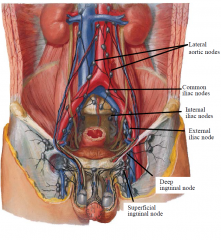
Pelvic organs drain to nodes around nearby vessels, which are named accordingly; external, internal & common iliac nodes and sacral nodes.
Exception: ovaries, uterine tubes and fundus of uterus drain with ovarian vessels to para-aortic nodes. Area of uterus near attachment of round ligament drains with round ligament to superficial inguinal nodes. Skin of Perineum plus lower parts of urethra, vagina & anal canal drain to superficial inguinal nodes. Most of deeper parts of perineum drain to internal iliac nodes. Exception: testes & epididymis drain to para-aortic nodes (by following testicular vessels). |
|
|
Innervation of Pelvis & Perineum
|
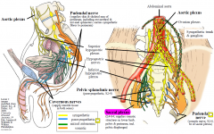
(Sympathetic nn descend to superior then L&R inferior hypogastric plexuses Parasympathetic nerve fibres ascend from inferior hypogastric plexus to reach hindgut)
|
|
|
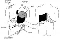
Visceral Afferents and Pain
|
Visceral afferents from trunk viscera travel with autonomic nerves Visceral afferents involved in visceral reflexes (eg, defaecation, micturition) travel with parasympathetic fibres (spinal cord level only) Visceral afferents that conduct pain travel with sympathetic nerves if the viscus is above the pelvic pain line and travel with parasympathetic nerves if the viscus is below the pelvic pain line
|
|
|
Pelvic Pain Line
|
Pelvic pain line – corresponds to the inferior limit of peritoneum, except for the GIT when it is midway along sigmoid colon
technically it goes down the pouches as well |
|
|
Above the pelvic Pain Line
|
Above pelvic pain line, visceral pain afferents travels with sympathetic nerves back to T1-L2 spinal cord, eg, heart T1-5; midgut T10; ureter T10/11-L1; pelvic viscera above pelvic pain line T11-L2 spinal cord
|
|
|
Below pelvic pain line
|
visceral pain travels with parasympathetic nerves to S2-4 spinal cord, eg, lower bladder, cervix, prostate
|
|
|
Visceral Pain Referral
|
|