Reading...
![]()
Play button
![]()
Play button
![]()
Use LEFT and RIGHT arrow keys to navigate between flashcards;
Use UP and DOWN arrow keys to flip the card;
H to show hint;
A reads text to speech;
38 Cards in this Set
- Front
- Back

ID:
|
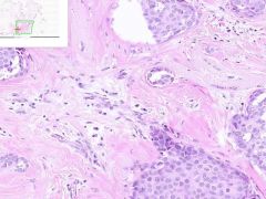
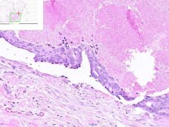
BREAST ADENOCARCINOMA
The ducts are lined by cytologically malignant cells. A few have large atypical nuclei, variably coarse chromatin, and prominent nucleoli. Mitotic figures are occasionally seen. In addition, multiple foci of infiltrating ductal carcinoma are also present. Notice the small poorly formed ducts infiltrating through the breast stroma and fat. |
|
ID:
|

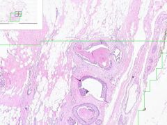
BREAST ADENOCARCINOMA, INTRADUCTAL WITH INVASION
Both intraductal and infiltrating ductal carcinoma are present in this digitized image. The intraductal carcinoma is growing in a comedo pattern, that is the central part of the involved duct is replaced by brightly eosinophilic necrotic debris. The ducts are lined by cytologically malignant cells. A few have large atypical nuclei, variably coarse chromatin, and prominent nucleoli. Mitotic figures are occasionally seen. In addition, multiple foci of infiltrating ductal carcinoma are also present. Notice the small poorly formed ducts infiltrating through the breast stroma and fat. |
|

ID:
|
BREAST ADENOCARCINOMA
The intraductal carcinoma is growing in a comedo pattern, that is the central part of the involved duct is replaced by brightly eosinophilic necrotic debris. The ducts are lined by cytologically malignant cells. A few have large atypical nuclei, variably coarse chromatin, and prominent nucleoli. Mitotic figures are occasionally seen. |
|
ID:
|

BREAST ADENOCARCINOMA
The intraductal carcinoma is growing in a comedo pattern, that is the central part of the involved duct is replaced by brightly eosinophilic necrotic debris. The ducts are lined by cytologically malignant cells. A few have large atypical nuclei, variably coarse chromatin, and prominent nucleoli. Mitotic figures are occasionally seen. |
|

ID:
|
BREAST ADENOCARCINOMA, INTRADUCTAL WITH INVASION
Both intraductal and infiltrating ductal carcinoma are present in this digitized image. The intraductal carcinoma is growing in a comedo pattern, that is the central part of the involved duct is replaced by brightly eosinophilic necrotic debris. The ducts are lined by cytologically malignant cells. A few have large atypical nuclei, variably coarse chromatin, and prominent nucleoli. Mitotic figures are occasionally seen. In addition, multiple foci of infiltrating ductal carcinoma are also present. Notice the small poorly formed ducts infiltrating through the breast stroma and fat. |
|

ID:
|
BREAST ADENOCARCINOMA
The ducts are lined by cytologically malignant cells. A few have large atypical nuclei, variably coarse chromatin, and prominent nucleoli. Mitotic figures are occasionally seen. In addition, multiple foci of infiltrating ductal carcinoma are also present. |
|

ID:
|

BREAST ADENOCARCINOMA, INTRADUCTAL WITH INVASION
Both intraductal and infiltrating ductal carcinoma are present in this digitized image. The intraductal carcinoma is growing in a comedo pattern, that is the central part of the involved duct is replaced by brightly eosinophilic necrotic debris. The ducts are lined by cytologically malignant cells. A few have large atypical nuclei, variably coarse chromatin, and prominent nucleoli. Mitotic figures are occasionally seen. In addition, multiple foci of infiltrating ductal carcinoma are also present. Notice the small poorly formed ducts infiltrating through the breast stroma and fat. |
|

ID:
|
BREAST ADENOCARCINOMA, INTRADUCTAL WITH INVASION
Both intraductal and infiltrating ductal carcinoma are present in this digitized image. The intraductal carcinoma is growing in a comedo pattern, that is the central part of the involved duct is replaced by brightly eosinophilic necrotic debris. The ducts are lined by cytologically malignant cells. A few have large atypical nuclei, variably coarse chromatin, and prominent nucleoli. Mitotic figures are occasionally seen. In addition, multiple foci of infiltrating ductal carcinoma are also present. |
|
ID:
|

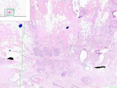
BREAST ADENOCARCINOMA
Normal looking ducts infiltrating into fat regions. Some micro collections of ductal cells. |
|

ID:
|
BREAST ADENOCARCINOMA
The intraductal carcinoma is growing in a comedo pattern, that is the central part of the involved duct is replaced by brightly eosinophilic necrotic debris. The ducts are lined by cytologically malignant cells. A few have large atypical nuclei, variably coarse chromatin, and prominent nucleoli. Mitotic figures are occasionally seen. In addition, multiple foci of infiltrating ductal carcinoma are also present. |
|

ID:
|
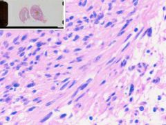
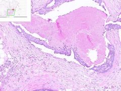
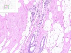
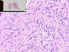
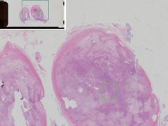
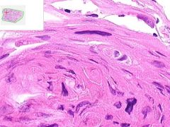
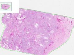
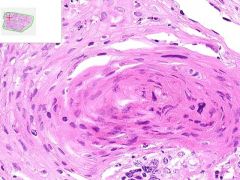
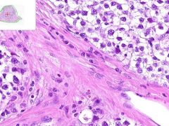
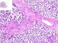
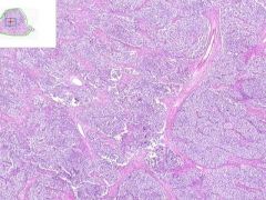
SCHWANNOMA
This tumor shows two typical patterns of growth. One pattern is that of compact spindle cells in a whirling pattern, referred to as Antoni-A tissue (pattern). The other appears as clusters of degenerative, vacuolated tumor cells, referred to as an Antoni-B tissue. Occasionally, in the spindle cell areas of tissue, cell nuclei can form a row of palisading nuclei, referred to as a Verocay body. Some nuclei show an increased density of chromatin, but typical cellular characteristics of malignancy are not seen in this tumor. |
|
ID:
|

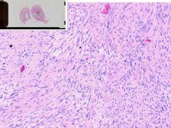
SCHWANNOMA
This tumor shows two typical patterns of growth. One pattern is that of compact spindle cells in a whirling pattern, referred to as Antoni-A tissue (pattern). |
|
ID:
|

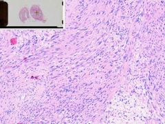
SCHWANNOMA
This tumor shows two typical patterns of growth. One pattern is that of compact spindle cells in a whirling pattern, referred to as Antoni-A tissue (pattern). |
|

ID:
|
SCHWANNOMA
One pattern is that of compact spindle cells in a whirling pattern, referred to as Antoni-A tissue (pattern). Occasionally, in the spindle cell areas of tissue, cell nuclei can form a row of palisading nuclei, referred to as a Verocay body. |
|
ID:
|

SCHWANNOMA
This tumor shows two typical patterns of growth. One pattern is that of compact spindle cells in a whirling pattern, referred to as Antoni-A tissue (pattern). The other appears as clusters of degenerative, vacuolated tumor cells, referred to as an Antoni-B tissue. |
|
ID:
|

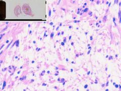
SCHWANNOMA
This tumor shows two typical patterns of growth. One pattern is that of compact spindle cells in a whirling pattern, referred to as Antoni-A tissue (pattern). The other appears as clusters of degenerative, vacuolated tumor cells, referred to as an Antoni-B tissue. Occasionally, in the spindle cell areas of tissue, cell nuclei can form a row of palisading nuclei, referred to as a Verocay body. Some nuclei show an increased density of chromatin, but typical cellular characteristics of malignancy are not seen in this tumor. |
|
ID:
|

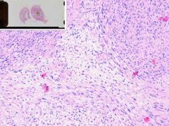
SCHWANNOMA
This tumor shows two typical patterns of growth. One pattern is that of compact spindle cells in a whirling pattern, referred to as Antoni-A tissue (pattern). The other appears as clusters of degenerative, vacuolated tumor cells, referred to as an Antoni-B tissue. Occasionally, in the spindle cell areas of tissue, cell nuclei can form a row of palisading nuclei, referred to as a Verocay body. Some nuclei show an increased density of chromatin, but typical cellular characteristics of malignancy are not seen in this tumor. |
|

ID:
|
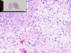
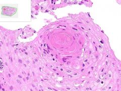
SCHWANNOMA
Antoni-A: compact spindle cells in a whirling pattern. Antoni-B: clusters of degenerative, vacuolated tumor cells. Occasionally, in the spindle cell areas of tissue, cell nuclei can form a row of palisading nuclei, referred to as a Verocay body. |
|

ID:
|
SQUAMOUS CELL CARCINOMA OF LUNG
These large epithelial cells appear in sheets with an intermittent swirling pattern. Occasional keratin pearls and intercellular bridges can be seen. |
|

ID:
|
SQUAMOUS CELL CARCINOMA OF LUNG
These large epithelial cells appear in sheets with an intermittent swirling pattern. Occasional keratin pearls can be seen in the swirl of cells. |
|

ID:
|
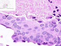
SQUAMOUS CELL CARCINOMA OF LUNG
The normal lung tissue in this section is entirely replaced by squamous cell carcinoma. These large epithelial cells appear in sheets with an intermittent swirling pattern. The cells have open-appearing nuclei and high N/C ratio. The tumor is moderate-poorly differentiated. Occasional keratin pearls can be seen in the swirl of cells, but intercellular bridges are more difficult to see. |
|

ID:
|
SQUAMOUS CELL CARCINOMA OF LUNG
The normal lung tissue in this section is entirely replaced by squamous cell carcinoma. These large epithelial cells appear in sheets with an intermittent swirling pattern. The cells have open-appearing nuclei and high N/C ratio. The tumor is moderate-poorly differentiated. Occasional keratin pearls can be seen in the swirl of cells, but intercellular bridges are more difficult to see. |
|
|
Which cancers tend to met. to the brain?
|
Breast
GI Kidney Lung Melanoma Ben Good's Kids Love Mom |
|
|
Which cancers tend to met. to the liver?
|
Breast
GI Lung pancreato-biliary Beer Gets Large Parties Beer Gets Lost Panties |
|
|
Which cancers tend to met. to the bone?
|
prostate
breast lung kidney thyroid testes "peanut butter lunch kids, thank teachers" |
|
ID:
|

SQUAMOUS CELL CARCINOMA OF LUNG
The normal lung tissue in this section is entirely replaced by squamous cell carcinoma. These large epithelial cells appear in sheets with an intermittent swirling pattern. The cells have open-appearing nuclei and high N/C ratio. The tumor is moderate-poorly differentiated. Occasional keratin pearls can be seen in the swirl of cells, but intercellular bridges are more difficult to see. |
|
ID
|

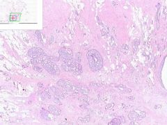
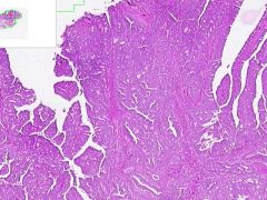
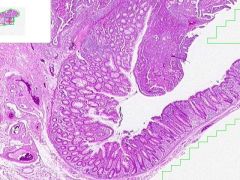
COLON ADENOCARCINOMA
Glands show haphazard architectural arrangement. The tumor becomes malignant when it invades into the submucosa and replaces normal colonic tissue. |
|

ID
|
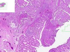
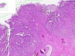
COLON ADENOCARCINOMA
Colon cancer with adjacent normal mucosa. The neoplastic tissue show haphazard architectural arrangement with atypical cells and staining. The tumor becomes malignant when it invades into the submucosa and replaces the normal colonic tissue. |
|

ID:
|
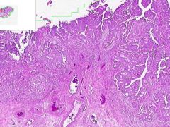
COLON ADENOCARCINOMA
Colon cancer with adjacent normal mucosa. The neoplastic tissue show haphazard architectural arrangement with atypical cells and staining. The tumor becomes malignant when it invades into the submucosa and replaces the normal colonic tissue. |
|
ID:
|

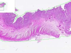
COLON ADENOCARCINOMA
The neoplastic tissue show haphazard architectural arrangement with atypical cells and staining. The tumor becomes malignant when it invades into the submucosa and replaces the normal colonic tissue. |
|

ID:
|
COLON ADENOCARCINOMA
The neoplastic tissue show haphazard architectural arrangement with atypical cells and staining. The tumor becomes malignant when it invades into the submucosa and replaces the normal colonic tissue. |
|
ID:
|

COLON ADENOCARCINOMA
Colon cancer with adjacent normal mucosa. The neoplastic tissue show haphazard architectural arrangement with atypical cells and staining. The tumor becomes malignant when it invades into the submucosa and replaces the normal colonic tissue. |
|

ID:
|
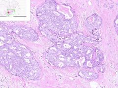
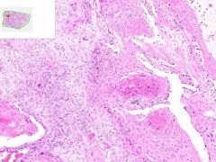
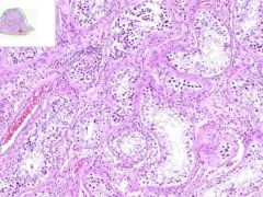
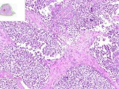
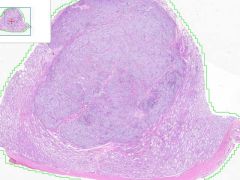
SEMINOMA
Abnormal cells are arranged into packets or compartments of different size. These compartments result from the division of the tumor by eosinophilic fibrous bands that intersect the lesion and contain infiltrating lymphocytes. Tumor cells have fairly abundant cytoplasm, prominent nucleoli and coarse, hyperchromatic chromatin. "Fried egg" appearance. The division into nests is most characteristic feature of seminomas. |
|

ID:
|
SEMINOMA
Abnormal cells are arranged into packets that are darker and more cellular than the non-neoplastic testicular tissue surrounding it. |
|

ID:
|
SEMINOMA
Abnormal cells are arranged into packets or compartments of different size. These compartments result from the division of the tumor by eosinophilic fibrous bands that intersect the lesion and contain infiltrating lymphocytes. Tumor cells have fairly abundant cytoplasm, prominent nucleoli and coarse, hyperchromatic chromatin. "Fried egg" appearance. The division into nests is most characteristic feature of seminomas. |
|

ID:
|
SEMINOMA
Abnormal cells are arranged into packets or compartments of different size. These compartments result from the division of the tumor by eosinophilic fibrous bands that intersect the lesion and contain infiltrating lymphocytes. Tumor cells have fairly abundant cytoplasm, prominent nucleoli and coarse, hyperchromatic chromatin. "Fried egg" appearance. The division into nests is most characteristic feature of seminomas. |
|
ID:
|

SEMINOMA
The seminoma appears as a nodule of tumor that is darker and more cellular than the non-neoplastic testicular tissue surrounding it. It compresses the adjacent tissue without a well defined capsule. The normal tissue has well defined seminiferous tubules lined by germinal epithelium, which could be seen at higher mag. The nest-like division by fibrotic bands is characteristic feature of seminoma. |