![]()
![]()
![]()
Use LEFT and RIGHT arrow keys to navigate between flashcards;
Use UP and DOWN arrow keys to flip the card;
H to show hint;
A reads text to speech;
148 Cards in this Set
- Front
- Back
|
contains most parasitic worms dealt with in veterinary medicine. occur in almost every body system and location
|
nematodes
|
|
|
which life cycle stage is infective in nematodes
|
L3
|
|
|
nematodes usually have ____ larval stages, which are sexually immature juveniles superficially resembling the adults
|
4
|
|
|
the L in the developmental cycle of nematodes stands for what
|
life stage
|
|
|
nematodes undergo incremental growth punctuated by malts of the _______, which covers the body surface. they go through these malts to develop into infective third stage larva
|
cuticle
|
|
|
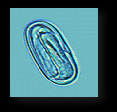
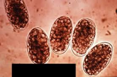
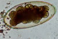
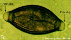
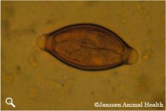
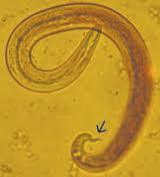
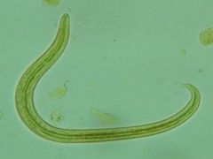
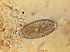
this is an esophageal worm, eggs are passed down the esophagus and out into the feces, have a unique paper clip shape, animal is infected by ingesting the eggs of this parasite. eggs observed on qualitative fecal flotation but can also be recovered from vomitus. Prepatent period for this roundworm is 6 months
|
Spirocerca lupi
|
|
|
spirocerca lupi
|
|
|
stomach worms of dogs and cats, usually firmly attached to mucosal surface of stomach where they suck blood. occasionally found in lumen of stomach or small intestine. may be viewed with an endoscope
|
physaloptera spp.
|
|
|
3 clinical signs of physaloptera spp
|
vomiting, diarrhea, dark tarry stools
|
|
|
features of adult physaloptera spp. (4)
|
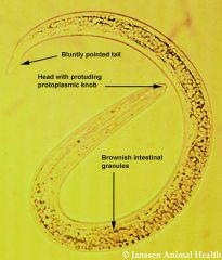
creamy, white, sometimes tightly coiled. 1.3-4.8cm long. often recovered in vomitus. can be confused with ascarids or roundworms
|
|
|
physaloptera spp.
|
|
|
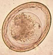
5 features of physaloptera spp. eggs
|
30-34um by 49-58um. contain larva when they are laid. can be recovered using a standard fecal flotation. prepatent period 56-83 days
|
|
|
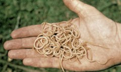
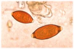
most frequently diagnosed nematodes in young puppies and kittens. may be found in the small intestine of dogs and cats in most areas of the world. all young puppies and kittens should be examined for this parasitic infection
|
ascarids
|
|
|
4 clinical signs of ascarids
|
vomiting, diarrhea, constipation, and other non-specific signs
|
|
|
5features of adult ascarids
|
3-18cm. do not attach to host, move around in small intestine, when passed in feces, usually tightly coiled. in vomit, looks like spaghetti
|
|
|
ascarids
|
|
|
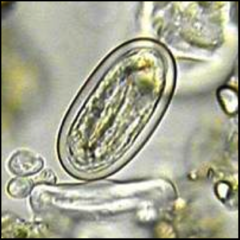
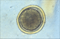
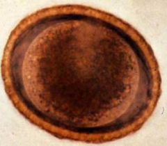
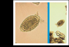
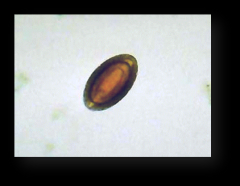
3 characteristics of toxocara species eggs
|
unembryonated, spherical, deeply pigmented center with rough, pitted outer shell
|
|
|
toxocara canis egg
|
|
|
this ova is generally round and thick walled (individual species may vary in color and texture). may remain viable in soil for years in the right conditions
|
ascarids
|
|
|
these are in small intestines where they mate. female produces unembryonated eggs that are passed in feces. eggs embryonate where they contain L2 larvae on the ground where they are ingested by the host, and then released from the egg. L2 larvae grow and migrate to various tissues in young host then migrate to host's lungs. coughed up and swallowed by host. grow to adulthood in small intestine and begin life cycle again
|
life cycle of roundworms
|
|
|
infective larvae remain dormant until host mates and produces hormones during pregnancy. larvae then become active once more and migrate through body. larvae (except T. cati) cross placental barrier to infect offspring
|
life cycle of roundworm in adult female host
|
|
|
3 most common clinical signs of roundworm infection
|
diarrhea, vomiting, pot-bellied appearance
|
|
|
2 IDs and treatments for roundworm
|
qualitative fecal flotation technique. anthelminics (dewormers)
|
|
|
kittens and puppies shed roundworm eggs as early as ___ weeks postpartum
|
3
|
|
|
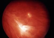
usually children <5 years. associated with high level of infection. larvae migrate through liver and lungs resulting in fever, hepatomegaly, and pneumonia. may recover, but will always have larvae in lungs and liver. no effective treatment, but there is supportive therapy
|
larval Migrans (VLM-visceral larva migrans)
|
|
|
occurs in older patients (7-8+ years). associated with low levels
|
OLM- ocular larva migrans
|
|
|
ocular larva migrans (OLM)
|
|
|
occurs younger than 5 years. associated with high levels. more commonly seen with Baylisascaris procyonis. related to migration of larvae and larval growth in brain and spinal cord
|
neural larva migrans (NLM)
|
|
|
neural larva migrans (NLM)
|
|
|
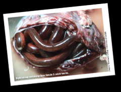
hosts: dogs and wild canids. ZOONOTIC! location in host: small intestine.
|
toxocara canis
|
|
|
life cycle of toxocara canis (4)
|
fecal-oral; ingestion of paratenic host; transmammary transmission; transplacental transmission
|
|
|
egg size of toxocara canis
|
75-90um
|
|
|
adults: problems due to somatic migration are rare. puppies: dull hair coat, abdominal distention, poor appetite, vomiting, loose stool. heavy infections-pneumonia and intestinal obstructions can occur. zoonosis: causative agents of visceral and ocular larva migrans
|
clinical disease of T. canis
|
|
|
host: cats. ZOONOTIC!. location: small intestine. life cycle: similar to T. canis. PPP- 8 weeks. transplacental and transmammary transmission does not occur. paratenic host transmission big role for adult cats. no migration occurs; larvae matures to adults in SI within 21 days or so. Eggs 65-75um in size
|
Toxocara cati
|
|
|
adults rarely show clinical signs of T. cati. kittens and puppies show these 2 signs
|
pot-bellied appearance. intermittent diarrhea
|
|
|
Toxocara cati
|
|
|
hosts: dogs, cats, foxes, and wolves. location: small intestine. life cycle: direct, or by use of paratenic host. Eggs are usually 75-85umx60-75um. oval with smooth, thick shell; colorless. no protein coat
|
Toxascaris leonina
|
|
|
3 common clinical signs rarely observed with Toxascaris leonina
|
diarrhea, vomiting, GI obstruction
|
|
|
Toxascaris leonina
|
|
|
host: raccoons, skunks, woodchucks, dogs. ZOONOTIC! location: small intestine. life cycle: direct development of adults in SI; no migration. very pathogenic zoonotic parasite that can infect humans. adult raccoons infected by ingestion of infected animals. in intermediate hosts larvae may migrate to eyes and brain causing CNS damage and death. raccoons find and eat dead animals, and larvae mature to adults in SI of raccoon
|
Baylisascaris procyonis
|
|
|
Baylisascaris procyonis
|
|
|
raccoons generally have no clinical signs of B. procyonis. occasionally obstruction and _________ can be seen with heavy worm burdens. if eggs ingested in other species eggs hatch into larvae and migrate. causative agent of neural larva migrans. although very rare, dogs can be a definitive host
|
diarrhea
|
|
|
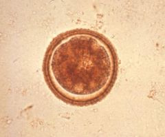
B. procyonis is seen on a ___________ fecal float. egg size is 63-88x53-68um. ellipsoidal. golden brown, thick shell
|
qualitative
|
|
|
this is an important cause of fatal nervous system disease, eye disease, and other problems in over 100 species of wild and domestic animals and people. eggs become infective within 30 days of being passed in feces. routine fecal exams are only method of diagnosing. cages and enclosures used to house or transport raccoons should not be used for other animals. decontaminated by flame or autoclaving. bleach and other common disinfectants do not work
|
B. procyonis
|
|
|
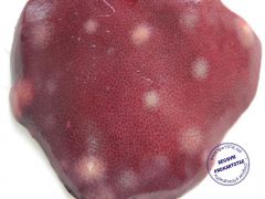
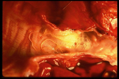
found in swine. ZOONOTIC! tracheal migration after ingestion of infective eggs. clinical disease: "thumps" and "milk spots", stunted growth, diarrhea, interference with nutrient absorption. possible perforation of the intestine
|
ascaris suum
|
|
|
Ascaris suum
|
|
|
Ascaris suum on liver
|
|
|
presented in horses. direct infection with tracheal migration. foals 3-9 months are most susceptible. foals begin to develop immunity by 6 months.
|
Parascaris equorum
|
|
|
clinical signs of Parascaris equorum (6)
|
coughing, eosinophilia, diarrhea, poor appetite, failure to thrive, colic
|
|
|
Parascaris equorum
|
|
|
4 steps to large animal parasite control
|
pregnant mares and sows should be dewormed prior to giving birth. also should be thoroughly washed and placed in clean pens prior to birthing. keep stalls clean after delivery. SANITATION!!
|
|
|
Ancylostoma caninum
|
canine hookworm
|
|
|
ancylostoma tubaeforme
|
feline hookworm
|
|
|
ancylostoma braziliense
|
canine and feline hookworm
|
|
|
uncinaria stenocephala
|
northern canine hookworm
|
|
|
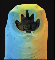
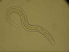
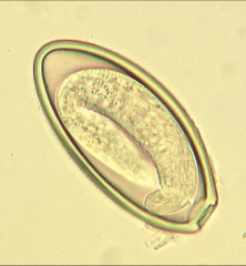
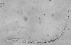
these attach to small intestine mucosa. have a frightful buccal cavity (3 pairs of teeth, feed on blood and secretes an anticoagulant from mouth. can cause significant anemia. can detach and move to different area). adult worms live 4-24 months in the intestin
|
hookworms
|
|
|
hookworm
|
|
|
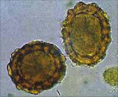
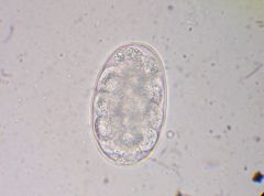
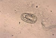
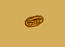
hookworm egg
|
|
|
4 features of Ancylostoma spp.
|
ellipsoid, smooth, thin shell, colorless
|
|
|
Uncinaria stenocephala
|
|
|
in both puppies and adults with hookworms, mild infections frequently present as ___________. 4 signs of this are
|
enteritis. loose stool, mild abdominal discomfort, decreased appetite, lethargy
|
|
|
severe infections are a great threat to puppies. 3 examples of these are
|
profound anemia, dark tarry stools, sudden death is possible
|
|
|
natural age resistance occurs between ____ & ____ months in dogs.
|
8-11
|
|
|
dermatitis is also seen in animals infected with hookworms by skin __________
|
penetration
|
|
|
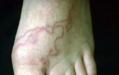
cutaneous larval migrans
|
|
|
puppies and kittens with severe anemia should be stabilized first; ______ ___________ possible. can also be treated with several anthelmintics
|
blood transfusion
|
|
|
nursing puppies should be treated at ___ weeks of age and every __ weeks thereafter until weaning
|
2; 2
|
|
|
treating the bitch with fenebndazole helps reduce ______________ transmission
|
transmammary
|
|
|
ways to control hookworms
|
remove feces frequently; sunlight, freezing, and some chemicals can kill larvae in environment
|
|
|
hookworms of ruminants. host: cattle, sheep, and goats. location in host: small intestine. mode of transmission: skin penetration, or ingestion of infective larvae. PPP 5-8 weeks. Eggs are dark, and granular; irregular, roughened surface
|
Bunostomum spp.
|
|
In ruminants, hookworm can cause this |
submandibular edema. aka bottle jaw
|
|
|
host: sheep, cattle, and goats. location in host: abomasum, small and large intestines. direct infection.
|
Cooperia spp., Haemonchus spp., Ostertagia spp. and Trichostrongylus spp. aka Trichostrongyles of cattle and sheep
|
|
|
Trichostrongyles of cattle and sheep
|
|
|
Affects over 50 species; not much differentiation. intestinal threadworms. eggs hatch in environment and enter one of two cycles: homogonic cyle & heterogonic cyle. larvae reach infective stage. penetrate and undergo tracheal migration to the small intestine. in dogs, the eggs hatch in the intestine, releasing first stage larvae observed in feces
|
Strongyloides spp.
|
|
|
Hosts for Strongyloides stercoralis.
|
dog, cat, human, fox
|
|
|
Strongyloides papillosus
|
ruminants
|
|
|
Strongyloides ransomi
|
swine
|
|
|
Strongyloides western
|
horses
|
|
|
hosts: dogs, cats, humans, and foxes. location of adult: small intestines. transmission: through skin and in mammary milk. Life cycle: PPP 8-14 days. parasitic males do not exist. only a pathogenic female is parasitic host. females produce embryonated eggs which hatch in intestine and can be found in feces. larvae go into free-living stage in environment before becoming infective 3rd stage larvae. L3s penetrate skin and cause migratory damage
|
Strongyloides stercoralis
|
|
|
Strongyloides stercoralis
|
|
|
dogs and cats infected with Strongyloides stercoralis will have moderate to severe _________. Require anthelmintic treatment on a regular basis
|
diarrhea
|
|
|
host: cattle. location of adult: small intestine. transmission: ingestion of infective larvae or penetration of skin by infective larvae. life cycle: female worm produces larvated eggs. eggs grow and malt; pass through feces to environment. eggs hatch; larvae become free-living (infective females) which can enter by skin penetration or ingestion. PPP 5-7 days. clinical signs: light infections may be asymptomatic. heavy infections: diarrhea, anorexia, weight loss, and blood & mucus in feces
|
Strongyloides papillosus
|
|
|
Strongyloides papillosus
|
|
|
host: pigs. location of adult: small intestine. transmission: transmammary, percutaneous. life cycle: closely resembles S. papillosus. PPP 3-7 days.
|
Strongyloides ransomi
|
|
|
clinical signs of S. ransomi (3)
|
diarrhea, anemia, weight loss
|
|
|
Strongyloides ransomi
|
|
|
host: horses. location of adult: small intestine. transmission: transmammary and skin penetration. life cycle: like other Strongyloides sp. PPP 5-7 days.
|
Strongyloides westeri
|
|
|
clinical signs of S. westeri (4)
|
diarrhea, weight loss, anemia, and poor appetite
|
|
|
Strongyloides westeri
|
|
|
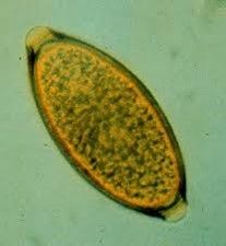
Strongyles of horses. large and small Strongyles (3)
|
Strongylus vulgaris, Strongylus edentaus, and Strongylus equinus
|
|
|
Type of Strongyle: most pathenogenic of the two types, includes Strongylus edentaus and Strongylus equinus |
large Strongyles
|
|
|
type of Strongyle: most pathogenic is Strongylus vulgaris. often associated with thrombi within the anterior mesenteric artery of horses |
small Strongyles
|
|
|
Life cycle of Strongyles (horses): eggs passed in feces and _____ in environment. infective stage larvae migrate up and down blades of _______ and then ingested. larvae swallowed and migrate through intestines to the mesenteric arteries and liver where they grow and malt into the next stage. larvae migrate back toward the large intestine and grow and molt on their way. when in the large intestine, the larvae enter the mucosa of the _______ ________ and mature to adults. |
hatch; grass; large intestine
|
|
|
clinical signs of Strongyles in horses (5)
|
colic, weight loss, lethargy, fever, and poor appetite
|
|
|
prevention methods of Strongyles in horses
|
pasture management, fecal egg counts/exams, and deworming based upon those results
|
|
|
Strongyles of horses
|
|
|
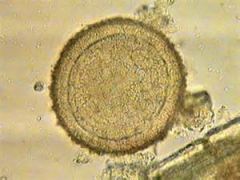
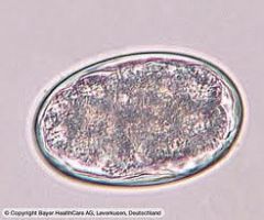
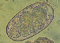
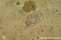
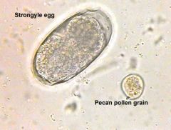
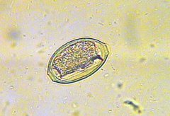
Strongyle egg
|
|
|
host: cattle, goats, and sheep. location in host: abomasum, small and large intestine. found worldwide. direct infection. Trichostrongyles with very large eggs
|
Nematodirus sp.
|
|
|
Nematodirus sp.
|
|
|
pinworm of horses. location of adult: cecum, colon, and rectum. transmission route: ingestion of infective larvae. diagnostic tests: scotch tape prep method. rare to find on fecal flotation; female attaches her eggs to anus, causing itching. gross parasites; adult worms protruding anus.
|
Oxyuris equi
|
|
|
Oxyuris equi
|
|
|
aka canine heartworm, although nematode has been known to parasitize cats and ferrets as well. long and slender. adults are found within right ventricle and pulmonary artery where they can cause obstruction. also where the mating process begins and offspring is called microfilaria that are released into the bloodstream. here they are ingested by female mosquitos. grow and molt until they reach infective stage, and then enter a new host when the mosquito feeds next
|
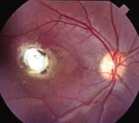
Dirofilaria immitis
|
|
|
the parasite is often recovered in a variety of different sites, such as the brain, anterior chamber of the eye, and SQ sites. PPP 6 months. treatment is available.
|
Heartworms
|
|
|
clinical signs of heartworms (3)
|
decrease in exercise tolerance, right-sided heart enlargement, abdominal ascites
|
|
|
cats are susceptible to this parasite but tend to be very resistant. takes greater exposure to the microfilaria from the mosquito. Cats do not show specific symptoms of infection. life cycle is same as dogs. cats do not produce microfilaria. no approved treatment in cats
|
Dirofilaria immitis in felines
|
|
|
Trichuris spp. found in canines |
Trichuris vulpis
|
|
|
Trichuris spp. found in cats (rarely seen)
|
Trichuris campanula/serrata
|
|
|
Trichuris spp. found in sheep
|
Trichuris ovis
|
|
|
Trichuris spp. found in swine
|
Trichuris suis
|
|
|
Trichuris spp. found in cattle
|
Trichuris discolor
|
|
|
location of Trichuris spp
|
cecum and colon
|
|
|
life cycle of this parasite: eggs passed in feces and develop into infective stage in about 1 months in environment. animal infected by ingestion of eggs (L1 larvae). eggs hatch and larvae migrate into wall of SI and remain in SI until they develop into adults and then migrate to colon. PPP is up to 3 months in dogs
|
Trichuris spp.
|
|
|
clinical signs of Trichuris spp. (6)
|
diarrhea, anemia, mucus-coated stools, no appetite, weight loss, dull haircoat
|
|
|
Trichuris spp.
|
|
|
Trichuris vulpis
|
|
|
Trichuris ovus
|
|
|
Trichuris suis
|
|
|
Can be seen on fecal float, however whipworms do not float very well, therefore it is imperative to let floats sit for full 20 minutes before viewing. due to long PPP and long resistance to the larvae, treat once a month for 3 months. eggs are very resistant in soil; most chemical disinfectants are useless. keep animals away from contaminated soil
|
Trichuris spp.
|
|
|
canine lungworm. adults found in trachea, lung parenchyma, and bronchioles. first stage larvae are immediately infective. larvae identified by dorsal spine with s-shaped tail. most commonly passed from the dam to her offspring as she cleans her pups. diagnosed by finding larvae on qualitative fecal float/Baermann technique. PPP 10 days. nodules at bifurcation of the trachea are usually found during necropsy
|
Oslerus (Filaroides) osleri
|
|
|
Oslerus osleri
|
|
|
Oslerus osleri larvae (L1)
|
|
|
feline lungworm. adult found in respiratory bronchioles and alveolar ducts. larvae are coughed up and then passed through the feces. diagnosis: finding larvae on qualitative fecal float, Baermann technique, tracheal wash. PPP 30 days. relatively uncommon in Indiana. seen more in southeastern US
|
Aelurostrongulus abstrusus
|
|
|
Aelurostrongulus abstrusus
|
|
|
Dictyocaulus spp. in cattle and deer
|
Dictyocaulus viviparus
|
|
|
Dictyocaulus spp. in sheep and goats
|
Dictyocaulus filaria
|
|
|
Dictyocaulus spp. in donkeys
|
Dictyocaulus arnfieldi
|
|
|
Dictyocaulus spp. what type of life cycle?
|
direct life cycle
|
|
|
Dictyocaulus spp. clinical disease: usually worse in young animals in their first year grazing. Ruminants present with these 5 signs
|
difficulty breathing, coughing with nasal discharge, harsh lung sounds, weight loss, possibly death
|
|
|
Dictyocaulus viviparus
|
|
|
Dictyocaulus filaria
|
|
|
Dictyocaulus arnfieldi
|
|
|
sheep and goats. adult located in bronchioles and lung parenchyma. infection via ingestion of infective larvae while grazing. PPP 6-10 weeks. sheep usually show no clinical disease. goats will have severe respiratory distress and susceptible to secondary bacterial infections. diagnosed finding larvae on Baermann technique. larvae tail has undulating tip and dorsal spine
|
Muellerius capillaris
|
|
|
Muellerius capillaris
|
|
|
giant kidney worm. host: carnivores, pigs, humans. location of adult: right kidney. can be up to 3 feet long. also can be found in peritoneal cavity. eggs passed in urine; found in urine sedimentation. 71-84 x 46-52um. barrel-shaped, yellow-brown. roughly pitted. clinical disease varies. most often asymptomatic. may have kidney infection or renal failure with hematuria. can cause peritonitis, hemorrhage, adhesions, and liver damage if found in body cavity. requires surgical removal of parasite. prevent animals from eating raw freshwater fish and frogs
|
Dioctophyma renale
|
|
|
Dictophyma renale
|
|
|
Dictophyma renale
|
|
|
bladder worm of canines and felines. location of adult: urinary bladder. eggs clear to yellow and have flattened bipolar ends. seen in urine sedimentation or in a fecal sample contaminated with urine. usually no clinical signs. no known specific treatment. keep pet from eating earthworms.
|
Capillaria spp. (Pearsonema)
|
|
|
Capillaria spp. in dogs and cats
|
Pearsonema plica
|
|
|
Capillaria spp. in cats
|
Pearsonema feliscati
|
|
|
Pearsonema plica
|
|
|
Pearsonema plica
|
|
|
host: swine and human. location: small intestine. indirect life cycle. infection occurs by ingestion of IH (dung beetle and grubs). PPP 2-3 months. clinical disease varies from no clinical signs to diarrhea and emaciation with abdominal pain. Eggs 67-110 x 40-65um. tri-laminar eggs. ellipsoidal; brown. do not float well and are frequently missed. no effective drug therapy. use nose rings to prevent rooting by pigs reared outside
|
Macracanthorhynchus hirudinaceous
|
|
|
Macracanthorhynchus hirudinaceous
|
|
|
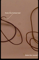
not a parasite of domestic animals. juveniles are parasitic to insects. adults are free-living near fresh water. very long worms (20-30cm)
|
Nematomorphs and Mermithids
|
|
|
yellow to brown. translucent anterior with dark ring. tail with 2 or 3 lobes
|
Nematorph adults
|
|
|
white, long, and thin
|
mermithids
|
|
|
Nematomorph adults
|
|
|
can occur as endo- and ectoparasites of animals. dorsoventrally flattened, large posterior sucker for attachment, anterior with smaller sucker and proboscis. nasal passages and sinuses of waterfowl and mammals (other countries). most feed on blood and can serve as a vector of disease. name and class?
|
leeches. class Hirudenea
|