![]()
![]()
![]()
Use LEFT and RIGHT arrow keys to navigate between flashcards;
Use UP and DOWN arrow keys to flip the card;
H to show hint;
A reads text to speech;
106 Cards in this Set
- Front
- Back
|
General characteristics of filarial worms |
- esophagus divided into anterior muscular and posterior glandular portions - life cycle indirect - males frequently have a spirally coiled tail - parasitic outside the enteric tract in body tissues - long thin worms with a small mouth and NO buccal capsule, pharynx, or lips - females produce microfilaria that are founding blood and connective tissue - migrate in DH - indirect life cycle (use blood sucking arthropod) |
|
|
Dirofilaria immitis |
DH: dogs, marine mammals, ferrets, cats IH: mosquitos adults: male- 12-22 cm female- 25-31cm Mf: 300-325 x 6-7 um PPP= 6 months
adults in right heart and pulmonary artery can migrate aberrantly to skin and eyes |
|
|
Dirofilaria immitis pathology |
- pulmonary arterial disease with inflammation and proliferative lesions --> arteritis - pulmonary thromboemboli - pulmonary hypertension--> can lead to right heart failure if prolonged |
|
|
Transmission of D. immitis |
1. mosquito ingest Mf and spread resultant L3 to new host 2. Mf can be transferred prenatally (not infective in babies but can be transmitted from them) |
|
|
Diagnosis of Dirofilaria immitis in dogs |
Knott's test- look for Mf in blood
antigen test- look for female uterine antigen
radiographs- right heart enlargement (reverse D)
serology
|
|
|
Dirofilaria immitis clinical signs in dogs |
- cough/dyspnea - exercise intolerance - weight loss - ascites - anemia (due to damage to blood vessels) - eosinophilia and thrombocytopenia - glomerulonephritis - proteinurea |
|
|
what are the steps in treatment of heart worm disease? |
FIRST MONTH 1. evaluate the health of the patient and determine severity 2. stabilize clinical cases 3. Start on monthly macrolytic lactone 4. administer prednisone at decreasing dose 5. give doxycycline to kill wolbachia 6. restrict exercise MONTH 2 1. continue ML MONTH 3 1. ML 2. first dose of adulticides (immiticide) 3. restrict exercise further MONTH 4 1. ML 2. Dose 2 and 3 of immiticide 3. prednisone if necessary 4. continue exercise restriction
|
|
|
What are the goals of administering macrolytic lactones in treatment of heart worm disease? |
1. kill larvae 2. prevent new infection 3. stunt maturation of immature adults 4. prevent female reproduction |
|
|
What is the procedure for administering immiticide to kill adult heart worms? |
- inject into epaxial muscles - 2.5 mg/kg at least 2 times 24 hours apart (3 times is best) - should kill adults 4 mo. and older
if doing 3 dose regimen give first dose 1 month before the next 2 |
|
|
Why is it best to administer 3 doses of immiticide during heart worm treatment? |
it increases efficacy - reduces the need for repeated treatments
it is safer because it decreases the chance of a mass die off and resultant embolism |
|
|
why would you give doxycycline during treatment of heart worms? |
to kill wobachia, a bacteria that lives inside the adult heart worms and can be released upon tremens. By administering doxycycline you can hopefully prevent bacterial infection |
|
|
What is possibly a major reason that heart worm infection is not as severe in cats as in dogs? |
Dirofilaria have much shorter lifespans in cats so are less likely to reproduce and there are fewer microfilaria |
|
|
What clinical signs are more common with cat heart worm infection than infection in dogs? |
ectopic infections (skin, eyes) and respiratory disease HARD |
|
|
How can Dirofilaria be diagnosed in cats? |
both and antigen and antibody (not in dogs) test
antigen detects worms after 5-8 months antibody detects after 3 months |
|
|
How does treatment of cat heart worm infection differ from dog? |
cats cannot be given immiticide so there really is not treatment- just supportive
therefore it is extremely important that they be kept on prevention all the time. |
|
|
Acanthocheilonema (Dipetalonema) reconditum |
filarial worm DH: dogs, cats, primates, raccoons IH: fleas, ticks, lice adults: male- 9-17 mm, female- 20-32 mm Mf= 250-280 um in dog PPP= 2 months
rarely cause clinical disease |
|
|
differences between Acanthocheilonema reconditum and Dirofilaria immitus |
A. reconditum is shorter and thinner A. reconditum has a hooked tial A. reconditum has a flattened/ rounded anterior end with no tapering while D. immitus tapers to a point |
|
|
Onchocerca |
filarial worm (nodular worm) DH: horses, cattle, people, dogs IH: biting flies adults: males- 40 cm females- 75 cm Mf: 250-350 mu PPP = 1 year
adults live in connective tissue Mf are in the skin |
|
|
Onchocerca cervicalis |
filarial worms DH: horse found in nuchal ligament |
|
|
What are the clinical signs of onchocerca infection in horses? |
pruritic dermatitis |
|
|
How would you diagnose an Onchocerca infection |
clinical signs with presence of worm or microfilaria
skin biopsy
history of exposure to biting gnats |
|
|
What are the species of Onchocerca in humans? Where are the adults found? |
filarial worms O. volvulus- subcutaneous tissues |
|
|
What are the clinical signs of Onchocerca infection in humans? |
- dermatitis - river blindness big problem in Africa |
|
|
Setaria |
filarial worm- peritoneal worm DH- cattle, horse IH: mosquitos adults: male- 40-80 mm female- 60-130 mm Mf: 140-230 um PPP= 8-10 months
|
|
|
Where are Setaria worms generally found? |
microfilaria are in the blood adults are in the peritoneum
- do not generally cause clinical signs unless they migrate aberrantly (adults to the eye, microfilaria to CNS) |
|
|
Stephanofilaria |
filarial worm DH- cattle IH- biting flies adults: 10 mm Mf: 45-60 um
adults and Mf in abdominal cutaneous tissue
|
|
|
Clinical signs of Stephanofilaria infection |
dermatitis on ventral abdomen |
|
|
Dracunculus |
filarial worm (guinea worm) DH: dogs, raccoons, people, other mammals IH: copepod (crustacean) adults: male- 40 mm female- 120 mm Mf- 500-760 um PPP= 1 year |
|
|
Dracunculus life cycle |
1. DH drinks water with copepod and L3 larvae 2. larvae emerge and migrate through lymphatics and circulation 3. larvae move to extremities and develop into adults 4. adults mate and male disappears 5. female moves to end of extremity and forms a sore 6. when sore gets wet female busts it open and releases eggs |
|
|
Dracunculus clinical signs |
painful lesion at the sight of emergence |
|
|
Dracunculus diagnosis |
lesion with protruding worm and long tailed larvae |
|
|
How can you tell whether a cutaneous lesion is caused by dracunculus or dirofilaria? |
dracunculus larva are long tailed |
|
|
Acanthocephala general characteristics |
"spiny headed worms" - elongate - pseudocoelomate - dioecious - no digestive tract - retractible proboscis with spines - adults live in digestive tract of vertebrates - indirect life cycle - may appear segmented but it is just wrinkles - eggs have thick tri-layered shell with visible spines |
|
|
What is unique about the reproduction of acanthocephala? |
after mating the male plugs the female reproductive tract with cement to prevent further matings |
|
|
Macracanthorhynchus hirudinaceus |
acanthocephalan DH: swine, people rarely IH: dung beetles and june bugs adults: male- 2-10cm female- 10-65 cm eggs: 67-110 x 40-65 um ppp= 2-3 months |
|
|
Clinical signs of Acanthocephalan infection |
diarrhea emaciation peritonitis pain anorexia |
|
|
What is the difference between M. hirudinaceus and M. ingens |
DH: - hirudinaceus: swine - ingens: raccoons, skunks IH: - hirudinaceus: dung beetles - ingens: millipedes
ingens has paratenic hosts: rodents, snakes, frogs
both are zoonotic |
|
|
What other type of worms can acanthocephalans, especially Moniliformis, be confused with? Why? |
tapeworms, they can have a wrinkly appearance that could suggest they are segmented- but they are not |
|
|
What are the 3 groups of worms within the phylum platyhelminthes? |
turbellaria trematoda cestoda |
|
|
general characteristics of platyhelminths |
- tripoblastic - bilaterally symmetrical - nerve ganglion in anterior end - dorsoventrally flattened - acoelomate - hermaphroditic |
|
|
Common characteristics of cestodes |
elongated (can be multiple feet) segmented body (proglottids-->stroblia) scolex for attachement no alimentary canal adults - parasites in small intestine of vertebrates eggs and/or proglottids passed in feces indirect life cycle |
|
|
What are general features of Trematodes that are different from cestodes? |
- leaf shaped: bilaterally symmetrical and dorsoventrally flattened - no segmentation - incomplete alimentary canal (no anus - suckers for attachmnet (no hooks) - all parasitic |
|
|
What are the 2 major orders of cestodes? |
diphyllobothridea
cyclophyllidea |
|
|
characteristics of cestode order diphyllobothridea |
aquatic 2 intermediate hosts eggs expelled from proglottids (just eggs not whole proglottids) minor veterinary importance |
|
|
characteristics of cestode order cyclophyllidea |
terrestrial usually just one intermediate host proglottids are passed in feces suckers for attachment important parasites of domestic animals |
|
|
dipylidium caninum |
cestode DH: dog, cat, human *** zoonotic IH: fleas, lice adults - 5 cm eggs: 40-50 um; egg packets: 200 um PPP- 2-3 weeks
2 pores in proglottid |
|
|
Dipylidium caninum diagnosis and prevention |
dx: - visualize proglottids or egg packets in feces - observe fleas - see stroblia in vomit
prevention: - use flea preventative |
|
|
Anoplocephala/ Paranoplocephala |
cestode DH: equine IH: pasture mites eggs: 50-80 um PPP- 1-2 months
eggs have pyriform apparatus (looks like wrench) proglottids have 2 pores |
|
|
important species of anoplocephala/ paranoplocephala |
P. mammilata - duodenum, adults: 3.5 cmx 4 mm
A. magna- ileum; adults: 80 cm x 2 cm
A. perfoliata- ileocecal junction; 5-8 cm x 1.5 cm |
|
|
Which is the only species of Anoplocephala/Paranoplocephala that causes clinical disease? What are the clinical signs? |
A. perfoilata- causes colic |
|
|
Moniezia spp. |
cestode DH: cattle, sheep, goats IH: pasture mites adults: 6 m eggs: 50-80 um PPP: 1-2 months
egg is triangular to square 2 pores in proglottid no clinical signs
in intestine |
|
|
What is the major difficulty in preventing infection with anoplocephala, paranoplocephala, or moniezia? |
the IH (pasture mites) are so numerous that there is no way to control them |
|
|
What cestodes have an arthropod intermediate host? |
- Dipylidium caninum - Anoplocephala - Paranoplocephala - Moniezia |
|
|
What is a cysticercus? |
tapeworm larvae in a cyst in IH - has envaginated scolex - usually in vertebrate
|
|
Taenia spp. general characteristics |
Cestode: Taeniidae DH: dogs and cats IH: mammal eggs: 35-40 um
adults have suckers and rostellum (hooks) shed proglottids with 1 pore |
|
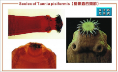
Taenia pisiformis |
Taeniidae DH: dog IH: rabbit- metacestode= cysticercus pisiformis
in small intestine of DH
metacestode is cyst w/ 1 egg in liver or peritoneal cavity of IH |
|
|
Taenia taeniaeformis |
taeniidae DH: cat IH: rodents- metacestode= cysticercus fasciolaris
In small intestine of DH
metacestode is an elongated cyst (strobilocercus) associated with neoplasms in liver of IH
|
|
|
Taenia hydatigena |
taeniidae DH: dog IH: swine, ruminants, deer metacestode: cysticercus teniuicollis --> Hepatitis cysticercosa
metacestode is in liver of IH and causes clinical sings there |
|
|
Taenia ovis |
taeniidae DH: dog IH: sheep and goats metacestode= cysticercus ovis
metacestode is in muscle of IH and causes clinical signs and condemnation of carcass
|
|
|
How do you diagnose Taeniidae infections? |
eggs on sugar flotation (not in egg packets) see stroblia or proglottids |
|
|
Taenia saginata |
Taeniidae DH: humans IH: cattle metacestode= cysticercus bovis--> beef measles
metacestode in muscles of IH humans get it from eating contaminated beef cows get it from eating human feces |
|
|
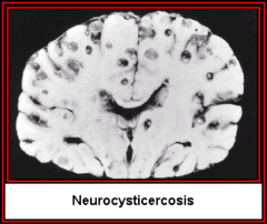
Taenia solium |
taeniidae DH: human IH: swine, human metacestode= cysticercus cellulosae--> pork measles
when humans are infected with egg (IH) the larvae migrate to the brain and cause encephalitis
no disease if humans are DH |
|
|
What would be required to differentiate an infection of Taenia spp. from Echinococcus spp.? |
to differentiate between the genera you need adult worms
to differentiate between species within the genus you need the cysts in the IH |
|
|
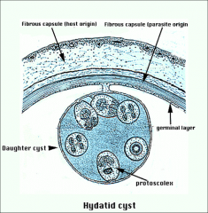
Echinococcus granulosus |
taeniidae DH: canines IH: ruminants, swine, horses, humans, elk metacestode= hydatid cyst (multiple metacestodes/ small cyst within larger cyst)
no clinical signs in DH; found in SI
IH will develop large cyst that acts like a metastatic tumor |
|
|
Echinococcus multilocularis |
DH: canines and felines IH: rodents, ruminants, swine, humans Metacestode= alveolar hydatid cyst
metacestodes around heart in IH almost always fatal in IH |
|
|
general characteristics of diphylobothridea tapeworms |
- no hooks or suckers on scolex - 2 intermediate hosts - eggs are expelled from a mid ventral pore in the proglottid - eggs are operculated - have bothria- grooves - minor veterinary importance |
|
|
what is the life cycle of diphylobothridean tapeworms? |
1. unembryonated eggs are shed in the feces 2. embryos develop when eggs are in water 3. egg hatches and releases coracidia (ciliated larvae) 4. coracidia is ingested by 1st IH 5. develops into procircoid in first IH 6. first IH is ingested by 2nd IH and larvae develops to pleocircoid 7. 2nd IH is ingested by DH or PH. 8. once in DH larvae develops to adult in small intestine |
|
|
Diphyllobothrium latum |
diphylobothridean tapeworm- " broad fish tapeworm" DH- dogs, bears, humans ** potentially zoonotic 1st IH: copepod 2nd IH: fish PPP= 6 weeks |
|
|
Spirometra spp. |
diphlobothridean tapeworm DH: cats, dogs, wild carnivores, raccoons 1st IH: copepod 2nd IH- water snakes, amphibians, birds, mammals PPP- 10 days
**potentially zoonotic |
|
|
how can you tell the difference between Diphyllobothrium datum and Spirometra spp. eggs |
D. latum eggs have a knot on the end opposite the operculum but Spirometra do not |
|
|
what is the difference in the lifecycle between D. latum and Spirometra spp.? |
Spirometra life cycle is more terrestrial with birds, small mammals, and amphibians as their 2nd IH |
|
|
What are 3 groups within the class Trematoda? |
1. aspidogastrea- parasites of mollusks, fish, turtles
2. monogenea- external parasites of fish with direct lifecycles
3. digenea- parasites of vertebrates with indirect life cycles |
|
|
Describe unique features of Digenetic trematode life cycles |
they are hermaphroditic Adults reproduce sexually in DH larvae reproduce asexually in IH
larvae are very host specific (like to the species level) while adults are less so |
|
|
Characteristics of digenetic trematodes |
- leaf shaped - no segmentation - incomplete alimentary canal (no anus) - attachment via suckers (acetabulum- attachment; oral- ingestion) - indirect life cycle: 1st IH is always a mollusk - hermaphroditic but can reproduce sexually - produce operculated eggs |
|
|
general life cycle of digenetic trematodes |
1. miracidum (ciliated larval stage) actively seeks out snail 2. asexual generations (sporocyst, redia) within snail 3. cercaria (in host) leaves snail and encysts in new host or environment as metacercaria (enviro) 4. (meta)cercariae reach DH and develop to adult |
|
|
How do digenetic trematode (meta)cercaria reach definitive host? |
- passive ingestion - predation - skin penetration
depends on species of worm |
|
|
What are the 3 liver flukes |
all digenetic trematodes - Fasciola hepatica- sheep (common) fluke - Fascioloides magna- deer fluke - Platynosomum concinnum- lizard poisoning fluke |
|
|
Fasciola hepatica |
digenetic trematode- common sheep fluke DH: ruminants and humans 1st IH: amphibious snail, 2nd= NONE adults: 2-3 x 1 cm eggs: 130-150 x 63-90 um PPP= 2-3 months
adults can live for 11 years migrate in liver and bile ducts
|
|
|
risk factors of Fasciola hepatica infection |
wet pastures transmission occur between february and july in southeast US |
|
|
treatment/ prevention of Fasciola hepatica infection |
- clorsulon - NO ivermectin- has no efficacy against flukes |
|
|
control of Fasciola hepatica infection |
- control DH access to water (drain or fence of ponds) - molluscicides - periodic anhelminthics |
|
|
Diagnosis of Fasciola hepatica infection |
geographic location transport history (were they recently where it is found) eggs on fecal floatation clinical signs imaging adults on necropsy |
|
|
Fascioloides magna |
Digenetic Trematode- deer fluke DH: deer 1st IH: amphibious snail; 2nd: NONE AH: sheep, goats, cattle, camelids, elk adults: 30-100 x 20-30 mm eggs: 110-170 x 75-96 um
disease only seen in AH metacercaria is infective for both DH and AH |
|
|
pathogenesis of Facioloides magna |
deer(DH)- form a cyst in liver with communicating duct to bile duct where eggs are released
AH- forms a cyst but can't form communication with bile duct so eggs cannot be released. In sheep and goats the larvae continue to migrate through the liver causing extensive damage |
|
|
Paragonimus spp. |
Trematode (pulmonary fluke) DH: mammals 1st IH: snail; 2nd: crayfish and crabs adults: 10 x 5 mm eggs- 75-118 x 48-65 um
zoonotic live in pairs in lung cysts |
|
|
Characteristics of blood flukes (schistomes) |
- diocious (males live in groove in female) - cercariae penetrate skin - eggs migrate through tissue to lumen of GI or bladder - major pathology from granulomatous inflammation around eggs |
|
|
Heterobilharzia americana |
DH: dog, raccoon, bobcat 1st IH: snail; 2nd: NONE adults: 4 mm eggs: 88 x74 um
adults found together in mesenteric veins penetrate skin as cercariae (motile- not in cysts)- no metacercaria (encysted) |
|
|
Clinical signs of H. americana infeciton |
diarrhea with mucous and blood vomiting anorexia/ weight loss polyuria/ polydypsia hepatic fibrosis |
|
|
Geographic distribution of Heterobilharzia americana |
From Florida to Texas along the Gulf Coast, as far north as Kansas |
|
|
Diagnosis of H. americana |
geographic distribution sedimentation of eggs using saline |
|
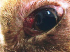
this dog presented with acute conjunctivitis. His new owners recently found him abandoned and covered with flies. What parasite is causing his problem? |
Onchocera lupi filarial worm |
|
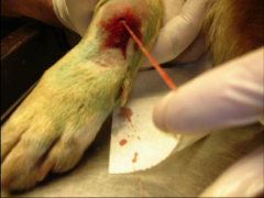
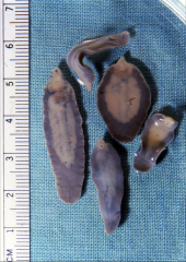
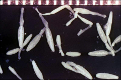
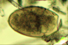
What are 2 possibilities of what this parasite could be. How would you tell them apart? |
Dracunculus insignis or Dirofilaria immitis
dracunculus produces long tailed larvae |
|
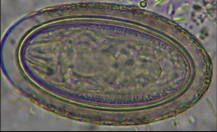
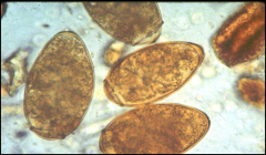
What is your best guess as to the type of parasite that produced this egg? What makes you think that? |
acanthocephala
it has a thick tri-layered shell |
|
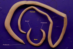
This worm was found in the digestive tract of a pig. What type of parasite is this? |
acanthocephala - has spiny probiscus - elongate - dioecious
Specifically because it was found in a pig it is Macracanthorhynuchus hirudinaceus |
|
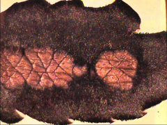
These worms were found in the liver from a deer. There are cysts with communications to the bile ducts. What parasites are these |
Fascioloides magna- deer is DH
|
|
These eggs were found in the feces of a dog that is suffering from respiratory problems. When you perform radiographs you see cysts in the lung tissue. What parasite is likely causing the problem? |
Paragonimus |
|

What type of worm is this? What are the individual structures that make it up called and what are they called collectively? |
Cestode (tapeworm) individual structures are proglottids multiple proglottids make up a storable |
|
This structure was found in the feces of a dog brought into your clinic. the dog is infested with fleas. What parasite is likely causing his problem? |
Dipylidium caninum (tapeworm) |
|
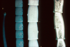
Based on this picture, what species of worm might this be? What host would you expect to find it in? |
Paranoplocephala mammilata: duodenum Anoplocephala magna: ileum Anoplocephala perfoliata: ileocecal junction
DH is horse
you can tell because of the shape and the piriform apparatus
|
|
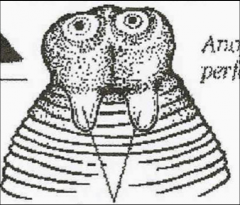
Which species of Anoplocephala/Paranoplocephala is this? How can you tell? |
Anoplocephala perfoliata
it has lappets- muscular extensions of the scolex |
|
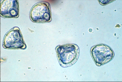
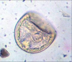
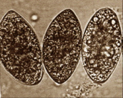
What species of worm produced this egg and what is its DH? |
Moniezia
DH: cattle, sheep, goats
you can tell because of the triangular/diamond shape |
|
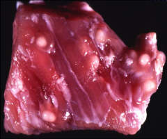
These lesions seen in pork at a slaughterhouse are characteristic of infection with what pathogen? Is it zoonotic? |
Taenia solium
pork measles
if humans are infected with egg (i.e. are the intermediate host) they will get encephalitis |
|
These adult worms were found in the small intestine of a dog. What species is it? what type of cystercercoid is formed in the IH? |
Echinococcus granulosus DH: dog IH: ruminants, swine, horses, elk - metacestode is in hydatid cyst (multiple cysts within a cyst) |
|
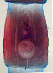
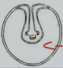
This proglottid was shed in the feces of a dog? What parasite likely produced it? How can you tell? |
Mesocestoides
the large circle in the center (parauterine organ) is characteristic of this parasite |
|
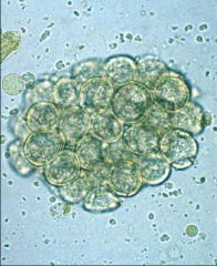
These eggs were found in the feces of a cat whose owner reports likes to eat frogs from the pond in their yard. What species of parasite do you suspect? Should you warn about zoonosis? |
Spirometra Yes, while unlikely there have been reports of zoonosis when exposed to infected frogs or snakes. |
|
Adult worms that produce this egg were found in the liver of a sheep. If the passed the egg what species do you suspect produced it? What if the sheep did not pass the egg? |
if sheep is DH: Fasciola hepatica if sheep is IH: Fascioloides magna- DH is deer |