![]()
![]()
![]()
Use LEFT and RIGHT arrow keys to navigate between flashcards;
Use UP and DOWN arrow keys to flip the card;
H to show hint;
A reads text to speech;
143 Cards in this Set
- Front
- Back
|
Common arthropod characteristics
|
- chitinous exoskeleton
- jointed appendages - segmentation (head, thorax, abdomen) - some species contain festoons at caudal most end - separate sexes: usually only observable in adults - larval stage has six legs, nymphs and adults have 8 |
|
|
How do arthropods cause disease?
|
direct: cause disease themselves
- paralysis, wounds, predisposure to secondary bacterial infection indirect: transmit a variety of infectious organisms |
|
|
Types of metamorphosis
|
1. simple (incomplete)
- egg--> larvae/nymph--> adult 2. complex (complete) - egg--> larvae--> pupa--> adult - there are varying numbers of molts during the immature stages depending on species |
|
|
2 groups within the phylum arthropoda
|
1. insecta (lice, fleas, assassin bugs, bed bugs, flys, beetles, ants)
2. arachnids (ticks, mites, spiders) |
|
|
Tick bite reaction pathogenesis
|
-inflammation and blood loss
- deep, painful bites (predispose to bacterial infections, myiasis) |
|
|
Tick bite reaction treatment
|
remove tick and clean wounds
supportive care (transfusions, antibiotics) use tick prevention! |
|
|
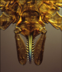
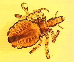
Differentiate between ticks and mites
|
Ticks
- larger than mites - attach substantially to the host with a tooth hypostome Mites - smaller than ticks - smooth hypos tome does not allow firm attachment - stigmata location varies or may be absent |
|
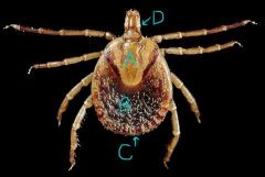
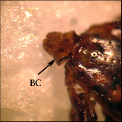
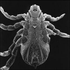
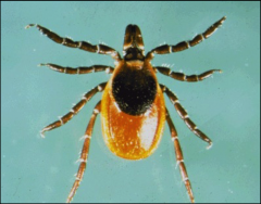
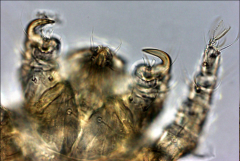
Label the structures on this tick. Is it a male or female? How can you tell?
|
A- scutum
B- abdomen C- festoons (not always present) D- capitulum This is a female; you can tell because she has a large abdomen, in males the scutum covers then entire dorsum |
|
|
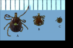
How can you tell the difference between a tick larvae and nymph or adult? why is this important?
|
larvae has 3 pairs of legs (6 legs total)
nymphs and adults have 4 pairs unless a disease is transmitted transovarially larvae will not be infected and be able to transmit disease |
|
|
What are some characteristics of ticks that make them good vectors
|
- persistent blood-suckers which attach firmly and feed slowly
- long life span - can be geographically widespread - resistant to environmental conditions - high reproductive potential - can demonstrate both transstadial and transovarial transmission - wide host range: wildlife, livestock, pets, people |
|
|
What are some pathogenic problems caused by ticks?
|
1. blood loss
2. secondary infection (act as a vector, create opportunity for other infectious agents, or release toxin) 3. predispose to myiasis |
|
|
Characteristics of soft ticks (family argasidae)
|
- no scutum
- soft, tough, leathery body - do not stay attached- take multiple small meals on a host - lay multiple small sets of eggs (400-500) - only larvae and nymphs are parasitic and they often only feed at night |
|
|
Characteristics of hard ticks (family ixodid)
|
- largest family of ticks
- have scutum - remain attached until engorged - once engorged females drop to ground to explode releasing eggs (3,000-8,000) - varying number of hosts (1-3) |
|
|
Otobius megnini
|
"spinose ear tick"- soft tick
- nymph and larvae parasitize many mammals - found in warm regions of the world (TX and FL in US) - transmit relapsing fever caused by Borrelia spp. (not burgdorferi): causes ear canal damage (deafness), anorexia, and recurrent fever |
|
|
Which stages of the tick life cycle can be sexed morphologically?
|
Adults
|
|
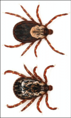
General characteristics of Dermacentor spp.
|
- ornate ticks with eyes
- basis capitulum is rectangular if viewed from above with stubby palps - has 11 festoons and small rectangular patterns on posterior abdomen giving it a peppery appearance - move very quickly and prefer haired areas of the body |
|
|
Dermacentor variabilis
|
" american dog tick"- hard tick
- Host: mammals and birds (wide range) - distribution: eastern US and west coast, hole in the middle because of competition with other ticks - life cycle: --3 host tick -- may take form 3 months to 2 years depending on environmental conditions - disease: -- principle vector of Rickettsia rickettsia (RMSF) --tick paralysis -- tularemia -- babesia canis (infrequent vector) -- cytauxzoon felis (infrequent vector) |
|
|
Rhipichepalus sanguineus
|
"brown dog tick"- hard tick
a. host: DOGS, others are possible b. distribution: tropical tick: in US mainly found in SW or indoors (can survive for years in cracks and crevices in buildings) c. disease - Babesia canis voglei - Ehrlichia canis |
|
|
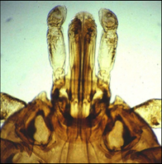
Rhipichepalus sanguineus morphology
|
similar to Dermacentor
- 11 festoons - small rectangular pattern on posterior abdomen basis capitulum is hexagonal in shape (can differentiate from Dermacentor with this |
|
|
Rhipicephalus annulatus
|
"cattle fever tick" - hard tick
a. host: mainly cattle; also deer, horses, sheep, etc b. distribution: US and Mexico (eradicated from US but is moving back up again on transported deer) c. Life cycle: 1 host tick- makes eradication easier d. disease: - Bbesia bigmina- causes severe anemia and death in cattle - reportable tick species |
|
|
Amblyomma spp. general characteristics
|
- ornate ticks with eyes
- long mouth parts (parallel) - 11 festoons |
|
|
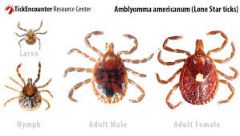
Amblyomma americanum
|
"lone star tick"- hard tick
a. host: wild and domestic animals, birds, and humans b. distribution: mainly southern US but is moving north on WTD c. life cycle: 3 host d. disease: - STARI (reaction to tick saliva, looks like RMSF) and red meat allergy - tularemia - Ehrlichia chaffeensis -Ehrlichia ewingii - Cytauxzoon felis e . Characteristics: - large silver spot on at apex of scutum of females - very aggressive - antigenic saliva |
|
|
Amblyomma maculatum
|
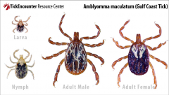
"Gulf coast tick"- hard tick
a. host: wild and domestic animals, birds, humans b. distribution: SE US and Gulf Coast area (moving north and west c. life cycle: 3 host d. disease: - Hepatozoon americanum (dog must eat tick) - Rickettsia parkeri (human pathogen) - Ehrlichia ruminatum (heart water)- is a potential vector but disease is not currently in US e. characteristics: - orante scutum: streaky black and white (could be confused with Dermacentor) - long mouth parts (different from Dermacentor) |
|
Ixodes spp. general characteristics
|
-hard ticks
- inornate ticks - reddish abdomen - anterior (rather than posterior) anal groove- only visible in nonengorged female - long mouth parts with fusiform shape (differentiate from Amblyomma spp.) - no festoons - nymphs are very dark black - adults are out in the winter, larvae and nymphs in the spring |
|
|
Ixodes scapularis
|
"black legged tick"- hard tick
a. host: wild and domestic animals, birds, and humans b. distribution: east, south and midwest: highest in upper midwest and New England c. Life cycle: 3 host - larvae on reptiles, birds, and small mammals - adults on larger mammals and humans d. disease: - Borrelia burgdorferi (lyme disease - Babesia microti - anoplasma phagocytophila e. Characteristics - severe bite - reptiles clear them of disease (which is why disease is less common from them in SE where there are more lizards) |
|
|
Which ticks are most likely to cause tick bite reactions?
|
Amblyomma- long mouth parts and antigenic saliva
Ixodes- long mouth parts |
|
|
Treatment for tick bites
|
- remove tick- slow and firmly pull out making sure to remove full hypostome- don't break it off
- supportive care- transfusions (if blood loss is severe), antibiotics |
|
|
Tick paralysis host and agent
|
host: cattle, sheep, horses, dogs, humans
agent: Dermacentor, Amblyomma, Ixodes |
|
|
Tick paralysis pathogenesis
|
potentially fatal reaction to a paralyzing neuromuscular toxin secreted in the saliva of a female tick late in feeding
acute ascending flaccid paralysis - starts with incoordination and can progress to complete paralysis |
|
|
Tick paralysis clinical signs
|
- headache, vomiting, malaise
- loss of motor function and reflexes - paralysis that begins in lower body and spreads - can result in respiratory failure and death the closer to the head the tick feeds the more severe the signs |
|
|
Tick paralysis diagnosis
|
- removal of the tick resulting in rapid disappearance of clinical signs
|
|
|
tick paralysis treatment
|
- remove tick, supportive care
- dip animal to remove any tick that might have been missed - I. holocyclus antitoxin (IgG antibody) |
|
|
mechanical transmission by arthropods
|
arthropod transfers pathogen from an infectious source to a susceptible host without reproduction or developmental stages of the pathogen occurring in the vector
|
|
|
Biological transmission by arthropods
|
arthropod transfers pathogen from an infectious source to a susceptible host and while in arthropod pathogen undergoes reproduction, developmental changes or both
|
|
|
Transstadial transmission
|
pathogen is maintained within thick as tick molts from one life stage to the next
- passed from larvae to nymph to adult |
|
|
Transovarial transmission
|
pathogen is maintained in tick population through vertical transmission transmitted to offspring via ova
|
|
|
Lyme Disease: Agent, vector, host, and distribution
|
agent: Borrelia burgdorferi
vector: Ixodes scapularis (eastern and middle US), Ixodes pacific us (western US)- nymphs most important for transfer Host: reservoir= white footed mouse; disease= cats, dogs, cattle, horses, and humans Geographical distribution: New England and mid atlantic, upper midwest, pacific coast (moving south) |
|
|
Lyme disease transmission (within tick)
|
transstadial
|
|
|
Lyme disease: human health
|
Most cases occur in spring and summer (nymphs most important in transfer because they are active at this time) Clinical signs:- acute: erythema chronicum migrans, flu-like symptoms - chronic: cardiac, neurologic, arthritis |
|
|
Lyme disease: animal health
|
most cases in spring and summer
clinical signs - acute: usually not recognized (95% asymptomatic) - chronic: fever, anorexia, depression, shifting leg lameness, proteinuria, muscle pain, joint pain |
|
|
Lyme disease diagnosis
|
- serological tests (may come back with false positive)
- PCR of tissue (doesn't stay long in blood) - bacterial cultures |
|
|
Lyme disease: treatment
|
positive serological test is not enough to justify treatment
- if no signs, negative PCR, negative culture ignore serological test - if no signs run PCR before treating - predictive value influences serological test interpretation |
|
|
Rickettsiae general characteristics
|
- intracellular gram negative bacteria
- causes nonspecific flu-like clinical signs - transstadial transmission - treatable with tetracyclines |
|
|
Rocky Mountain Spotted Fever: Agent, vector, host, and distribution
|
Agent: Rickettsia rickettsia
Vector: Dermacentor variabilis and andersoni Host: wild rodents, humans, dogs Distribution: Mainly in Eastern US |
|
|
RMSF: human health
|
a. most frequent and deadliest tick borne disease in US (especially eastern)
b. most cases in spring and summer c. Clinical signs - nonspecific flu-like symptoms (fever, aches) - generalized petechial rash (once this appears and spread problem is serious) - death due to renal failure and encephalitis |
|
|
RMSF: transmission and pathogenesis
|
transmission: transstadial and transovarial
pathogenesis: invades endothelial cells of small blood vessels and forms pores making them leaky |
|
|
RMSF: animal health (dogs mainly)
|
Clinical signs:
- severe generalized pain all over body - nonspecific flu-like symptoms |
|
|
Canine ehrlichiosis: Agent, host, vector, distribution
|
Agent: Ehrlichia canis (rickettsial bacteria)
Host: dogs (especially GSD) and wild canids Vector: Rhipicephalus sanguineus (tropical tick) Distribution: cosmopolitan (in cities all over the world |
|
|
Canine Ehrlichiosis pathogenesis
|
- infects monocytes
- replicates in morulae - stimulates inflammatory and response resulting in avasculitis |
|
|
Canine Ehrlichiosis: clinical signs
|
- acute: fever, anorexia, lethargy, depression, lymphadenopathy, thrombocytopenia
- chronic: pancytopenia, pyrexia, ocular abnormalities, limb edema, hemorrhage, weight loss, hypovolemic shock, death |
|
|
Canine Ehrlichiosis: diagnosis
|
- clinical signs
- PCR (acute), serology (chronic) - CBC looking for leukopenia - blood smear looking for morulae in monocytes (negative does not mean negative for disease) |
|
|
Canine Ehrlichiosis treatment
|
- doxycycline or minocycline- don't treat unless showing clinical signs or definitive blood smear/PCR result (don't just go off of a positive serological test
|
|
|
Ehrlichia chaffeensis
|
- pathogen related to E. canis that is transmitted by Ambylomma americium
- causes very mild to absent disease |
|
|
Canine granulocytic ehrlichiosis: agent, host, vector, distribution
|
Agent: Ehrlichia ewingii
Host: canids Vector: Amblyomma americanum Distribution: mainly southern US |
|
|
Canine Granulocytic ehrlichiosis: pathogenesis and clinical signs
|
pathogenesis:
- invades granulocytes (neutrophils and eosinophils) Clinical signs - often asymptomatic - mild fever, anorexia, lethargy - mild pancytopenia - chronic- polyarthritis |
|
|
Equine granulocytic ehrlichiosis: agent, host, vector, distribution
|
agent: Anaplasma phagocytophilum (formerly E. equi)
Host: equids and canines Vector: Ixodes spp (scapularis and pacificus) distribution: midatlantic and upper midwest but moving south- most common in late fall and early spring |
|
|
Equine granulocytic ehrlichiosis: pathogenesis and clinical signs |
pathogenesis: invades granulocytes, zoonotic clinical signs: - mild disease - fever, lethargy - edema - pancytopenia - petechiae |
|
|
What is the disease caused by each of the following Babesia spp. and what is their vector?
a. babesia microti b. Babesia canis and gibsoni c. Babesia cabali and equi d. Babesia bigenima and bovis |
a. morbidity in human in combo with lyme; Ixodes scapularis
b. hemolytic anemia in dogs, Rhipicephalus sanguineus c. hemolytic anemia in horses; Dermacentor and Rhipicephalus d. hemolytic anemia in cattle; Rhipicephalus annulatus |
|
|
testing options for tick borne diseases
|
1. serological tests- best in chronic disease, not always reliable (if no signs don't treat a positive)
2. PCR- best in acute cases 3. blood smear- only for certain diseases (cytauxzoon and Babesia) |
|
|
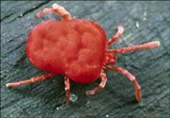
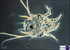
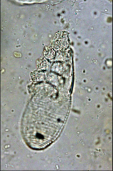
General characteristics of mites
|
- 6 legged larvae, 8 legged nymphs and adults
- smaller than ticks, often microscopic - membranous body - may be covered in fine hairs - generally hypostome is unarmed and hidden |
|
|
Mite diagnosis
|
collecting and identifying mites
- capture with comb or brush - skin scrapings and crusts (must go deep enough to get blood - skin biopsy - fecal (if animal is biting at itself it could ingest some mites) |
|
|
What are 3 types of mites?
|
- long- legged, active mites
- mange, itch, fur, and hair-clasping mites - oribatid mites |
|
|
Dermanyssus galinae description
|
red fowl mite
host: all birds -nocturnal, live in cracks and crevices of tree branches during the day |
|
|
Dermanyssus galinae clinical signs and treatment
|
clinical signs:
- swarm over roosting or nesting birds at night causing severe irritation - severe bites in humans and other animals when birds are not available treatment/control - don't use twigs as perches, if you do heat in oven first - supportive care |
|
|
Family Trombiculidae general description
|
chiggers
- adults and nymphs are free living, larva are parasitic stage - host: humans, birds, mammals, reptiles, amphibians |
|
|
Family Trombiculidae pathogenesis and clinical signs
|
pathogenesis:
- firmly attach and digest a long channel-like tunnel through the dermis using proteolytic salivary enzymes - don't burrow clinical signs - severe irritation and dermatitis due to reaction to enzymes |
|
|
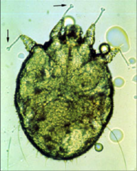
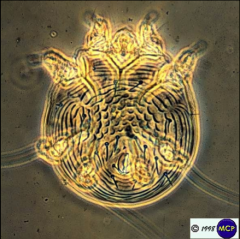
Family Sarcoptidae general description
|
- burrow into skin
- diagnose by deep skin scraping - short legs with bell-shaped nasal suckers on long, unjointed pedicles |
|
|
sarcoptes scabiei: host and transmission
|
hosts: animals and humans
- can be very host specific transmission - larvae and nymphs come to surface of skin and are transferred by contact |
|
|
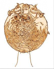
Sarcoptes scabiei adult morphology
|
- oval with rounded, short mouthparts
- legs 3 and 4 are very short - tarsal suckers on long, unjointed pedicles (legs 1 and 2 of females, 1, 2, and 4 of males) - dorsum covered with spines - terminal anus |
|
|
Sarcoptes scabiei pathogenesis and clinical signs
|
pathogenesis:
- burrow into skin and carry out lifecycle clinical signs: pruritic lesions of tunnels, dandruff, secondary infections - reportable in livestock |
|
|
Sarcoptes scabiei diagnosis and treatment
|
Dx:
- very very difficult - skin scraping, must get deep enough to see blood tx: - ivermectin or selamectin - may require more than one round |
|
|
Notoedres cati host and transmission
|
head mange of cats (sarcoptidae family)
- host: cats and rabbits, zoonotic - transmission- direct contact |
|
|
Notoedres cati adult morphology
|
- oval with rounded, short mouthparts (rounder and smaller than S. scabi)
- legs 3 and 4 are very short - tarsal suckers on long, unjointed pedicles - lack dorsal spines - terminal anus |
|
|
Notoedres cati pathogenesis and clinical signs
|
pathogenesis:
- burrows into skin and reproduces - starts at tips of ears, spread to face, head, paws and hindquarters clinical signs - alopecia with marked hyperkeratosis and abundant epidermal flakes |
|
|
Notoedres cati diagnosis
|
- skin scrapping
- mites are very easy to find (much easier than S. scabiei |
|
|
Knemidokeptes mutans general description
|
- claw like tarsi in females
- small and round with small mouth parts - tiny legs (almost like they aren't there) - transmitted by direct contact |
|
|
Family Psoroptidae general characteristics
|
Scab mites
- do NOT burrow : pierce skin and cause exudation of serum forming a scab - longer legs that extend beyond margin - bell shaped tarsal suckers on stalks of some or all legs |
|
|
Psoroptes cuniculi
|
DH: rabbits, goats, and horses
cause psoroptic mange - long sharp mouth parts - long and segmented pedicles tx: - don't clean ears of rabbit or use frontline (it will kill them ) - just treat with ivermectin |
|
|
Psoroptes ovis
|
DH: sheep and cattle
cause psoroptic mange- - long sharp mouth parts and segmented pedicles - reportable |
|
|
Psoroptes ovis clinical signs
|
worse in winter
- fleece loss on backs and sides of body (huge patches just slough off) - pustules form on thickened hardened, bare skin |
|
|
Psoroptes spp. diagnosis
|
shallow skin scrapping
visually can be seen with hand lens under characteristic scabs |
|
|
Chorioptes bovis morphology
|
oval
compressed laterally blunt mouth parts long legs large tarsal suckers on short, unjointed pedicles |
|
|
Chorioptes bovis disease
|
foot, tail, leg, and scrotal chorioptic mange
host: cattle, sheep, goats, horses, rabbits decreases quality as semen because testicles become too hot |
|
|
Otodectes cynotis disease
|
ear mites--> ear mange
host: dogs, cats, ferrets direct transmission- very contagious clinical signs - deep in external ear canal causing considerable irritation - head shaking - scratching ears - ears filled with dark, greasy, malodorous exudate - can penetrate tympanic membrane and cause neurologic disease |
|
|
Otodectes cynotis morphology
|
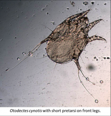
short, unjointed pedicles
tarsal suckers on legs |
|
|
Otodectes cynotis life cycle
|
1. larva hatch and feed on ear wax and skin oils for about 1 week
2. adults live on surface of ear canal skin |
|
|
family demodicidae general characteristics
|
- small, cigar shaped mites
- usually in hair follicles and sebaceous glands - vermiform shaped with long, transversely striated abdomen and short stubby legs - most species are non or mildly pathogenic - generally very host specific |
|
|
Demodex diagnosis
|
- deep skin scrapings
- lesions are non pruritic (differentiate demodectic vs. sarcoptic mange) |
|
|
Demodex canis disease
|
local (puppies)
- transmitted from mom to pups - localized lesions that regress naturally generalized - in adult dogs that are immunocompromised - pustules and hair loss all over body (erythematous lesions) |
|
|
Demodex treatment
|
generalized form- acaricide and antibiotic treatment in dogs
- need to identify underlying immunosuppressive problem |
|
|
Cheyletiella yasguri morphology
|
large and ovoid shaped
large, smooth dorsal scutum abdominal striations large palpal claws comb-like tarsal appendage |
|
|
Cheyletiella yasguri disease
|
"walking dandruff"- live on the skin surface of dogs, cats, and rabbits- cause dermal irritation- exfoliative dermatitis
highly contagious |
|
|
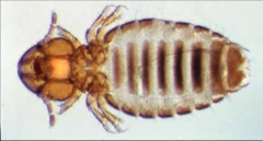
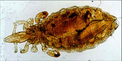
lice general characteristics
|
Class insect (adults with 6 legs)
- compressed dorsoventrally - wingless - recurved claws for grasping hairs and feathers - small thorax with short stout legs - lay 1 egg at a time - incomplete metamorphosis |
|
|
lice life cycle
|
- very host/organ specific- claw size correlates with body location
- direct transmission - may be worse in winter/spring when animals huddle |
|
|
2 orders of lice
|
mallophaga- chewing/biting lice
anoplura- sucking lice |
|
|
order mallophaga
|
chewing/biting lice
- only order that parasitizes birds (also on mammals) - broad heads (as wide as or wider than thorax) - pigmented mandibles - long, filiform antannae (3-5 segments) - feed on skin, debris, hair, and feathers causing skin irritation |
|
|
order anoplura
|
sucking lice
- narrow head, not as wide as thorax - visible antennae - large, recurved claws - sucking mouthparts- feed on host blood and tissue - cause anemia and hypoproteinemia in mammals - efficient vectors of many diseases |
|
|
Anoplura lice treatment
|
because they suck blood you can treat with injectable antiparasitics
|
|
|
diseases vectored by Anoplura lice
|
- Rickettsia prowazekii (typhus)
- Borrelia spp (relapsing fever) - francisella tularensis (tularemia) |
|
|
Genera of Mallophaga lice
|
Damalinia spp. - large animals
Trichodectes spp - dogs Felicola subrostratus- cats |
|
|
genera of Anoplura lice
|
Haematopinus spp
linognathus spp |
|
|
Haemotopinus suis
|
hog lice
- largest louse (6 mm) - found most frequently in the folds of skin behind the ears and between the legs - blood sucking activity results in much irritation and discomfort - more common in winter |
|
|
Fleas: general characteristics
|
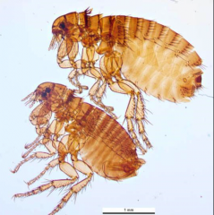
- blood suckers
- laterally compressed - wingless - large rear legs for jumping (can jump up to 33 cm) - may or may not have eyes - heavily pigmented comb spines (genal on head, prenatal on 1st thoracic segment) |
|
|
flea general life cycle
|
adults are parasitic
1. lay eggs in fur of animal 2. eggs fall off 3. worm like larvae hatch out and feed on dried blood or other debris 4. third larval stage will pupate 5. pupae will hatch and seek out host for blood meal 6. molt into adult |
|
|
what is the major difference between fleas and lice
|
life cycle
- fleas have complete metamorphosis and lice have incomplete - only adult fleas are on host, entire louse life cycle is spent on host - fleas are not host specific while lice are very host specific |
|
|
Flea disease
|
- irritating bites
- blood loss and anemia - saliva allergy--> flea bite dermatitis can act as intermediate host for helminth and protozoan parasites - diplydiium caninum (flea tapeworm) can vector disease microorganisms |
|
|
what are some diseases vectored by fleas
|
- yersinia pestis (bubonic plague)
- Francisella tularensis (tularemia) - rickettsia typhi (typhus) - Bartonella henselae (cat scratch bacterium) |
|
|
Ctenocephalides spp. general characteristics
|
- genal and pronotal combs are present
- well- developed eyes - anterior abdominal term each with one row of setae - genal comb horizontal with sharp curved teeth |
|
|
Ctenophalides felis
|
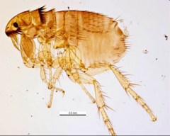
"cat flea"
- can infect dogs and cats - 1st genal tooth same length as other teeth - anterior margin of head is low - head is twice as long as it is high and is pointed - can transmit Bartonella henselae |
|
|
how can you tell Ctenophalides felis from canis?
|
the length of general and pronotal combs
- 1st comb is same length as other in felis - 1st comb is shorter in canis |
|
|
Transmission of fleas
|
direct contact or in the same environment as fleas (they can jump very high to reach host)
|
|
|
Flea infestation clinical signs
|
flea dirt, anemia, dermatitis
|
|
|
Flea diagnosis
|
visible fleas - flea comb
flea dirt- wet papertowl |
|
|
flea infestation treatment
|
1. adulticide (capstar)
2. IGR (frontline, sentinel) - stop hatching/molting 3. environmental control- vacuum |
|
|
What are 2 key points of vulnerability in mosquito life cycle?
|
molting/metamorphosis
reproduction |
|
|
Order Diptera, Family Culicidae
|
mosquitos
- aquatic breeders- still water - females take blood meals, males feed on plants - important vectors of disease |
|
|
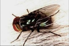
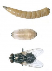
Order Diptera general characteristics
|
two winged, true flies
- 1 pair of wings if winged - large compound eyes - pests, disease vectors, some have obligately parasitic larvae - complete metamorphosis - larva are called maggots |
|
|
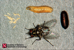
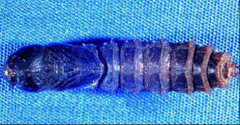
myiasis
|
"fly-strike"
- suborder cyclorrapha family Calliphoridae (blow flies, bottle flies, screwworm flies) - dipteran larvae (maggots) developing in the tissue or organs of a vertebrate host - facultative: free living larvae can adapt to parasitic dependence - obligatory: larvae must have host to develop |
|
|
Species that perform facultative myiasis
|
house fly (Musca domestic)
blowfly bottle fly (Calliphora spp.) flesh flies (Sarcophagidae) |
|
|
Species that perform obligatory Myiasis
|
primary screwworm (Cochliomyia hominivorax)
|
|
|
Cochliomyia hominivorax
|
Family calliphoridae: "primary screwworm"
- adults: shiny, greenish blue with orange head - larvae: wood screw shaped- obligate myiasis - eradicated in US but risk of reentry with globalization and climate change - reportable |
|
|
Musca autumnalis life cycle
|
family Muscidae (face fly)
- crawl on or around faces of livestock, feeding on ocular discharge that they induce - won't follow host indoors (differentiate from house and stable flies (same family) - breeds in fresh cow manure - life cycle complete in 14 days in warm weather |
|
|
Musca autumnalis morphology
|
adults:
- 7-8 mm long (larger than house fly) - 4 dark stripes on thorax - female has black sided abdomen pupae: - white and found in cow manure |
|
|
Musca autumnalis disease
|
no direct disease but...
biological vector for Thelazia nematodes- eye worm in cattle mechanical vector for keratoconjunctivitis organism |
|
|
Stomoxys calcitrans morphology
|
family: Muscadae "stable flies"
- slightly larger than house fly - 4 longitudinal stripes on thorax - broader abdomen than house fly with checkerboard dark spots on the dorm of the abdomen |
|
|
Stomoxys calcitrans life cycle
|
- breeds in horse and cow manure
- 30 day generation time - both sexes feed on blood |
|
|
Stomoxys calcitrans disease
|
- viscous biters of many animals and humans
- not a major vector of disease |
|
|
How can you differentiate Muscoid fly species?
|
tracheal trunk transection morphology of maggots- tracheal trunk is used to passively absorb oxygen so they can breathe while imbedded in host
|
|
|
Tabanid flies
|
Deer and horse flies
- vector EIA - very large - other flies take advantage of their large wounds for myiasis - larvae are predatory - cocoon like larvae (looks more like moth than fly) |
|
|
Aedes spp.
|
black body with white stripes
require stagnant water for reproduction Aedes albopictus- asian tiger mosquito - vectors WNV and dengue - active throughout the day - only female eats blood Aedes aegypti- yellow fever mosquito - vectors yellow and dengue fever - females feed on blood, males on plants - active during the day |
|
|
Culex pipens
|
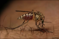
common house mosquito
- breeds in standing water - vectors WEE, EEE, WNV and heartworms |
|
|
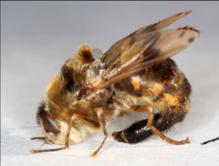
Bot flies
|
several families of flies whose larvae live as parasites in the bodies of mammals
|
|
|
Bot fly families
|
Cuterebra
hypoderma gasterophilus Oestrus |
|
|
How do you distinguish between bees and bot flies?
|
- number of wings: flies have 2 bees have 4
- antennae: flies don't have them - mouth parts |
|
|
Cuterebra sp. life cycle
|
parasites of rabbits and rodents
- adults lay eggs in rabbit and rodent nests/trails - Host brushes up against eggs -larvae hatch and enter animals through orifices - usually located SQ in cervical region (oral and nasal are also common) august, sept, oct most common |
|
|
Cuterebra sp. disease
|
- not usually associated with significant clinical signs
- secondary infections--> bacteria - can migrate to brain |
|
|
Cuterebra sp. ID and removal
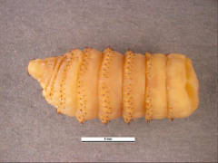
|
ID by black larvae with thick concentration of spines
removal: widen breathing hole and remove larvae with forceps - wound heals slowly |
|
Gasterophilus morphology |
"stomach bots" adults: ovipositor curve around under the body larva: yellowish with distinct rows of spurs |
|
|
Gasterophilus life cycle/ pathogenesis |
1. eggs laid on forelegs or shoulders and hatch when horse puts muzzle or tongue on hairs 2. larvae burrow into mouth, gums, and tongue or attach to GI mucosa 3. does not cause serious problems (horse can maintain large populations) |
|
|
Gasterophilus treatment |
- can coax larva out of burrow with wet sponge - treat with ivermectin past in the fall after adults are dead and when larvae are still vulnerable in stomach (treatment can be done year round in the south) |
|
|
Hypoerderma life cycle |
"cattle grub" 1. eggs laid on leg skin of cattle 2. larvae hatch and burrow into skin 3. migrate through connective tissue a. to esophagus then back through skin and fall to the ground to pupate b. to spinal cord (from december to may) very long life cycle |
|
|
Hypoderma clinical signs |
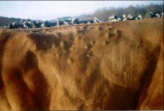
grubby back in humans- creeping myiasis (migrate to CNSS while "trying to find the top of the cow" and cause blindness and/or paralysis |
|
|
Hypoderma treatment |
systemic insecticides in many different forms don't treat between december and may because the larva are in the spinal cord and will die and remain there. |