![]()
![]()
![]()
Use LEFT and RIGHT arrow keys to navigate between flashcards;
Use UP and DOWN arrow keys to flip the card;
H to show hint;
A reads text to speech;
55 Cards in this Set
- Front
- Back
|
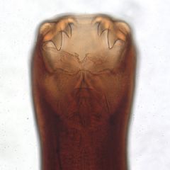
Ancylostoma caninum
|
ancylostomoidea
D.H.- dogs and wild canidae; PH: rodents adults: male- 11-13 mm female- 14-21 mm eggs: 60X40 um PPP:2-3 weeks |
|
|
A. caninum routes of transmission
|
transmammary
ingestion of L3 or PH skin penetration |
|
|
A. caninum clinical signs
|
- diarrhea (melena because they infect SI)
- anemia - hypoproteinemia - listlessness - emaciation - deathinfection can be peracute, acute, chronic, or secondary |
|
|
A. caninum treatment and prevention
|
treatment:
- supportive care (fluids, keep warm, blood transfusion - paraciticide (pyrantel, fenbendazole) Prevention: - pyrantel (2,4,6,8) - off label treatment of mom around time of delivery |
|
|
General characteristics of strongyles
|
- large, scleratized buccal capsule
- males have copulatory bursa - eggs are thin shelled and morulated - often live in large intestine of DH - often migratory in DH - mouth surrounded by corona radiata - direct life cycle |
|
This parasite was found in the small intestine of a dog. What species is it most likely?
|
Ancylostoma caninum
|
|
|
Common characteristics of hookworms
|
- males have caudal copulatory bursa
- large, heavily sclerotized buccal capsule with cutting plates, teeth, or lancets - feed by sucking blood - anterior end curved dorsally - eggs are thin shelled and morulated - live in small intestine of host - most species migrate in domestic host |
|
|
Strongylus vulgaris
|
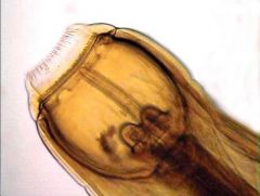
strongyle
D.H.- horses eggs: 70-100 x 40-50 um PPP: 6 months has 2 rounded teeth on bottom L3 is ingested from environmentadults in large intestine, L4 larvae migrate through cranial mesenteric artery to the aorta and back to the colon |
|
|
Cyathostomes
|
strongyle
D.H.- horse eggs: 70-100x 40-50 um (same as large strongyles) more prevalent than large strongyles adults live in large intestine and there is no migration in host |
|
|
Cyathostomes pathogenicity
|
larvae encyst in nodules in large intestine and emerge en masse in the spring.When so many emerge at once this can lead to damage to the GI epithelium and - diarrhea - weight loss - depression
|
|
|
Haemonchus contortus
|
Trichostongylus
DH: ruminants eggs: 60-108 um adults: male- 10-20 mm female: 18-30 mmadults have a single tooth and cervical papilla female has barber pole appearance, vulvar flap adults in abomasum of DH |
|
|
Haemonchus clinical signs
|
- edema--> bottle jaw
- anemia - often no diarrhea |
|

Name the species of parasite and the host it is most likely found in.
|
Haemonchus contortus- ruminants (sheep)
|
|
|
Common characteristics of metastrongyloidea
|
- reduced buccal capsule
- males with reduced copulatory bursa - adults found in respiratory tissues of DH - usually shed motile larvae rather than eggs - life cycle usually indirect - migratory in definitive host |
|
|
Aelurostrongylus
|
metastongyle
DH: cats IH: snails and slugs PH: amphibians, reptiles, birds, rodents L1: 350-400 um, kinked tail, NO dorsal spineAdults in bronchioles and alveolar ducts (possibly parenchyma) of DH |
|
|
Aelurostrongylus diagnostic techniques
|
- larvae in feces
- tracheal wash - radiographs (looks like feline asthma) |
|
|
Oxyuroidea General characteristics
|
"pinworms"
- medium sized worms with slender sharp pointed tails - muscular bulb on end of esophagus - no buccal capsule - very host specific - live in posterior portion of large intestine - eggs thin shelled, asymmetrical, and operculated at one end - direct life cycle with no migration |
|
|
Oxyuris
|
oxyuroidea
DH: horses adult: males: 9-12mm; females 3-6 cm eggs: 80-100x 40-50um PPP- 5 months causes peritis ani: barbering of hair around tail, rubbing of tail head on structures, hair loss |
|
|
Oxyuris methods of diagnosing
|
-clinical signs
-scotch tape test - fecal float (not very reliable) |
|
Identify this parasite. What distinguishing feature led you to this identification?
|
Oxyuris
- thin pin-like tail |
|
|
Ascarioidea general characteristics
|
The True Roundworms
- Large, stout worms of the small intestine - 3 lips around mouth opening (no buccal capsule) - eggs have thick albuminous shell - most species migrate in DH |
|
|
Toxocara canis
|
Ascarid
DH: dogsPH: rodents, birds adults: males- 4-10cm; females 5-18 cm eggs: 90x75 um highly prolific adults in small intestine larvae migrate extensively- liver--> lungs --> small intestine- liver --> lungs--> pulmonary artery --> arrest in other tissue until female (host) becomes pregnant |
|
|
Toxocara canis transmission
|
transplacental is very important
possibly transmammary ingestion of infective egg or infected PH |
|
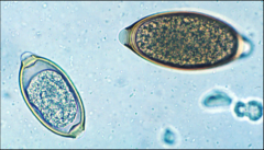
These eggs were found in the feces of a dog brought into your clinic. What parasite is it infected with? What should you warn the owner about?
|
Toxocara canis
- visceral larval migrans (zoonotic) |
|
|
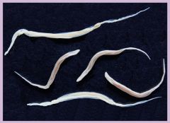
Trichuris vulpis
|
Trichinelloidea
DH: dogs adults: 4-8 cmeggs: 70-90 x 32-41 um adults in large intestine L1 eggs ingested and hatch as they are digestedeggs can survive for up to 5 years in environment |
|
|
Trichuris vulpis clinical signs
|
disease is generally mild to asymptomatic but can result in:
- bloody mucoid diarrhea - weight loss - dehydration - death |
|
|
Trichinelloidea common characteristics
|
Whipworm
- anterior end more slender than posterior end - very reduced or absent lips and buccal capsule- very long thin esophagus embedded in stichosome - male and female each have only one gonad - eggs have 2 polar plugs - L1 is infectious stage |
|
Identify these two eggs from a dog's fecal sample
|
1. larger one is Trichuris vulpis
2. smaller one is a capillarid Both are Trichinelloidea eggs |
|
|
General characteristics of filarial worms
|
- esophagus divided into anterior muscular and posterior glandular portions
- life cycle indirect - males frequently have a spirally coiled tail - parasitic outside the enteric tract in body tissues - long thin worms with a small mouth and NO buccal capsule, pharynx, or lips - females produce microfilaria that are found in blood and connective tissue - migrate in DH - indirect life cycle (use blood sucking arthropod) |
|
|
Dirofilaria immitis
|
DH: dogs, marine mammals, ferrets, cats
IH: mosquitos adults: male- 12-22 cm female- 25-31cm Mf: 300-325 x 6-7 um PPP= 6 months adults in right heart and pulmonary artery can migrate aberrantly to skin and eyes |
|
|
Dirofilaria immitis pathology
|
- pulmonary arterial disease with inflammation and proliferative lesions --> arteritis
- pulmonary thromboemboli - pulmonary hypertension--> can lead to right heart failure if prolonged |
|
|
Transmission of D. immitis
|
1. mosquito ingest Mf and spread resultant L3 to new host
2. Mf can be transferred prenatally (not infective in babies but can be transmitted from them) |
|
|
Diagnosis of Dirofilaria immitis in dogs
|
Knott's test- look for Mf in blood
antigen test- look for female uterine antigen radiographs- right heart enlargement (reverse D) serology |
|
|
Dirofilaria immitis clinical signs in dogs
|
- cough/dyspnea
- exercise intolerance - weight loss - ascites - anemia (due to damage to blood vessels) - eosinophilia and thrombocytopenia - glomerulonephritis - proteinurea |
|
|
what are the steps in treatment of heart worm disease?
|
FIRST MONTH
1. evaluate the health of the patient and determine severity 2. stabilize clinical cases 3. Start on monthly macrolytic lactone 4. administer prednisone at decreasing dose 5. give doxycycline to kill wolbachia 6. restrict exercise MONTH 2 1. continue ML MONTH 3 1. ML 2. first dose of adulticides (immiticide) 3. restrict exercise further MONTH 4 1. ML 2. Dose 2 and 3 of immiticide 3. prednisone if necessary 4. continue exercise restriction |
|
|
What are the goals of administering macrolytic lactones in treatment of heart worm disease?
|
1. kill larvae
2. prevent new infection 3. stunt maturation of immature adults 4. prevent female reproduction |
|
|
Why is it best to administer 3 doses of immiticide during heart worm treatment?
|
it increases efficacy - reduces the need for repeated treatments
it is safer because it decreases the chance of a mass die off and resultant embolism |
|
|
why would you give doxycycline during treatment of heart worms?
|
to kill wobachia, a bacteria that lives inside the adult heart worms and can be released upon tremens. By administering doxycycline you can hopefully prevent bacterial infection
|
|
|
What is possibly a major reason that heart worm infection is not as severe in cats as in dogs?
|
Dirofilaria have much shorter lifespans in cats so are less likely to reproduce and there are fewer microfilaria
|
|
|
What clinical signs are more common with cat heart worm infection than infection in dogs?
|
ectopic infections (skin, eyes) and respiratory disease HARD
|
|
|
How can Dirofilaria be diagnosed in cats?
|
both and antigen and antibody (not in dogs) testantigen detects worms after 5-8 months
antibody detects after 3 months |
|
|
How does treatment of cat heart worm infection differ from dog?
|
cats cannot be given immiticide so there really is not treatment
- just supportivetherefore it is extremely important that they be kept on prevention all the time. |
|
|
Common characteristics of cestodes
|
elongated (can be multiple feet)
segmented body (proglottids-->stroblia) scolex for attachementno alimentary canal adults - parasites in small intestine of vertebrates eggs and/or proglottids passed in feces indirect life cycle |
|
|
dipylidium caninum
|
cestode
DH: dog, cat, human *** zoonotic IH: fleas, lice adults - 5 cmeggs: 40-50 um; egg packets: 200 um PPP- 2-3 weeks 2 pores in proglottid |
|
|
Dipylidium caninum diagnosis and prevention
|
dx:
- visualize proglottids or egg packets in feces - observe fleas - see stroblia in vomit prevention:- use flea preventative |
|
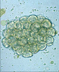
This structure was found in the feces of a dog brought into your clinic. the dog is infested with fleas. What parasite is likely causing his problem?
|
Dipylidium caninum (tapeworm)
|
|
|
Taenia pisiformis
|
Taeniidae
DH: dog IH: rabbit - metacestode= cysticercus pisiformisin small intestine of DH metacestode is cyst w/ 1 egg in liver or peritoneal cavity of IH |
|
|
How do you diagnose Taeniidae infections?
|
eggs on sugar flotation (not in egg packets)see stroblia or proglottids
|
|
|
Characteristics of digenetic trematodes
|
- leaf shaped
- no segmentation - incomplete alimentary canal (no anus) - attachment via suckers (acetabulum- attachment; oral- ingestion) - indirect life cycle: 1st IH is always a mollusk - hermaphroditic but can reproduce sexually - produce operculated eggs |
|
|
general life cycle of digenetic trematodes
|
1. miracidum (ciliated larval stage) actively seeks out snail
2. asexual generations (sporocyst, redia) within snail 3. cercaria (in host) leaves snail and encysts in new host or environment as metacercaria (enviro) 4. (meta)cercariae reach DH and develop to adult |
|
|
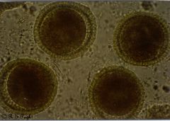
Fasciola hepatica
|
digenetic trematode- common sheep fluke
DH: ruminants and humans 1st IH: amphibious snail, 2nd= NONE adults: 2-3 x 1 cmeggs: 130-150 x 63-90 umPPP= 2-3 months adults can live for 11 years migrate in liver and bile ducts |
|
|
risk factors of Fasciola hepatica infection
|
wet pastures
transmission occurs between february and july in southeast US |
|
|
treatment/ prevention of Fasciola hepatica infection
|
- clorsulon
- NO ivermectin- has no efficacy against flukes |
|
|
control of Fasciola hepatica infection
|
-control DH access to water (drain or fence of ponds)
- molluscicides - periodic anhelminthics |
|
|
Diagnosis of Fasciola hepatica infection
|
geographic location
transport history (were they recently where it is found) eggs on fecal floatation clinical signs imagingadults on necropsy |