Reading...
![]()
Play button
![]()
Play button
![]()
Use LEFT and RIGHT arrow keys to navigate between flashcards;
Use UP and DOWN arrow keys to flip the card;
H to show hint;
A reads text to speech;
91 Cards in this Set
- Front
- Back
|
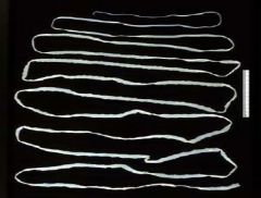
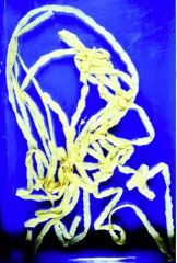
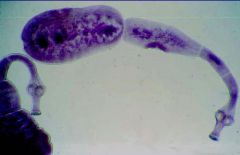
Taenia saginata adult
|
|
|
How do we acquire Taenia saganata?
|
By eating raw meat
|
|
|
Adult Taenia saginata
|
|
|
What worms are in the phylum nematoda?
|
Roundworms
"Nematodes" -Pinworm, Whipworm, Ascaris+VLM, Hookworm+CLM |
|
|
What worms are in the phylum Platyhelminthes?
|
Flatworms
-Class Cestoidea (segmented flatworms)- "Cestodes" -Class Trematoda (non-segmented flatworms)-"Trematodes" |
|
|
What are Cestodes? What are the important species and what do they cause?
|
Cestodes - All are flat, segmented worms and adults are obligate parasites on the intestinal tract
Taenia saginata (beef tapeworm) Taenia solium (pork tapeworm) ->Cysticercosis Echinococcus granulosus (dog tapeworm) -> Hydatid disease |
|
|
Where does the adult Taenia saginata live? Where does the larva live?
|
Adult: Humans
Larva: Cow |
|
|
How long can Taenia saginata grow to?
|
30 feet
|
|
|
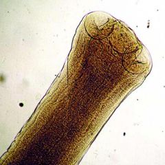
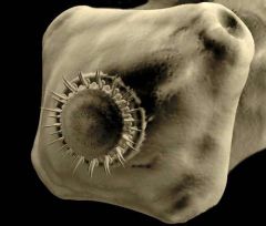
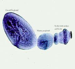
What is the scolex?
|
The scolex is at one end of the tapeworm. it is what attaches to the intestinal villus. There are suckers and sometimes there are hooklets. The rest of the worm dangles through the rest of the intestine, moving both passively and actively
|
|
|
What are cysticerci?
|
White cysts that can be seen throughout the heart of a cow. The disease is called cysticercosis. These cysts are the larval stage of Taenia saginata
|
|
|
What is the strobila?
|
The body of the tapeworm, composed of individual segments (proglottids)
|
|
|
What are proglottides?
|
Individual segments of the strobilia. They are anatomically independent, but linked togehter.
|
|
|
Describe the nervous system of tapeworms
|
A nervous system runs through the entire worm, allowing for locomotion. It moves against the peristaltic wave of the intestine.
|
|
|
What is the tegument?
|
The worm doesn’t have a mouth and intestinal tract. It absorbs nutrients through the tegument. The tegument covers the entire surface of the worm. It is an ATP rich ATPase. It is involved in active transport. The tegument is on the surface, just like your intestinal surface it is composed of microvilli. Under that there is active and passive transport of nutrients into the worm. The tegument is also involved in protecting itself against host digestive enzymes. If we disrupt the ATPase with a drug the worm can no longer prevent itself fro being digested and it is digested in the gut tract.
|
|
|
Explain tapeworm growth
|
As the worm grows the new proglottids are grown at the head end. They first emerge as immature proglottids. As they move out they mature and then become gravid. Gravid means that they have copulated in some fashion and they are full of infectious eggs.
|
|
|
Taenia saginata scolex
|
|
|
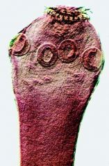
Describe the scolex of Taenia saginata
|
There are 4 suckers
|
|
|
Describe the sex organs of Taenia saginata
|
There are testes, ovaries, and a copulatory bursa. When proglottides rub against each other they copulate, fertilizing a proglottid. Then the eggs have to go through a process of differentiation and emryonation which occurs inside the intestine.
|
|
|
Explain the life cycle of Taenia saginata
|
-Adult worm is in humans
-At the terminal end of the worm, gravid proglottids break off and exit in stool. -Cows ingest proglottid segment -The eggs are digested and it releases an immature larval state called an oncosphere. -That oncosphere stage exits the egg and invades the mucosa of the gut of the cow and enters the circulatory system of the cow. -Disseminates throughout the cow -We slaughter the cow and eat the muscle -That muscle has cysticerci in it, containing another form of a juvenile larval state in it. -Hatches in stomach, releasing scolex -Scolex attaches to intestinal villus and grows -Fully grown in 3 months |
|
|
What is the definitive host? intermediate host?
|
Human. Cow
|
|
|
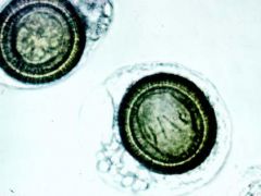
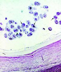
Taenia eggs (cannot differentiate between species on egg morphology alone)
|
|
|
What is a hexacanth larva?
|
The larva inside of the onchosphere. This is the larva that invades the cow's circulatory system and migrates throughout the body
|
|
|
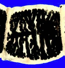
How do you differentiate species of Taenia tapeworm?
|
You can distinguish species by looking at mature proglottids. This is Taenia saginata. India ink has been injected in the copulatory bursae. It fills the uterus and uterine branches. If there are more than 12 branches, it is Taenia saginata.
|
|
|
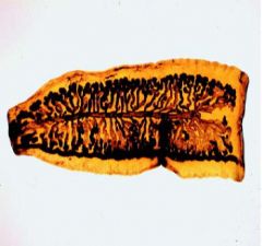
Gravid proglottid of Taenium saginata
|
|
|
What is the pathogenesis of T. saginata?
|
None
|
|
|
What clinical disease is assocaited with T. saginata?
|
None
|
|
|
How do you diagnose T. saginata infection?
|
1. Find eggs or proglottids in stool
2. Identify species based on proglottid morphology, after formalin and India ink 3. Identify scolex |
|
|
What is the drug of choice for T. saginata infection? How does it work?
|
Praziquantel
It increases permeability of flatworm tegument to Ca2+ ions, causing muscle tetany and worm detachment. |
|
|
How do you prevent transmission of T. saginata?
|
1. Sanitary disposal of human feces
2. Prevent cows from coming into contact with human feces 3. Freeze and/or cook all beef until well done 4. Federal meat inspection programs |
|
|
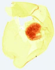
Whole cystircercus of Taenia solium
|
|
|
What is the pork tapeworm?
|
Taenia solium
|
|
|
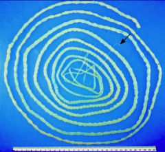
Adult Taenia solium
|
|
|
Taenia solium scolex
|
|
|
How long can Taenia solium grow?
|
20 feet
|
|
|
Taenia solium scolex
|
|
|
Gravid proglottid of Taenia solium
|
|
|
How many branches are there on the Taenia solium proglottid?
|
Less than 12
|
|
|
Describe the life cycle of Taenia solium
|
Humans harbor the adult worm. Proglottids are shed. They pass out in the feces. The eggs are ingested. Humans ingest undercooked infected pig meat. The cysticerci then release the larvae. The scolex everts from the larval cyst and grabs onto the intestinal villus. Adults mature over the course of time.
|
|
|
How do you diagnose Taenia solium infection?
|
1. Find eggs or proglottids in stool
2. Identify species based on proglottid morphology. 3. Identify scolex 4. Stool PCR or ELISA (not readily available) |
|
|
What is the pathogenesis of Taenia solium infection?
|
None
|
|
|
What is the clinical disease associated with Taenia solium?
|
None
|
|
|
What is the treatment for Taenia solium infection? How does it work?
|
1. Praziquantel
-Increases permeability of flatworm tegument to Ca2+ ions, causing muscle tetany and worm detachment 2. Niclosamide -Not absorbed systemically -Uncouples cestode oxidative phosphorylation, preventing ATP production -Parasite is then digested by host enzymes |
|
|
What drug should you use if you want to make a tapeworm diagnosis?
|
Praziquantel
|
|
|
How do you prevent and control Taenia solium infections?
|
1. Sanitary disposal of human feces
2. Sanitary practices on pig farms: seperate disposal of human feces from pigs' range 3. Cooking and/or freezing pork products thoroughly 4. Federal pork inspection programs 5. Treat pigs (oxfendazole) or vaccinate pigs |
|
|
What is the definitive host for T. solium? Intermediate?
|
Human. Pig(Human)
We can ingest the eggs from our own worm and get a T. solium infection. |
|
|
|
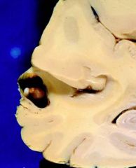
Cysticercus in brain
|
|
|
Cysticercus in brain
|
|
|
Multiple intracerebral cysts/Neurocysticerci
|
|
|
Multiple intracerebral cysts/Neurocysticerci
|
|
|
When does edema occur with neurocysticerci?
|
-Initially the cyst modulates the host response so there is no edema
-Immunologically silent for 1-2 years -Larva ages and starts to degrade -Plasma cells and lymphocytes infiltrate and start to invade -Edema develops -Symptoms are caused |
|
|
How do humans acquire cysticercosis?
|
-Food contaminated with human feces in endemic locations - another person's worm
-Bedsheets -Autoinoculation (~15%) - one's own worm -Reverse peristalsis or vomiting- bile contains eggs |
|
|
Explain the life cycle of cysticerosis
|
-Adult worms release proglottids
-Gravid proglottids release eggs -Invade gut mucosa and get into circulatory system -Disseminate -Go to skeletal muscle, eye, brain -Humans have no predators so cycle ends |
|
|
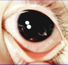
Cysticercus floating freely in anterior chamber
|
|
|
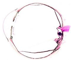
Cysticercus near optic nerve
|
|
|
Radiogram of lower leg with numerous calcified cystercerci of T. solium
|
|
|
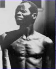
This is someone who must have ingested proglottid after proglottid. The skin lesions could be cysticerci.
|
|
|
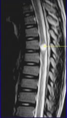
This is cystercerci in the spine, causing paralysis of the lower extremities.
|
|
|
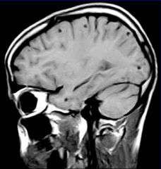
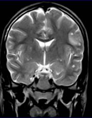
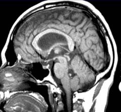
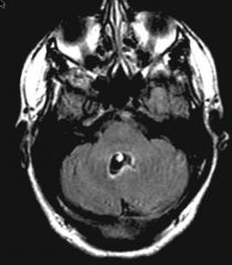
This is cysticercus involving the cerebello-pontine angle, visible on MRI. The cyst occupies almost the entire 4th ventricle here. If it were big enough it could obstruct flow, causing hydrocephalus and then ultimately death.
|
|
|
This is cysticercus involving the cerebello-pontine angle, visible on MRI. The cyst occupies almost the entire 4th ventricle here. If it were big enough it could obstruct flow, causing hydrocephalus and then ultimately death.
|
|
|
How does T. solium module the immune system?
|
-Taeniastatin
-protease inhibitor -Paramyocin -Inhibits complement -Other proteases: -Degrade IL-12, immunoglobulin, and IFN |
|
|
When do neurocysticerci undergo calcification?
|
2-5 years
|
|
|
What are the symptoms of a neurocyst?
|
Symptoms vary based on cyst:
Number: Single or multiple Size: Giant or small State: cysts are living, degenerating, or dead and calcified Neurological effects: Seizures CSF obstruction Hydrocephalus Arachnoiditis Mass effect Focal neurologic deficits |
|
|
What is the pathogenesis of neurocysticerci?
|
-Space-occupying lesions
-Local immunologic reactions |
|
|
What clinical diseases are associated with neurocysticerci?
|
-Visions impairment/blindness
-Seizures/death -Obstructive hydrocephalus/Coma/Death -Focal neurologic deficits that depend upon location of mass and area affected |
|
|
Describe the distribution of neurocysticercocic and Taeniasis
|
The distribution is fairly global
|
|
|
Describe the clinical epidemiology of cysticercoci
|
-Est. 50 million people with intestinal taeniasis, world-wide
-20% have cysticercosis, at least half will be symptomatic -Leading cause of adult-onset seizures worldwide (~40%) -Leading cause of epilepsy among children in endemic areas -In US: Est 1000 new cases per year (no mandatory reporting) -Immigrants account for >95% annually -Travelers account for ~3% -Autochthonous transmission: rare. Typically within families where one member harbors adult tapeworm |
|
|
How do you diagnose neurocysticerci?
|
*Must differentiate between cysticercosis and other possible lesions (benign cysts, solid tumors, etc.)*
1. Biopsy whenever possible 2. Physical (palpation) and X-ray evidence 3. Enzyme-linked immunoblot serological test, can be as high as 98% sensitive, 100% specific 4. MRI - how most cases are found in the US |
|
|
How do you treat neurocysticerci?
|
1. Surgical removal of cystercercus when appropriate
2. Steroids during time of neurological symptoms 3. Anticonvulstants 4. Antiemetics if patient has intestinal taeniasis 5. Antiparasitic antibiotics: Praziquantel or albendazole + steroids + anticonvulsants for multiple or symptomatic cysticerci, or for inoperable cysts |
|
|
What is Echinococcus granulosus? What does it cause?
|
Dog tapeworm
Causes hydatid disease in humans |
|
|
Where does the adult Echinococcus granulosus live?
|
In the dog
|
|
|
What is the intermediate host for Echinococcus granulosus?
|
Sheep/Human
|
|
|
Where does Echinococcus granulosus disease occur?
|
Wherever sheep husbandry occurs
|
|
|
Adult of Echinococcus granulosus
|
|
|
Describe the adult of Echinococcus granulosus
|
-There is a scolex and typically only 3 proglottids
-About 1cm -Scolex has succkers and hooklets -Proglottids are produced from the neck region -Worm covered in tegument -Received nutrients through skin |
|
|
Describe egg production by Echinococcus granulosus
|
There are several hundred eggs per gravid proglottide and there is typically 1 gravid proglottid per worm
|
|
|
Adult Echinococcus Granulosus
|
|
|
Describe the normal life cycle of Echinococcus granulosus
|
-The sheep eat where the dog poops and ingest the eggs.
-Larva invade through sheep mucosa and goes through the portal circulation -Lodges in sheep liver -Liver develops a large hydatid cyst -Inside the hydadid cyst is the infectous larval stage, called a protoscolex. -Dog eats the sheep viscera -Protoscolex enters dog gut lumen and attaches to microvilli -Adult grows in dog over several months -Humans can ingest the eggs and develop hydatid disease |
|
|
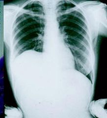
Radiograph of upper body showing elevation in right lobe of liver due to large hydatid cyst
|
|
|
Describe the distribution of hydatid cysts
|
Liver: 63%
Lungs: 25% Muscles: 5% Bone marrow: 3% (Fatal) Kidney: 2% Spleen: 1% Brain: 1% (Fatal) |
|
|
|
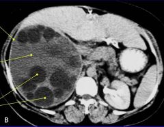
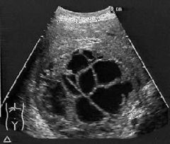
Hydatid cyst of liver. The dark black circles are daughter cysts. The daughter cysts have smaller daughter cysts all the way to the brood capsule. The brood capsule contains 8 protoscolexes.
The light gray is the hydatid fluid. This is highly allergenic, causing an immediate hypersensitivity reaction. |
|
|
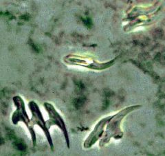
What is hydatid sand?
|
Hydatid sand is composed of the hooklets of degraded portions of daughter cysts that have ruptured.
|
|
|
Explain how hydatid cysts form
|
When the organism nvvades through the circulatory system and lodges in the liver the first thing that the protoscolex does is around itself it secretes a hyaline membrane that is acellular.
The exterior portion is immunologically silence, so it can grow over 10-20 years to be this size. On the inner surface is a germinal layer from which one cell is capable of producing a new hydatid cyst. It is an omnipotent geminal layer. If you happen to rupture this in any faction, if a germinal cell gets out it can cause local spread and new hydatid cysts to form. The membrane composed of the hyaline and the geminal layer is an integral part of the parastie. |
|
|
This showed daughter cysts inside of a large hydatid cyst. This lets us know we are dealing with an echinococcal disease.
|
|
|
As you get to a smaller level you see the hyaline membrane surrounding the cyst. There is the germinal layer from which everything inside arises and then there is the host reaction which is pretty much silent. The infectious stage is the protoscolex inside of the brood capsules.
|
|
|
Hydatid sand
|
|
|
Describe the pathogenesis of Echinococcus granulosus
|
-When intact, hydatid cysts are immunologically and often clinically silent, especially in the liver
-In other organs (e.g., brain, lung, bone marrow), hydatid cyst is a space-occupying lesion -It may leak or rupture, seeding/metastasizing adjacent areas -When hydatid cysts ruptures, allergic reactivity and anaphylaxis often ensue. This may be fatal |
|
|
How do you diagnose Echinococcus granulosus?
|
A. Direct
1. NO BIOPSY 2. Can remove surgically. Find "hydatid sand" on microscopic examination of fluid from hydatid cyst B. Indirect 1. ELISA-based serology 2. Imaging: MRI, CAT scan, X-ray, US 3. Accurate case history (ownership of dogs, living on a sheep farm, etc.) |
|
|
How do you treat Echinococcus granulosus?
|
-Surgical whenever possible
-PAIR (puncture, aspirate, inject, reaspirate) technique for liver lesions -Pharmacologic has less than 50% success |
|
|
What is the drug of choice for Echinococcus granulosus? How does it work?
|
Albendazole (for up to 6 months)
It prevents microtubule polymerization, blocking glucose absorption, starving the worm |
|
|
How do you prevent and control Echinococcus granulosus?
|
-Regularly treat all sheepherding dogs with niclosamide. This drug kills the adult parasite (by inhibiting ATPase)
-Avoid feeding hydatid cyst material (sheep offal) to dogs -Public health education of sheep farmers |