![]()
![]()
![]()
Use LEFT and RIGHT arrow keys to navigate between flashcards;
Use UP and DOWN arrow keys to flip the card;
H to show hint;
A reads text to speech;
134 Cards in this Set
- Front
- Back
|
Filarial nematodes -- Which are lymphatic (2)?? Cutaneous (3)? Body cavity (1)? -- Which stage makes a primitive larva and what is it called?? How can they circulate in blood, what does this correspond to? |
* Lymphatic: Wucheria and Brugia (both are also spread by mosquito) -- Cutaneous: loa loa, oncocerca, mansonella --- Body cavity: mansonella (perstans and ozzardi) -- Female adult worms makes primitive larva called a microfilaria -- Circulates c (nocturnal / diurnal) periodicity corresponding to vector's feeding schedule
|
|
|
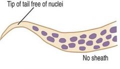
Filarial nematodes -- Which have no sheath? No tail nuclei? **mneumonic** What characteristics are important in dx'ing (3)? Which are spread by mosquitoes (2)?? Treatment (2)? |
Mansonella perstans and Oncocerca volvulus have no sheath or periodicity (***MO PBS [periodicity/ blood / steath]***) --- Wucheria bancrofti and O volvulus have no tail nuclei (***WO tail nuclei*** W after M as T(tail) after S(heath)***) --- Id'd on size, sheath and tail structure --- W bancrofti and B malayi spread by mosquito (also affect lymph and cause elephantiasis) --- Tx'd c diethylcarbamazine (DEC) and ivermectin |
|
|
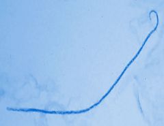
Wucheria bancrofti -- intermediate hosts and vectors? Where do adult worms live and what dz caused? Periodicity? 2 histo characteristics of microfilariae? What procedure used to dx? |
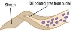
Culex, aedes and anopheles sp are hosts and vectors --- Adult worms in lymph --> elephantiasis --- Nocturnal periodicity -- Microfilariae lightly-stained sheath and pointed tail --- Present in small #'s, so must use sensitive procedures (Knott concentration, thick films. etc) |
|
|
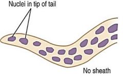
Brugia malayi --- What dz caused? What is vector and intermiate host? Sx? Periodicity? Histo of microfilariae? 3 |
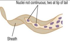
Malayan filariasis -- Anopheles, aedes, armigere, and mansonia mosquitoes are vectors and intermediate hosts -- Pts get elephantiasis of legs -- Nocturnal periodicity -- Sheathed tail c 2 distinct nuclei in tip of tail, which stains bright pink |
|
|
Brugia timori -- Host? Periodicity? Sx (2)?? Histo? |
* Humans are the only host -- Nocturnal periodicity -- Elephantiasis c abscess formation -- Nuclei go to tip of tail, but do not stain c Giemsa
|
|
|
Loa loa -- Where endemic? Vector? Nickname and explanation of why called this? Another characteristic sx and pathophys? Periodicity? Histo (2)?? Dx? Tx? |
* Endemic to West and Central Africa -- Human infx by Chrysops (deer) fly -- Eye worm (cornea) -- Calabar swellings, transient inflam 2/2 adult worms moving in tissues -- Diurnal periodicity -- Sheathed microfilariae c nuclei that go to end of tapered tail -- Dx diurnal blood films -- Tx surgical removal when in eye
|
|
|
Onchocerca volvulus -- Vector? Sx? Where found on body? Histo? Dx? |
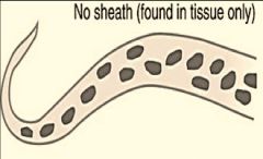
Vector is blackfly genus Simulium -- Adult worms embedded in fibrous nodules -- Location on body depends on parasite strand, but found in skin -- Have no sheath and anucleate tail -- Dx'd c skin snips teased in water or saline |
|
|
Mansonella ozzardi -- Vectors (2)? What special about these vectors? Sx? Histo (2)? Dx? Periodicity? |
Vectors Culicoides sucking midge flies or Simulium blackflies -- Vectors too small for screening nets -- Asx, or hypersensitivity-like rxn -- No sheath and lots of nuclei that do not extend to tail -- Dx: blood and skin bx's -- Nonperiodic
|
|
|
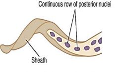
Mansonella perstans -- Vector? Reservoir host? Where do adult worms settle? Sx? Histo (2)?? Dx? Periodicity? |
Vector Culicoides fly -- Primates reservoir hosts -- Settle around eye -- Cause calabar swellings Body filled c nuclei that go all the way to tip of tail, has no sheath -- Draw blood at any time bc -- No periodicity
|
|
|
Mansonella streptocerca -- Vector? Host (2)? What tissues infected (2) Histo of microfilariae (2)? |
Vector small midges in genus Culicoides -- Hosts monkeys and humans -- in skin and blood -- Microfilariae unsheathed, c nuclei extending to tip of tail c shepherd's crook bent tail |
|
|
Dirofilaria immitis -- Nickname? Where adult worms reside? Where microfilariae reside? Vector? Life cycle in humans? Sx? Dx? |
aka dog heartworm -- Adult worms in right heart of dogs -- Microfilariae in blood -- Vector is infective larva from mosquito bite --Worms do not reach maturity in humans and microfilariae cannot be found in blood -- Cause hemoptsis, angina -- Coin lesions c dead / dying worms |
|
|
Malaria (general) -- Genus? Vector? tertian fever(3)? quartan fever (1)? -- Tx? 3 -- Which have hypnozoites (2) and mneumonic -- What dz protects against all malarial dz? |
Genus plasmodium -- Vector female Anopheles mosquito -- Ovale, vivax, and faliciparum (tertian fever, every 48 hrs) and malariae (quartan fever, every 72 hrs) -- Tx: quinine, chloroquine, primaquine -- Only P vivax and P ovale can have relapse, though all can have recrudescence (*** P and O in the word hyPnOzoites, f and m are not***) -- G6PD protects against all malarial spp
|
|
|
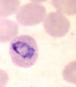
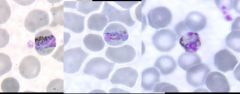
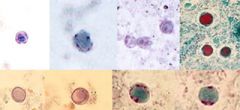
Malaria (general) -- Histo of ring form (early trophozoite)? 3 -- Histo of developing trophozoite? |
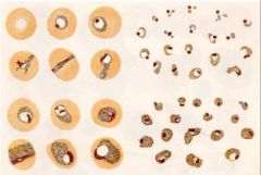
Ring form (early trophozoite) has blue cytoplasmic circle connected c red chromatin dot, with a vacuole inside the ring -- Varies by spp, but usually has pigment from hemoglobin metabolism
|
|
|
Malaria (general) -- Histo of immature schizonts? 2 -- Histo of mature schizonts? 2 -- Which 2 malarial spp are very similar to one another morphologically? |
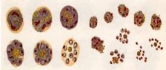
Have pigment granules and occupy more space in RBC as it grows -- Have merozoites, whose # and size depend on spp -- Vivax and ovale very similar to each other (both have Schuffner stippling and cause RBC enlargement)
|
|
|
Malaria (general) -- 1) Histo of microgametocytes? 2 -- 2) Histo of macrogametocytes? 3 |
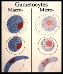
1) Round shape c large diffuse chromatin mass --- 2) Round to oval c compact chromatin mass and pigment
|
|
|
Malaria - schizogony (pre-erythrocytic stage) -- How is plasmodium introduced into humans? -- Where do they first go when in body? -- What forms in this first place? -- Then transforms to what? How multiplies, and what forms? -- Mneumonic (for forms) |
Anopheles mosquito injects sporozoites (infective stage) -- Transported by blood to the liver (hepatocytes) -- There, they transform into trophozoites -- At maturation, transforms to a schizont -- Schizont does asexual multiplication (schizogony), forming merozoites -- ***SpoT SchiMero (Sporozoite, Troph, Schizont, Merozoite) ***
|
|
|
Malaria - erythrocytic cycle -- What first attaches to RBC? -- What it transforms into? -- Then forms what with what inside? What happens after this stage? What happens after numerous erythrocytic cycles? 2 -- Mneumonic |
Merozoites burst from schizont in liver and enter RBC -- There, they transform into trophozoites -- Trophozoites transform into schizont with merozoites inside -- Merozoites burst from RBCs to infect more RBCs -- After many cycles, merozoites entering RBCs form macrogametocytes (female) and microgametocytes (male)
* ***SchiMero To Schimero (Schizont / Merozoite, Troph, Schizont / Merozoite)*** |
|
|
Malaria - sporogeny -- Occurs in what host? What life cycle stage (2) enters what organism? What 2 things form? What happens at this point? What results and what does it transform into? This gets surrounded by a capsule, forming a what? After meiosis, what is formed? Then asexually reproduces to form what, and where does this go? Mneumonic |
Occurs in invertebrate host (Anopheles mosquito) -- Microgametocytes + macrogametocytes enter female mosquito gut after feeds on human blood -- Macrogemetes and Microgametes form -- Microgamete fertilizes macrogamete -- Forms a zygote, forming a Ookinete -- Capsule made around ookinete, forming an oocyst -- After meiosis, forms a sporoblast -- Sporoblast divides asexually into sporozoites, which then go to mosquito's salivary gland to infect humans * ***MaMi, ZoO Eat Cyst BlaZo*** (Macro- and Microgametes, Zygote, OokinEte, OoCyst, sporoBlast, SporoZoites)
|
|
|
Plasmodium vivax -- Nickname? Incubation period? Periodicity? -- Chronic infx? -- Characteristic microscopic finding? -- What kind of RBC infected? How does infection change morphology? -- Name when found at edge of RBC? -- Describe the trophozoite? -- Describe the schizont? (what do they have inside them and what do these develop into??) -- What special blood group is protected? |

aka Benign Tertian malaria, (fever every 48 hours) -- Hypnozoites relapse after mo to yrs -- All forms Schuffners dots (image) -- Young RBCs infected, become large and distorted -- Called accole or applique crescent mass at outer RBC edge -- Trophs irreg ameboid -- Schizonts multiple chromatin bodies develop to 12-24 merozoites -- Duffy neg protected |
|
|
Plasmodium ovale -- Infects what kind of RBC? How does it change morphology? -- Describe schizont in RBC? 2 -- How does ring form compare to P. ovale? -- Periodicity? -- Chronic dz? |
Infects young/immature RBC, and look big and oval with ragged and irreg cell walls -- Schizonts circular c rosette of merozoites (~8 avg) -- Ring form large and thick -- 48 hour paroxysms -- Relapses by reactivation of hypnozoites from liver
|
|
|
Plasmodium malariae * Infects what kind of RBC? Change of morphology? * Morphologic characteristic (special dots)? * Describe troph (2) * Describe schizont * Periodicity? * Chronic dz? |
* Infects mature RBCs, which appear normal sized * Has Ziemann's dots, but not Schuffner's dots * Trophozoite c banded, bar or round shape * Schizont c rosettes / irreg clusters of merozoites * Cyclic paroxysms every 72 hours * No true relapses, but may have recrudescence, a low grade parasitemia for >20 yrs
|
|
|
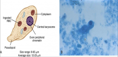
Plasmodium falciparum * Infects what kind of RBC? Change of morphology? * Characteristic dots? * Morphology? 3 * Describe troph * Describe schizont * Describe gamete * Which forms are seen in the PB? 2 * Periodicity? * Chronic dz? * Which blood group protected? * Px (from a PB dx?) |
* Infects all ages of RBC, which are normal sized * Can have Mauer's dots, but not Schuffner's or Ziemann's dots * Headphone config c multiple small delicate rings c 1-2 small chromatin dots c accole/applique * Trophozoite has "heavy" ring form * Schizont c up to 32 merozoites in clusters * Gametocytes banana / crescent shaped * Only young ring forms and gametocytes seen in PB * 36-48 hr paroxysms * No hyponozoites * Hemoglobin S trait protected * Seeing schizonts in PB means very grave prognosis
|
|
|
Plasmodium knowlesi * Nickname? * Vector? * Periodicity? * Morphology? * Dx? |
* aka Simian malaria (bc in monkeys) * Vector is Anopheles leucosphyrus in SE Asia * 24-hour cycles (quotidian) * No Schuffner's dots (very similar to P. malariae under scope) * Dx made by molecular detection methods
|
|
|
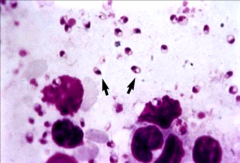
Babesiosis (Babesia microti / [rarely] divergens) * Name of dz in cattle? Sx in humans? * Vector? * Which kind of (post-surgical) pts have worst dz? * Which stage(s) present in human RBCs? * Morphology (2) and RBC shape? |
* Causes Red Water Fever in cattle, nonperiodic fever (also anemia and leukopenia )in humans * Ixodes tick is vector * Splenectomy pts get worst dz * Only trophozoite stage present in human RBCs * Have ring form c tiny chromatin dot and also Maltese crosses in normal sized/shaped RBCs
|
|
|
Phylum: Apicomplexa * What 2 life cycles characteristics do all genera share (and what are included, generally, in each?) |
All genera have life cycle that has: 1. Sporogeny (sexual cycle) in a definitive host c gametocytes, zygotes, oocysts, oocytes, and sporozoites 2. Schizogeny (asexual cycle) in intermediate host c merozoites, trophozoites, schizonts, and gametocytes |
|
|
Cryptosporidium (parvum and hominis) * How contracted? and in what age group? * Sx? * What is diagnostic stage? Infective stage? * Specimen of choice? What 2 stains? * Technique used? |
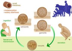
* Contracted from infected water sources (swimming pools) in kiddos in daycare * All cases have some acute diarrhea * Oocysts are Diagnostic and infective stages * Specimen of choice is Stool or Duodenal bx with Modified acid-fast or Auramine Rhodamine stains * may use Sheather's sugar floatation technique
|
|
|
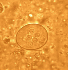
Isospora (belli) * Which stages are in human GI tract? 2 * What is the infective stage? * Morphology of infective stage (2) * Sx? * Tx? |
* Schizogony and sporogony in GI tract * Infective stage is when mature oocysts ingested * Weakly acid-fast elliptical eggs c 1 (unsporulated) or 2 (sporulated) sporocysts * Pt gets diarrhea * Tx is TMP-SMX
|
|
|
Cyclospora (cayetanensis) * Who affected? (2) * How does infection spread? * Diagnostic and infective stages? * Histo on acid fast? * Sx? |
* In Peruvian children and Asian travelers * Contracted by drinking contaminated water * Diagnostic stage is oocyst (c 2 sporocysts, each having 2 sporozoites); infective stage is mature oocysts * Oocysts light pink to deep red c bubbly granules on acid fast * Pts get explosive diarrhea for 1-3 weeks (no tx)
|
|
|
Sarcocystis * What kind of host are humans? 2 * In what tissue found? (2) What structure is made? * Sx? * How transmitted? 2 * Diagnostic stage and infective stage? Morphology? * Specimen of choice? 2 |
* Humans are accidental or intermediate hosts * Found in intestine and muscle (where long Mieschers tubules are made) * Asymptomatic (even in immunocompromised) * Transmitted by eating flesh c Mieschers tubules or in fecally contaminated food * Diagnostic and infective stages are oocysts; eggs are hour-glass shaped c 2 sporocysts * Specimen of choice is either Poop with oocysts seen or Gi/muscle mx c Miescher's tubules
|
|
|
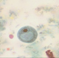
Blastocystis hominis * Pathogenic? * How stained? * Morphology? 2 |
* Originally thought to be nonpathogenic yeast (seen in asymptomatic AIDS pts) * Best seen c trichrome * Has membrane-bound central (fluid-filled) body c 2-4 nuclei in cytoplasm
|
|
|
Microsporidia * Where does it have to live to survive? 2 genetics facts? * With what organism is co-infective? * What is the MC organism? what are 2 sx? * What do microsporidium or nosema cause? * How is dz spread? 4 * How are cells infected? * Morphology? * Dx? 4 * Tx? 2 |
* Obligate intercellular parasite; has 70s ribosomes (like bacteria) and smallest genome of any eukaryote * Double infx c Cryptosporidium common * MC org is Enterocytozoon bieneusi, causing chronic diarrhea and malabsorption * Other organisms cause keratoconjunctivitis * Spread in feces, urine, death of host or eating infected meat * Infective spores inject sproplasm into host cell * Have spores that extrude polar filaments (tubules) that inject sporoplasm (infective material) into host cell * Dx by IFA or acid fast (standard techniques not effective), Chromotrope 2R or Hot Gram Chromotrope technique by light microscopy * No tx, though Fumagillin may help against intestinal variant
|
|
|
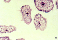
Toxoplasma gondii * How spread? 3 * Definitive host? * What seen in feces? * Sx in most pts? 2 ways of presenting in pregnancy? in AIDS pts? * What 2 forms seen in humans and morphology? * Infective stage? * Gold standard specimen c what test? * 2 Meanings of low IgM and what to do for this? * Tx? * Prevention? |
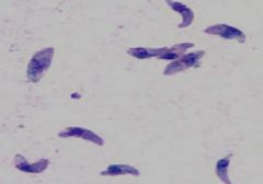
* Spread by ingestion of oocysts in cat feces, raw meat, milk, or transplacentally * Definitive host is cat * Oocysts found in feces * Most pts are asymptomatic; fetal loss if primary infx in early gestation fetus and fetal CNS infx if primary infx in late pregnancy; AIDS pts can get cerebral form * Tachyzoites ([image]crescent-shaped, extracellular; seen in acute infx) and bradyzoites (resting form seen in muscle and brain, lots of tachys packed in a histiocyte) * Best specimen is serum, c Sabin-Feldman dye test (need live organisms) or Antibodies * if IgM low positive, may be new infx or infx in last 2 yrs, need to draw again in 2 weeks * Tx is pyrimethamine and trisulfapyrimidine combo * Change cat box daily
|
|
|
Myxozoa * What does it usually infect? * Sx? * 2 spp? * What do spores look like and what are they made of? |
* Usually a fish parasite * Gi sx (esp in AIDS pts) * Henneguya salminicola and Myxobolus plectroplites * Spores look like sperm and are made of chitin
|
|
|
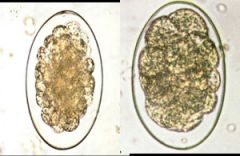
Ascaris lumbricoides * How acquired? * How does it initially invade body? 2 * Where does it travel to and what develops? * How can it cause death? * Then what does it do? (name of syndrome and sx?) * Where do adult worms reside and 3 sx? * Dx and (2) descriptions? What is best technique? * Tx? |
* Acquired by egg ingestion * Eggs hatch in duodenum, where larvae penetrate intestinal mucosa * May cause death from intestinal obstruction (worms are big!!) * Then goes by blood to alveoli (larval migrans), where causes coughing and asthmatic breathing and possibly Loefflers syndrome (c lots of yellow sputum) * Adult worms live in small intestine and cause colicky abdominal pain * Dx by rough, bile-stained eggs (fertilized or infertile) in feces, best to use sedimentation c formalin-ether * Tx c albendazole or mebendazole
|
|
|
Enterobius vermicularis (pinworm) * Common? * How acquired? * What first happens and where do worms migrate to? * Who and where to is the next migration? * What happens at night? * Where can worms go (2)? * Sx? * What kind of infx can occur in girls? * Dx? * Morphology? 2 |
* MCC helminth infx in USA * Acquired by ingesting eggs (hand to mouth) * Eggs hatch in small intestines and migrate to ileocecal area to develop into adult worms * Female worms migrate to anus * At night, females migrate out of anus and deposit sticky eggs on perianal skin * Babies can either go to mouth (by hand-mouth) or back into anus (retroinfection) * Cause intense anal itching * May cause vulvovaginitis in girls * Dx c scotch tape test * Eggs usually embryonated and are flat on one side and have translucent shell
|
|
|
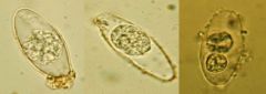
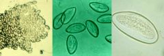
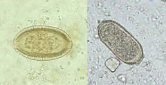
Trichuris trichura (Whipworm) * Common? * How acquired? * Worm morphology? * How invades? * Egg morphology? * A possible severe sx? * Dx? |
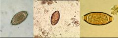
* 1/2 million infected (mostly kids) worldwide in places c poor sanitation * Acquired by ingesting eggs containing first stage larvae * Worms have narrow anterior end * Anterior end is threaded into intestinal mucosa * Barrel-shaped unembryonated eggs c polar corks at each end * May cause rectal prolapse * Dx by finding eggs in poo
|
|
|
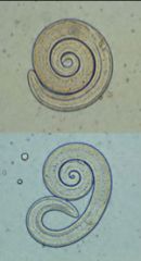
Ancyclostoma duodenale (Old World) and Necator americanus (New World) [Hookworm] * How to differentiate the 2 spp? * Larval buccal cavity and genital primordium? * What are the 3 phases? * How acquired to cause infx? Sx at that time? * How can be acquired that doesn't cause lung migration? * Life cycle? 5 and 3 important stage names * Infective stage and appearance? |
* 1st spp has teeth, 2nd has cuttin plates * Long buccal cavity and indistinct genital primordium in larvae * Have Cutaneous, pulmonary and intestinal phases * 3rd stage filariform larvae penetrate skin exposed to contaminated soil, causing "ground itch" * No lung migration if ingested * Larvae enter venules and lymph and goto heart, then go to lungs and are swallowed and go to small intestine (where develops into 4th stage larvae), where feed off intestinal blood for 5 wks and have sex making first-stage rhabditiform larvae, where develop into 3rd stage filariform larvae * Infective stage is 3rd-stage filariform larvae, which looks snake-like
|
|
|
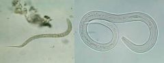
Strongyloides stercoralis (threadworm) * Where endemic in US? Where MC climate? * Larval buccal cavity and genital primordium? * What are 2 stages? * 3 life cycles and description of each * 3 phases of sx? * What can cause a frequently fatal infx? * Dx? (what finding, what apparatus, and what test) * Tx? 2 |
* Endemic rural SE states and Appalachia; MC in tropical / subtropical climates * Short buccal groove and prominent genital primordium * Has parasitic and free-living stage * Direct life cycles is similar to hookworm life cycle; indirect life cycle (in tropics) is a free-living cycle where rhabditiform larvae passed in feces and males and females live in soil; autoinfective life cycle when 1st stage rhabditiform larvae prematurely molt to filariform larvae that immediately reinfects host in sm intestine * Hyperinfection can occur in autoinfective life cycle where autoinfective filariform larvae are uncontrolled and massive * Cutaneous phase c small marks at point of infection and larval currens (snake-like tacts) caused by migration; Pulmonary phase; intestinal phase that can have intestinal perforation * Dx c larvae in feces c Baemann apparatus and Entero-test c duodenal contents * Tx c albendazole and ivermectin
|
|
|
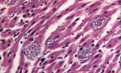
Trichinella spiralis * How acquired? (2 sources c what feature?) * Where do adult worms live, and what do they make and what does this do (2)? * How can it persist for years? * Sx? 3 * Dx? 3 |
* Acquired from eating pork or bear meat c encysted larvae * Adult worms in small intestinal mucosa; make larvae that enter blood and invade skeletal muscle * May encapsulate in muscle tissue and persist for years * Sx include eosinophilia, myositis, circumorbital edema * Dx c encapsulated larva in bx of sk muscle digested c artificial gastric juices, also bentonite flocculation, and IFA (has strong Ab response)
|
|
|
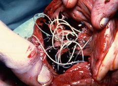
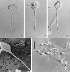
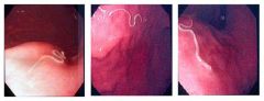
Dracunculus medinensis ("guinea worm") * Where do adult worms live? * When does infx become apparent? * How transmitted? 2 phases * Dx? * Tx? 2 |
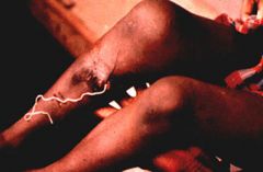
Adult worms live in suQ tissue -- Becomes apparent when female worm makes blister on skin surface which ruptures c H2O contact releasing motile larvae swarms -- Transmitted first when a copepod eats larva emitted by female worm; then humans eat the infected copepods -- Dx c appearance of female worm -- Tx by worm removal and drinking clean water |
|
|
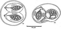
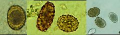
Capillariasis (Capillaria philippinensis) * How transmitted? * What is definitive host? * What 3 forms exist (different spp)? * What can autoinfection lead to? * 3 forms of sx? lethal dz? * Describe eggs? 2* Dx? |
* Transmitted by eating raw fish * Definitive host is fish-eating birds * Intestinal, hepatic and pulmonary spp exist * Autoinfection can cause hyperinfection (massive # of adult worms) * Can have intestinal, hepatic and pulmonary sx * Eggs have 2 polar plugs and striated shell * Dx c bx of respective organ causing sx
|
|
|
Anisakiasis * Transmission? * Sx? 3 * Tx? |
* Transmitted by eating raw fish * Cause eosinophilic granuloma (mimicking Crohn's disease), intestinal obstruction, coughing up worms * Tx c endoscopic worm removal
|
|
|
Entamoeba histolytica -- How is infection acquired? -- What lesion seen in intestinal mucosa? -- What extra-intestinal site affected and characteristic finding? -- Describe trophozoite karyosome, chromatin, and cytoplasmic finding? -- Describe cyst? -- Tx? 2 |
* Acquired by eating cysts * Flask-shaped ulcers in intestinal mucosa * May form liver abscesses filled c anchovy paste * Troph c central karyosome c even chromatin along nuclear membrane * Cyst has no more than 4 nuclei and smooth ends * Metronidazole and iodoquinole
|
|
|
Entamoeba coli -- Describe karyosome and chromatin in trophozoite -- How many nuclei in cyst? -- How to differentiate from E histolytica? |
* Troph c eccentric karyosome, irregular clumped nuclear chromatin * Cysts c up to 8 nuclei * Does not ingest RBCs
|
|
|
Entamoeba hartmanii -- Describe chromatin and karyosome (similar to what and how to differentiate (2)?) |
Central karyosome c even chromatin around membrane, but smaller than E histolytica and does not ingest RBCs |
|
|
Iodamoeba butschlii -- What found in cytoplasm? -- Stain? |
Prominent glycogen vacuole in cyst that stains c iodine |
|
|
Naegleria fowleri -- What part of brain affected?-- What seen on CSF wet mount? -- How cultured? -- What dz caused? |
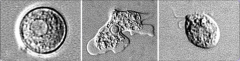
* Affects frontal lobes of brain * Trophozoites seen on wet mount * Cultured c lawn of E coli, which it ingests and leaves a trail -- * Causes Primary Amoebic Meningoencephalitis (PAM)
|
|
|
Acanthamoeba -- What 2 dzs? -- Dx? |
* Causes Granulomatous Amoebic Encephalitis (GAE) and ocular keratitis from infected contact lens solution * Diagnosed by finding organism on CSF and tissue samples
|
|
|
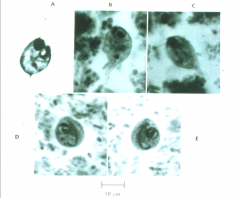
Giardia lablia (intestinalis) -- How acquired? -- What special about location? What special sampling technique can be used (and what other org can be similarly sampled?) -- Motility? -- Describe trophozoite? -- Describe cyst? (2) -- Are cysts durable (explain [2])? |
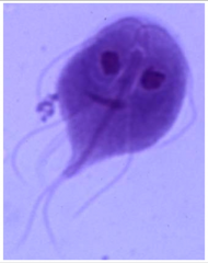
* Acquired by ingesting mature cysts, causing steatorrhea in hikers and daycare kids * Only protozoan found in duodenum and jejunum, so (like strongyloides stercoralis) can use the swallowed string technique to sample * Falling leaf motility * Troph is kite-shaped c central axostyle Cyst c 4 nuclei c 4 median bodies around axostyle * Cyst can live 3 months in H2O and resistant to chlorine
|
|
|
Dientamoeba fragilis -- Coinfection c what organism? Sx? Describe cyst -- Describe trophozoite -- Tx |

* Coinfection c Enterobius vermicularis common * Alternating periods of diarrhea and constipation * No cyst stage * Trophozoite c 2 nuclei c fractured karyosome * Tx c iodoquinol
|
|
|
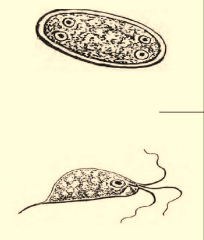
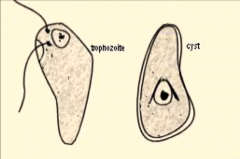
Chilomastix mesnili -- Sx Describe trophozoite (2)? Describe cyst? (3) |
* Not pathogenic to humans * Troph has 3 flagella at anterior end c cytosomal fibril that looks like a safety pin * Cyst looks like a nipple c a single nucleus and also the safety pin structure
|
|
|
Enteromonas hominis == Describe troph (3)? |
Troph c single nucleus c large central karyosome and no peripheral chromatin |
|
|
Retortamonas intestinalis -- Describe trophozoite? Describe cyst? 2 -- Sx? in what pts? |
* Troph c 2 anterior flagella * Cyst is pear-shaped c birds-beak fibrils around nucleus * Newborn girls at risk of getting respiratory disease and conjunctivitis from infected mothers at birth
|
|
|
Balantidium coli -- Sx? -- What special about it? -- Motility |
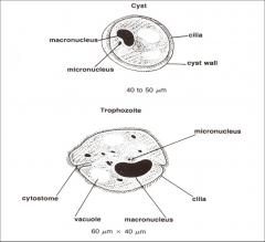
* Usually asymptomatic * Only ciliated parasite of humans * Boring motility
|
|
|
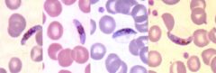
Leishmania -- Which stage found in humans? in vectors? What cells infected in humans and how does organism appear and how stains c PAS? -- Vector? (2) |
Amastigotes seen in humans, promastigotes found in arthropod vectors Monocytes and macrophages in humans infected c amastigotes, which have bar-like kinetoplast, PAS-negative (vs histoplasma) Vector is Phlebotomus sandfly (Old World) and Lutzomyia sandfly (New World) |
|
|
Cutaneous Leishmaniasis -- What species of leishmania? 4 (2 per world...) Nickname? |
Caused by Leishmania tropica / major in Old World, and L mexicana / brasiliensis in New World -- Nicknamed "Oriental sore"
|
|
|
Visceral Leishmaniasis -- What species of leishmania? (2 [1 per world]) -- What organs affected? (3) |
Caused by L donovani (Old World), or L infantum / chagasi (New World) -- Affects liver, spleen. and bone marrow
|
|
|
Chagas disease -- Parasite name? Reservoir? -- Vector? and how spread? -- Sx? 2 -- What stage found in organs? - What stage present in blood in early dz? -- Culture media? |
* Trypanosoma cruzi with rodent reservoir spread when reduviid (kissing) bug sheds T cruzi in feces and is rubbed into wound -- * Eventually causes heart failure and achalasia -- * Amastigotes found in organs -- * Trypomastigotes found in peripheral blood early in dz -- * Can be cultured on Novy-MacNeal-Nicolle (NNN) media
|
|
|
African trypanosomiasis -- Vector? -- Sx? (3 stages) -- 2 species? and which causes the worst disease but is more limited in distribution? |
Vector is tsetse fly * Causes chancre at site of bite, then hemolymphatic stage (fever, LNopathy), then the Sleeping sickness (encephalitis, somnolence) * Severe dz caused by T brucei rhodesiense but limited to SE and E Africa, whereas T brucei gambiense more widespread but does not cause severe dz |
|
|
Which 3 organisms detectable c modified acid fast stain? |
Cryptosporidium, cyclospora, and isospora |
|
|
4 Characteristics of arthropods |
Jointed appendages, chitinized exoskeleton, a hemocele, bilateral symmetry |
|
|
Ticks What 2 types and difference bwt them? |
Hard tick (capitulum visible on dorsal side) and soft ticks (capitulum not visible) |
|
|
Fleas How many legs and characteristics? |
3 pairs of powerful hairy legs designed for jumping and claw-like feet |
|
|
Monthly flea prevention medication? |
Lufenuron |
|
|
Flies How many body parts? How many legs? Wings? |
1 pair of antennae, head, thorax, segmented abdomen 2 pairs of wings and 3 pairs of legs |
|
|
Myiasis |
Infestation with fly larvae |
|
|
Facultative myiasis - 2 examples |
Feed on dead and decaying animal tissue - blowflies and fleshflies |
|
|
Obligatory myiasis - 1 example |
Species of fly c an obligate need to invade living tissue - Dermatobia hominis, the human Botfly |
|
|
Lice How many body parts? How is mouth adapted? How many wings? Legs? |
Have head. thorax, and abdomen, mouth adapted to pierce skin and suck blood No wings, 3 pairs of legs c claw-like feet |
|
|
Head louse (name?) Body louse (Name?) Crab louse (name) |
Pediculus humanus capitis Pediculus humanus humanus Phthiris pubis |
|
|
Pediculosis definition |
Infestation with lice |
|
|
Nit definition - how long does it last in this stage? |
Louse egg - stays in this stage for 24-27 days before passing into 3 nymph stages |
|
|
3 dz's assoc c lice |
Typhus, trench fever, and relapsing fever |
|
|
Sarcoptes scabei - nickname -- where does it live? |
aka the itch mite - burrows in epidermis of interdigital folds |
|
|
Crusted/ Norwegian scabies - in what kind of pt? - describe infx? |
Seen in immunocompromised pts - have generalized dermatitis c thousands of mites |
|
|
Demodex folliculorum - nickname -- where lives? |
- aka the itch mite - lives in hair follicles and sebaceous glands |
|
|
Hymenoptera - what kinds of insects? |
Group that includes bees, wasps, and ants |
|
|
Lactrodectus (Black widow spider) - what kind of rxn seen in bite? -- possible tx? |
Spider bite that can cause severe muscle spasms - antivenom is available |
|
|
Loxosceles (brown recluse spider) - what kind of rxn to skin bite? -- possible tx? |
Spider bite that causes necrotizing skin lesions that can continue to spread for weeks - tx is conservative and tissue should not be debrided for 3-6 wks |
|
|
Lepidopterism - definition -- 1 famous spp? |
Dermatitis caused by secretion of butterflies and moths -- Hylesia alinda moth caused dermatitis outbreak in Cozumel, Mex |
|
|
Erucism -- definition - 1 famous spp |
Dermatitis caused by contact c poison hairs of catepillars - Hemilevca mala (buck moth catebillars) causes cases in Louisiana |
|
|
Bed bugs - 2 spp |
Includes Cimex lectularius (cosmopolitan) and Cimex hemipterus (in tropics) |
|
|
Cestodes - nickname -- fun fact about their sexuality - how do they acquire nutrients? |
Includes tapeworms and ribbonworms - all are hermaphroditic - absorb nutrients through external tegument (surface), bc have no mouth or digestive tract |
|
|
Cestodes - all have what 3 body parts? |
All have a scolex, strobila, and proglottid |
|
|
Cestode - scolex Where found on worm? Function? Has what 2 structures? |
Seen on anterior end of worm Used to attach to intestinal wall Has suckers and hooks |
|
|
Rostellum |
Elongate protrusible thing in center of circular row of hooks in scolex of cestodes |
|
|
Cestodes - strobila - definition -- how do segments form? |
The entire body of an adult tapeworm - segments form from budding off posterior end of scolex in an area of germinal tissue |
|
|
Cestodes - proglottid - definition -- contain what reproductive structures when mature? |
One of the segments of a tapeworm - each contains male and female reproductive organs when mature |
|
|
Onchosphere / hexacanth embryo - definition |
Eggs of a cestode when has 6 tiny hooklets |
|
|
Coracidium - def -- in what spp? |
A ciliated hexacanth embryo in cestodes - D latum eggs develop to this stage and hatch in fresh water |
|
|
Cysticercoid - def -- has what structure? |
Larval stage of some tapeworms - has a small bladder-like structure c no fluid in which scolex is enclosed |
|
|
Cysticercus - nickname -- has what structure? |
- aka "bladder worm" in tenia spp - A thin-walled, fluid-filled bladder-like cyst that encloses a scolex |
|
|
Tenia saginata - nickname - intermiate host - definitive host - are eggs infective in humans? |
aka the beef tapeworm - intermediate host: cow -- definitive host: human - eggs are not infective in humans |
|
|
Tenia saginata - describe proglottid - describe scolex |
Gravid proglottid c >13 lateral uterine branches Scolex c unarmed rostellum and smooth surface |
|
|
Tenia solium - intermediate host -- definitive host - infective stage |
Intermediate host is pig Definitive host is humans Infective stage is cysticercus (encysted organisms) |
|
|
Tenia solium - describe proglottid - describe scolex |
Proglottid has <13 lateral uterine branches Scolex c armed rostellum (has multiple tiny hair-like hooks) and 4 suckers |
|
|
Cysticercosis - spp? -- cause -- finding -- intermediate host? |
In T solium, caused by eating eggs, which results in cysts c larvae in brain - humans are the intermediate hosts |
|
|
Hymenolepis nana - nickname - common? |
aka Dwarf tapeworm - MCC human tapeworm in US |
|
|
Hymenolepis nana - Definitive host - intermediate host |
Definitive host is man and rodents No intermediate host |
|
|
Hymenolepis nana - how acquired? - describe egg |
Acquired by eating eggs in feces from infected mice on water and food - egg c inner and outer shell c space in between (inner shell has 2 polar bow-like thickenings) |
|
|
Diphyllobothrium latum - definitive host -- intermediate hosts (2) - infective stage |
Definitive host: man Intermediate host: first a Copepod (in procercoid stage), and then freshwater fish (in plerocercoid stage) Infective stage: plerocercoid larva |
|
|
Diphyllobothrium latum Describe proglottid |
Is wider than long, with genital pore in center and uterus coiled in a rosette pattern |
|
|
Diphyllobothrium latum Describe Scolex |
An almond-shaped structure c 2 grooves and no true suckers which must embryonate in water |
|
|
Dipylidium caninum -- Intermediate host -- Infective stage -- Diagnostic stage |
Intermediate host is fleas on dogs and cats Infective stage is cysticercoid larva on infected fleas -- Diagnostic stage is pumpkin seed proglottid |
|
|
Echinococcus granulosus - definitive host -- intermediate host -- diagnostic stage |
Definitive host: Herd dogs and wolves Intermediate host: Sheep Diagnostic stage: Hyatid cyst in liver or lungs |
|
|
Echinococcus granulosus describe hyatid cyst |
Vesicular structure in liver or lungs c fluid that has protoscolices of potential tapeworms |
|
|
Echinococcus granulosus describe Hyatid sand |
Granular material material made of the free scolices, hooklets and daughter cysts |
|
|
Sparganosis 3 ways may be acquired |
Can be acquired by: -- Drinking water contaminated c copepods infected c procercoid -- Eating raw snakes or tadpoles infected c procercoid larval stage -- Rubbing raw frog or snake flesh in eyes |
|
|
Coenurus disease - name of worm - similar to what dz? |
Larval stage of Multicepts multiceps
- travels to brain or spinal cord producing dz similar to cysticercosis |
|
|
Hermaphroditic flukes - where do they live? |
Flukes c both sets of reproductive organs that live in the intestines |
|
|
Dioecious flukes - have how many reproductive organs? - where do they live? |
Unisexual (male and female) - live in the blood vessels of definitive host |
|
|
Trematodes (flukes) - what are the 3 larval forms? |
Larval forms include miracidium, cercaria and metacercaria |
|
|
Miracircadium - def - what does it infect |
Ciliated form of flukes c variant morphology from spp to spp - infects freshwater molluscs |
|
|
Fluke cercaria - def -- where do they come from? -- where do they invade and in which host? |
Free-living tailed form of flukes - released from snails into water -- invade tissues of second intermediate host; though can infect humans directly (schistosomes) |
|
|
Metacercaria |
Cercarial form of fluke after losing its tail |
|
|
Which spp of fluke does not have an operculum? |
All spp of fluke, except schistosomes have an operculum through which miracidium escapes |
|
|
Fasciolopsis buscki - nickname - - has what special characteristic |
aka Large intestinal fluke - is the largest and most pathogenic of the human intestinal flukes |
|
|
Fasciolopsis buski - what are the primary hosts? -- how acquired? - where does it cause dz? lethal? |
Pigs and humans are primary hosts - acquired by eating aquatic vegetation, on which metacercaria are encysted -- causes dz in duodenum and bile ducts, can be fatal |
|
|
Fasciola hepatica - geographic location -- how acquired -- definitive host |
Seen in sheep / cattle raising parts of world -- Acquired by ingestion of aquatic vegetation -- Sheep are the definitive host (human infx is a zoonotic dz) |
|
|
Fasciola hepatica - sx - describe egg |
Causes bile duct obstruction (from fibrosis) Eggs are large, c inconspicuous operculum and are unembryonated when deposited |
|
|
Clonorchis sinensis - nickname - how acquired |
aka Chinese or Oriental liver fluke Acquired by ingestion of metacercariae on the scales of freshwater fish |
|
|
Clonorchis sinensis Sx --- describe egg |
Causes biliary fibrosis and possibly cholangiocarcinoma -- small eggs c seated operculum, and a small knob at abopercular end |
|
|
Paragonimus westermani - nickname - how acquired |
aka the Oriental Lung Fluke - acquired by eating raw or undercooked infected shell fish |
|
|
Paragonimus westermani - how does it invade into lungs? -- sx |
Migrates through the diaphragm into lungs - causes hemoptysis |
|
|
Paragonimus westermani - describe egg |
Egg is large and unembryonated c thick abopercular end which has no knob |
|
|
Kayamata fever |
Systemic hypersensitivity rsn to schistosoma in tissue caused by circulating immune complexes |
|
|
Schistosomes - where do cercariae come from and what do they look like? -- how do they invade body? |
Snails make a fork-tailed cercariae that directly penetrates the skin to cause infx in humans |
|
|
Schistosoma mansoni - geographic location -- characteristic sx finding (2) -- where do adult worms liver (2) |
Found in the Nile Delta -- causes pipestem fibrosis and cirrhosis -- adult worms live in the portal system of liver and small venules of the inferior mesentery |
|
|
Schistosoma mansoni - describe egg |
Shell of the egg has a prominent lateral spine |
|
|
Schistosoma japonicum - where do adult worms live? -- describe egg |
Adult worms live in mesenteric veins and liver
Eggs are embryonated, have no operculum, and have a small inconspicuous spine |
|
|
Schistosoma hematobium - sx -- may cause what? |
Cause hematuria and may lead to bladder and ureteral SCC |
|
|
Schistosoma hematobium - where do adult worms live? - how to collect sample? |
Adult worms live in the venous plexus of the bladder -- best sample is the last bit of urine taken at mid-day |
|
|
Schistosoma hematobium - describe egg |
Eggs are thin, embryonated, and have a terminal spine |
|
|
Schistosoma intercalatum - describe egg |
Eggs c terminal spine, but are found in the feces and are larger than the eggs of the other spp that has eggs c terminal spine |