Reading...
![]()
Play button
![]()
Play button
![]()
Use LEFT and RIGHT arrow keys to navigate between flashcards;
Use UP and DOWN arrow keys to flip the card;
H to show hint;
A reads text to speech;
38 Cards in this Set
- Front
- Back
|
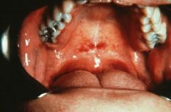
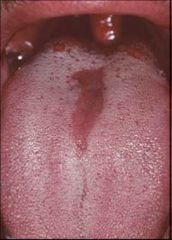
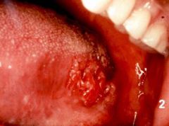
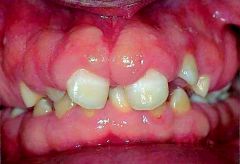
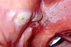
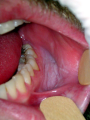
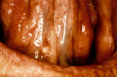
(this lesion does not rub off)
see below for differential diagnoses PRACTICE TESTS: (determine your differential list in the allotted time and modify your differential list based on the "patient" history) 1) GIVE 8 DIFFERENTIALS IN 2 MINUTES (patient does not smoke or use beetle nuts) 2) GIVE 6 DIFFERENTIALS IN 1MIN 30SEC (THIS LESION IS NOT HEREDITARY) |
Leukoedema – can leukoedema really look like this?
White sponge nevus Chronic hyperplastic candidiasis surgical scar – this DD is a stretch; it would be a weird scar (could be scars from aphthous major) frictional keratosis plaque lichen planus – take out this DD because it looks a lot more like reticular LP reticular lichen planus lichenoid drug reaction lupus erythematosus Isn't this considered a red/vascular lesion?? - lupus can cause annular leukoplakia areas, erythematous erosions and chronic ulcerations (according to book) snuff dipper's leukoplakia oral submucous fibrosis squamous cell carcinoma Additional Suggested Differential Diagnoses (add here if you think of any): Graft versus host disease (Dr. C considers GVHD to be erosive) Idiopathic leukoplakia Chronic ulcerative stomatitis (autoimmune) - (Dr. C considers this to be erosive) Contact stomatitis from cinnamon or amalgam (allergic) Erosive lichen planus (autoimmune) (Dr. C considers this to be erosive) |
|
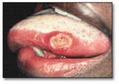
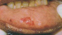
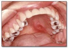
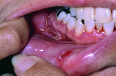
(long term ulcer)
1) GIVE 8 DIFFERENTIALS IN 2 MINUTES (NEGATIVE FOR FUNGAL INFECTIONS) |
Chancre (primary syphilis): Isn't this short term ulcer? No.
Gonorrhea Tuberculosis Actinomycosis North American Blastomycosis Cryptococcosis Histoplasmosis Coccidioidomycosis- Chronic traumatic ulcer Aphthous major Lupus erythematosus Squamous cell carcinoma Carcinoma-in-situ Behcet's Syndrome Additional Suggested Differential Diagnoses (add here if you think of any): nectrotizing sialometaplasia (pg 341 in book, most common at soft/hard palate junction, can also occur on tongue, retromolar pad) - Dr. C wants us to only consider it a palatal lesion Secondary syphilis mucus patch "Remember folks, it's a bacteria and not a viiiiirus, duh." Tertiary syphilis (gumma) Lymphoma: Non-Hodgkins: Note that Hodgkin's lymphoma rarely identifies in the soft tissue of oral cavity unless it's in stage 4 widely disseminated |
|
Instant ID
|
Nevus of Ota
|
|
1) GIVE 6 DIFFERENTIALS IN 1MIN 30SECS
|
Hereditary hemorrhagic telangiectasia
Infectious mononucleosis Measles (paramyxovirus) - no, I think Dr. C is considering measles to cause vesicular lesions - palatal petechiae are an additional symptom of measles (besides the vesicular lesions) Scarlet fever (streptococcus pyogenes) URI Suction Trauma Scurvey Thrombocytopenia, examples: Idiopathic thrombocytopenic purpura Autoimmune thrombocytopenic purpura chemotherapy induced thrombocytopenia Leukemia Additional Suggested Differential Diagnoses (add here if you think of any): hemophilia |
|
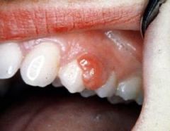
GIVE 5 DIFFERENTIALS IN 1 MINUTE 15 SECONDS
|
focal fibrous hyperplasia
pyogenic granuloma peripheral giant cell granuloma peripheral ossifying fibroma peripheral odontogenic fibroma focal hyperplastic gingivitis fibrosarcoma Additional Suggested Differential Diagnoses (add here if you think of any): Capillary hemangioma - maybe Traumatic Neuroma Granulation Tissue- can resemble pyogenic granuloma Malignant fibrous histiocytoma Neurofibroma Neurilemmoma |
|
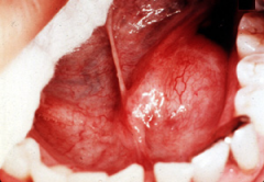
Instant ID
|
median rhomboid glossitis
|
|
DDx
|
Acinic cell carcinoma (this is the actual diagnosis)
Adenoid cystic carcinoma Mucoepidermoid carcinoma Pleomorphic adenoma Papillary cystadenoma lymphomatosum Oncocytoma Malignant mixed cell tumor Additional Suggested Differential Diagnoses (add here if you think of any): Hemangioma Hodgkin and Non-Hodgkin Lymphoma - just a guess |
|
GIVE TWO LISTS OF DIFFERENTIALS:
1) Consider these to be short term ulcers (GIVE 4 DIFFERENTIALS IN 1 MINUTE) 2) Consider these to be ulcers that were originally vesicular lesions that have already ruptured (GIVE 4 DIFFERENTIALS IN 1 MIN) |
1) short term ulcers
Cyclic neutropenia Acute trauma erythema multiforme Pyostomatitis vegetans Aphthous minor Proposed differential diagnoses and comments (in italics) 2) uclers that were originally vesicular lesions Recurrent intraoral herpes Acute primary herpetic gingivostomatitis Herpangina Measles (koplik spots) Herpes zoster (chicken pox) |
|
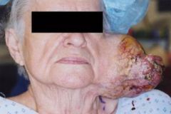
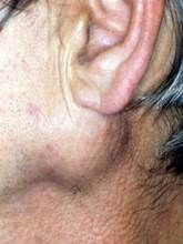
Try to list 1 hereditary condition, 2 infectious conditions and 4 neoplasms IN 1 MINUTE 45 SECONDS
|
Cervical lymphoepithelial cyst
Non-specific lymphadenitis Tuberculosis infection Cat scratch fever infection Cysticercosis Carotid body tumor Metastatic carcinoma Hodgkin lymphoma Non-Hodgkin lymphoma Lymphangioma Nasopharyngeal carcinoma Additional Suggested Differential Diagnoses (add here if you think of any): Submandibular gland sialadenitis (according to OMFS textbook) Sarcoidosis (according to OMFS textbook) Plunging ranula (according to OMFS textbook) |
|
DDx
DECIDE what category you think this lesion belongs to first. |
- I thought of this as an ulcerated lesion –
Gumma (tertiary syphilis) Tuberculosis Actinomycosis Histoplasmosis North American Blastomycosis Coccidioidomycosis Cryptococcosis Chronic traumatic ulcer Squamous cell carcinoma IF THIS WAS A "red lesion," then here are some potential differentials. BUT, What else could it be? Erythroplakia |
|
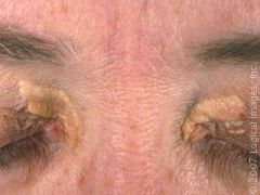
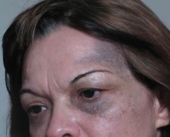
Instant ID
|
xanthelasma
|
|
TRY TO LIST 8 differentials IN TWO MINUTES and only have 3 neoplasms
|
Proposed differential diagnoses (add comments in italics):
Polycystic disease of the parotid Non-specific parotitis (example from book: after abdominal surgery) Cytomegalovirus infection Cysticercosis Epidemic parotitis (mumps) - not sure about this one; I think mumps is more generalized Obstructive sialolithiasis Sialadenosis - not sure about this one because I don't know what this looks like; is it more generalized? Amyloidosis Sarcoidosis Pleomorphic adenoma Monomorphic adenoma (more likely basal cell adenoma variant) Papillary cystadenoma lymphomatosum Oncocytoma Mucoepidermoid carcinoma Adenoid cystic carcinoma Acinic cell carcinoma Malignant mixed tumor Additional Suggested Differential Diagnoses (add here if you think of any): Polymorphous Low Grade Adenocarcinoma? - this generally occurs in minor salivary glands and rarely arises in major glands. |
|
DDx
|
Proposed differential diagnoses (add comments in italics):
Ludwig's angina Cysticercosis Dermoid cyst Ranula Mucous retention cyst Obstructive sialolithiasis in sublingual gland Chronic sclerosing sialadenitis Pleomorphic adenoma- but more likely to see this in Parotid? yes Oncocytoma Adenoid cystic carcinoma Osseous and cartilaginous choristoma Additional suggested differentials (list any that you can think of): Oral lymphoepithelial cyst - not sure about this one for this particular lesion |
|
1) GIVE 5 DIFFERENTIALS IN 1 MINUTE and 15 SECONDS (this male patient does not have a history of seizures)
|
Proposed differential diagnoses (add comments in italics):
Hereditary gingival fibromatosis Idiopathic gingival hyperplasia Non-specific hyperplastic gingivitis Plaque induced gingivitis - would likely be more red than this Puberty induced gingival hyperplasia Pregnancy induced gingival hyperplasia Diabetes induced gingival hyperplasia Drug induced gingival hyperplasia (cyclosporin, nifedipine, phenytoin- also verapamil, primidone) Leukemia - would likely be more red than this Multiple irritation fibromas (focal fibrous hyperplasia) - just a guess Additional suggested differentials: PRACTICE TEST: Patient Hx ELIMINATES pregnancy induced gingival hyperplasia and phenytoin induced hyperplasia |
|
|
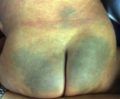
Proposed differential diagnoses (add comments in italics):
Hemangioma Ecchymosis Lupus erythematosus Erythroplakia Squamous cell carcinoma Idiopathic Additional suggested differentials: granulation tissue? |
|
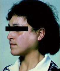
Instant ID
|
Sturge-Weber
bonus points: Encephalotrigeminal angiomatosis |
|
DDx
Remember: decide what category it is in first :) |
Epidermolysis bullosa
Erythema multiforme Pemphigus vulgaris Mucous membrane pemphigoid - Oral mucous membrane pemphigoid - Bullous pemphigoid - Cicatricial pemphigoid Erosive lichen planus Graft versus host disease Chronic ulcerative stomatitis Lupus erythematosus |
|
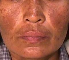
Instant ID
|
melasma
|
|
DDx
|
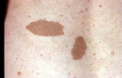
Ephelis
Actinic lentigo Lentigo simplex Cafe-au-lait spot Junctional nevus Other suggested differentials: Compound nevus Interdermal nevus Melanotic macule; This only occurs in the mouth. The equivalent on the skin is the ephelis |
|
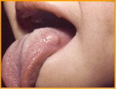
DDx
|
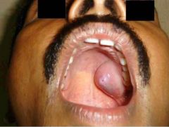
Neuroma of MEN IIB
Lingual thyroid nodule - on Dr. C's list, but not sure if it would look like this. Also this is located at the posterior tongue. Oral lymphoepithelial cyst Cysticercosis Amyloid nodule Neurilemoma Granular cell tumor Leiomyoma Rhabdomyoma Mucoepidermoid carcinoma Other suggested differentials: Most mesenchymal bumps and lumps can appear on the tongue (as mentioned in class) except for lipoma and osteoma. He mentioned that he will narrow some of the lumps and bumps out by giving us hints in the question. |
|
DDx
|
Physiologic pigmentation
Smoker's melanosis Addison's disease Post-inflammatory hyperpigmentation Drug related hyperpigmentation due to AZT, anti-malarial drugs, minocycline |
|
Instant ID
|
mongolian spot
|
|
DDx
|
Cysticercosis
Dental abscess Traumatic neuroma Kaposi sarcoma Burkitt lymphoma Pleomorphic adenoma Mucoepidermoid carcinoma Adenoid cystic carcinoma Polymorphous low grade adenocarcinoma Other suggested differentials: exostoses Basically any lesion that has fibrous tissue, nuerons, and blood vessels can be put in this differential: angioma/angiosarcoma neurofibroma solitary fibrosarcoma, etc. |
|
|
Multiple neurofibromatosis
Cysticercosis Focal fibrous hyperplasia (irritation fibroma) Mucocele - doesn't really look like it, but fits the category. I agree... Mucous retention cyst Fibroma Neurofibroma Lipoma/Liposarcoma Leiomyoma/Leiomyosarcoma Rhabdomyoma/Rhabdomyosarcoma Mucoepidermoid carcinoma Polymorphous low grade adenocarcinoma Other suggested differentials: I originally thought of this as a papillary lesion BUT many people have thought of this as a lump. My initial instinct was that it was an irritation fibroma, which I thought was a papillary lesion. BUT, papillary lesions are actually "papillary" according to Dr. C because of their histological traits. So, now I think this is a LUMP/BUMP category. Thanks for the clarification from many astute members of our class :) Very helpful :) |
|
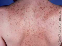
Instant ID
|
cafe au lait spots with some ephelides
|
|
|
White sponge nevus
Chronic hyperplastic candidiasis Frictional keratosis Surgical scar/graft Oral submucous fibrosis (usually on gingiva only?) - no, it usually affects soft tissues of buccal mucosa, lips, soft palate, and occasionally the pharynx Lichenoid drug reaction Plaque lichen planus Idiopathic Leukoplakia Squamous cell carcinoma Other suggested differentials (list any that you think of): Did anyone see this as a papillary lesion? Something resembling Proliferative verrucous leukplakia? Can someone clarify? Parts of it DO look like it could be papillary, if something like this comes up, he'll hopefully guide us in one direction or another. : ) |
|
(This lesion has been present for more than 3 weeks)
DDx |
Tertiary syphillis - gumma
Tuberculosis infection associated ulcer Actinomycosis infection associated ulcer Gonorrhea associated ulcer Deep fungal infection - North American Blastomycosis - Histoplasmosis - Cryptococcosis - Coccidioidomycosis Necrotizing sialometaplasia Chronic traumatic ulcer - finger nail trauma (Factitious injury) Burkitt lymphoma Non-hodgkin lymphoma Mucoepidermoid carcinoma Adenoid cystic carcinoma Squamous cell carcinoma Other suggested differentials (list any that you think of): Primary and second syphillis - not sure if these can be on the palate Agranulocytosis |
|
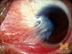
Instant ID
|
pterygium
|
|
DDx
|
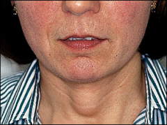
Polycystic disease
Epidemic parotitis Bacterial parotitis (ex. after an abdominal surgery [according to the book]) CMV infection Obstructive sialolithiasis Sialadenosis (obesity, bulimia, alcoholism, diabetes) Sjogren Syndrome Pleomorphic adenoma Malignant mixed tumor Monomorphic adenoma (basal cell adenoma variant) Mucoepidermoid carcinoma Adenoid cystic carcinoma Acinic cell carcinoma Papillary cystadenoma lymphomatosum Oncocytoma |
|
(this does not rub off)
|
Leukoedema
White sponge nevus Chronic hyperplastic candidiasis - could be Surgical scar/graft Oral submucous fibrosis Snuff dipper's leukoplakia Lichenoid drug reaction - maybe? Plaque lichen planus Idiopathic leukoplakia Other suggested differentials: Nicotine stomatitis? - only on the palate |
|
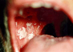
Patient has a white lesion that rubs off.
DDx |
Acute pseudomembranous candidiasis
Bacterial mucous patch Materia alba Chemical burn from mouth wash, denture paste Aspirin burn Radiation mucositis Collapsed bullae - not likely |
|
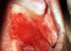
(please focus on the most obvious and largest lesion)
DDx |
Atrophic candidiasis
Radiation mucositis Xerostomia Lichenoid mucositis (ex. lichenoid drug reaction) Allergy Erythroplakia Squamous cell carcinoma Other suggested differentials: Ecchymoses |
|
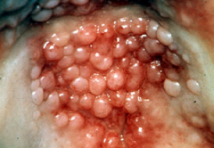
DDx
|
Inflammatory papillary hyperplasia
Condyloma acuminatum Squamous papilloma Verruca vulgaris Focal epithelial hyperplasia Other suggested differentials: Nicotine stomatits? it's a white lesion that doesn't rub off but it's supposed to have papules too. |
|
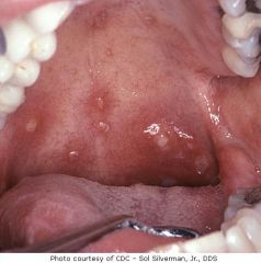
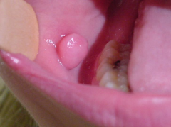
CASE: Patient reports no recollection of how long he has had this single, nodular lesion. Patient has never eaten raw pork.
GIVE 6 differentials in 1.5 minutes :) |
Oral lymphoepithelial cyst
Amyloid nodule Dermoid cyst Lipoma/Liposarcoma Leiomyoma/Leiomyosarcoma Rhabdomyoma/Rhabdomyosarcoma Solitary fibrous tumor Neurofibroma Osseous and cartilaginous choristoma Other suggested differentials: Cysticercosis (ruled out by the patient history) Ranula? |
|
LIST 5 differentials in 1 minute and 15 seconds...
|
Thyroglossal tract/duct cyst
Cysticercosis Goiter Hashimoto's thyroiditis Graves disease Thyroid cancer (ex. papillary carcinoma, medullary) Dermoid cyst |
|
LIST 6 differentials and only three of them can be neoplasms
|
Cervical lymphoepithelial cyst
Non-specific lymphadenitis (ex. from a dental infection) Tuberculosis infection Cat scratch fever infection Cysticercosis Carotid body tumor Metastatic carcinoma Hodgkin lymphoma Non-Hodgkin lymphoma Lymphangioma Nasopharyngeal carcinoma Lipoma/liposarcoma Rhabdomyoma/Rhabdomyosarcoma Leiomyoma/Leiomyosarcoma Other suggested differentials: Branchial cleft cyst Non-specific lymphadenitis |
|
Patient also has multiple bullae in the contralateral vestibule. Lesions in right vestibule, left vestibule (not shown) and lip mucosa are related.
DDx |
Epidermolysis bullosa
Mucous membrane pemphigoid - Oral mucous membrane pemphigoid - Cicatricial pemphigoid - Bullous pemphigoid Pemphigus vulgaris Chronic ulcerative stomatitis Graft versus host disease Erythema multiforme Erosive lichen planus Other suggested differentials: Lichenoid Drug Rxn? |
|
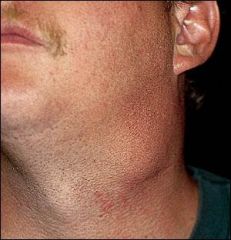
Pt chief complaint: "Woof woof!"
|
Cervical lymphoepithelial cyst
Non-specific lymphadenitis (ex. from a dental infection) Tuberculosis infection Cat scratch fever infection-got owned by cat Cysticercosis Carotid body tumor Metastatic carcinoma Hodgkin lymphoma Non-Hodgkin lymphoma Lymphangioma Nasopharyngeal carcinoma Lipoma/liposarcoma Rhabdomyoma/Rhabdomyosarcoma Leiomyoma/Leiomyosarcoma Other suggested differentials: Branchial cleft cyst this is the same as lymphoepithelial cyst Non-specific lymphadenitis |