Reading...
![]()
Play button
![]()
Play button
![]()
Use LEFT and RIGHT arrow keys to navigate between flashcards;
Use UP and DOWN arrow keys to flip the card;
H to show hint;
A reads text to speech;
25 Cards in this Set
- Front
- Back
|
What innervates the deltoid muscle? What is deltoid muscle's function?
|
C5 Axillary Nerve
Shoulder flexion, extension, and abduction |
|
|
What innervates the biceps? What is its function?
|
C5-C6 musculocutaneous nerve
Forearm flexion at elbow, supinator of forearm |
|
|
What nerve is responsible for the biceps reflex?
|
Primarily C5, some C6
(even a slightly diminished reflex indicates pathology) |
|
|
Describe dermatome of C5. What is C5?
|

lateral arm
C5 = axillary nerve |
|
|
Muscle test for C6?
Name the muscles and the nerves |
Neither test is pure;
- wrist extensor group innervated by C6-C7, radial nerve - three muscles: extensor carpi radialis longus and brevis (C6) and extensor carpi radialis (C7) - biceps innervated by C5-C6 (musculocutaneous nerve) |
|
|
C6 reflex?
|
Brachioradialis (only C6) - tested proximal to the wrist where the muscle becomes tendinous just before it inserts into the radius
Biceps reflex (C5 - C6) - |
|
|
Sensation testing for C6
|
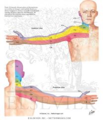
thumb and lateral arm
|
|
|
Triceps muscle testing: nerve?
|
C7, radial nerve
|
|
|
Wrist flexor group: nerve?
|
C7, median and ulnar nerves
wrist flexor group composed of two muscles: flexor carpi radialis (median nerve) and flexor carpi ulnaris (ulnar nerve) Flexor carpi radialis (C7) is more important of these two muscles, since it actually powers most of wrist flexion) Flexor carpi ulnaris, which is primarily innervated by C8, is less powerful |
|
|
Finger extensors: which nerve?
|
C7, Radial nerve
Finger extension performed by three muscles - extensor digitorum communis, extensor digiti indicis, and extensor digiti minimi All above groups have some C8 innervation (though predominantly C7) |
|
|
Triceps reflex: which nerve?
|
C7, tap its tendon where it crosses the olecranon fossa at the elbow
|
|
|
Sensation distribution of C7
|

Middle finger (occasionally, middle finger sensation also supplied by C6 and C8
|
|
|
Muscle testing of C8
|
Finger flexors
- two muscles which flex fingers are flexor digitorum superficialis (PIP joint) and flexor digitorum profundis (DIP joint) |
|
|
Sensation distribution of C8
|

pinkie finger
|
|
|
Finger Abductors: nerve?
|
ulnar nerve, T1
1) dorsal interossei and 2) abductor digiti quinti |
|
|
Sensation of T1
|

Medial arm: medial brachial cutaneous nerve
medial side of the upper half of the forearm |
|
|
Muscle group of L4
|
Tibialis anterior: L4, deep peroneal nerve
test: offer resistance to dorsiflexion and inversion by pushing against the dorsal and medial aspects of the head and the first metatarsal bone. |
|
|
Patellar reflex: what nerve?
|
L4
deep tendon reflex, mediated through nerves from L2-L4, but mostly through L4 |
|
|
Sensation of L4
|

covers medial side of the leg.
knee represents the division between the L3 dermatome (above) and the L4 dermatome (below) |
|
|
L5 muscle
|
Extensor hallucis Longus: deep peroneal nerve
Gluteus medius: superior gluteal nerve --> abduct leg as patient is lying on side Extensor digitorum Longus and Brevis: deep peroneal nerve --> flex toes |
|
|
Reflex of L5
|
diffiicult to obtain tibialis posterior muscle L5 reflex
|
|
|
Sensation of L5
|
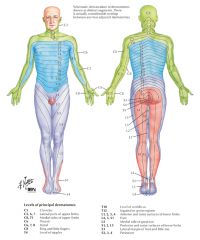
covers lateral leg and dorsum of foot
|
|
|
Muscles of S1
|
Peroneus longus and Brevis: S1, superficial peroneal nerve --> secure patient's ankle, have him plantarflex and evert his foot, and oppose his motion by pushing against the head of the fifth metatarsal with the palm of your hand.
Gastrocnemius-Soleus Muscles: S1, S2, Tibial nerve --> no legitimate manual test for it Gluteus maximus: S1, inferior gluteal nerve --> have patient lie prone on the exam table with knees flexed and hip extended. Resist hip extension and palpate the gluteus maximus for tone. |
|
|
Reflex of S1
|
Achilles tendon reflex: put achilles tendon into a slight stretch by gently dorsiflexing the foot. Then strike the tendon to induce a sudden, involuntary plantarflexion of the foot,.
|
|
|
Sensation of S1
|

covers lateral malleolus and lateral side and plantar surface of the foot.
|