![]()
![]()
![]()
Use LEFT and RIGHT arrow keys to navigate between flashcards;
Use UP and DOWN arrow keys to flip the card;
H to show hint;
A reads text to speech;
29 Cards in this Set
- Front
- Back
|
What is the purpose of extraoral imaging? |
|
|
|
What are other reasons for extraoral radiography? |
|
|
|
What do orthodontists use extraoral radographs for? |
|
|
|
What do Prosthodotists use extraoral radographs for? |
|
|
|
What do oral surgeons use extraoral radographs for? |
|
|
|
What are the types of Extraoral Radiographs of the Maxillofacial Region? |
|
|
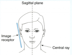
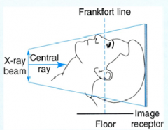
Which EO Radiograph is this? What does it examine? |
- Body and ramus - Coronoid process - Condyle |
|
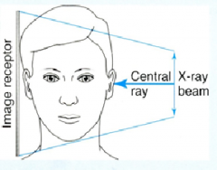
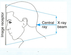
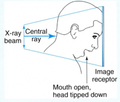
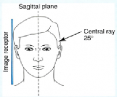
Which EO Radiograph is this? What does it examine? |
|
|
|
|
|

|
|
|
|
|
|
|
|
|
|
|
|
|
Why is a screen film used in conjunction with intensifying screens? |
to maintain low dosage |
|
|
How must extra oral films be handled? |
carefully, without plastic or latex gloves (cause static), on the edges (cause finger prints), in appropriate lighting because (not packaged separately so will ruin all films exposed to light) |
|
|
What is the intensifying screen? what does it do? |
|
|
|
What are the characteristics of the intensifying screens? |
|
|
|
What does sensitivity and image sharpness depend on? |
- large crystals produce a less sharp image
-Thicker emulsion = faster speed screen = less radiation but also less sharp image
|
|
|
How is the correct side of the film identified? |
|
|
|
What should be checked for when inspecting the cassettes and screens? |
|
|
|
What are grids? How are they used? What do they require? |
|
|
|
What are exposure factors based on? |
|
|
|
What are three types of new techonolgy? |
Tomography - Simultaneous movement of the x-ray source and the image receptor to image structures within a selected plane of tissue Computed tomography (CT scan) - Patient lies on a table with head in scanner x-ray beam rotates 360 degrees around the head Cone beam computed tomography - Patient seated or standing upright while machine rotates around head |
|
|
How many exposures does the Cone Beam Computed Tomography expose within the 360 degrees? What does the computer created from the images? What are the images for? |
|
|
|
What is the Cone beam computed tomography? What does it acquire and how? |
x-rays taken with a cone-shaped x-ray beam acquires 3-D information by the source of radiation rotating around the head of the patient |
|
|
What are the various sizes of Field of view? |
Large medium and small |
|
|
What are the three planes of the body that the CBCT images? |
|
|
|
What are advantages of the CBCT? |
- 42 mSv for maxilla and 75 mSv for mandible - pandoramic exposure is 7 mSv
|
|
|
What are disadvantages of CBCT digital radiography? |
|