![]()
![]()
![]()
Use LEFT and RIGHT arrow keys to navigate between flashcards;
Use UP and DOWN arrow keys to flip the card;
H to show hint;
A reads text to speech;
14 Cards in this Set
- Front
- Back
|
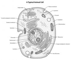
Label and drawn an animal cell
|
|
|
|
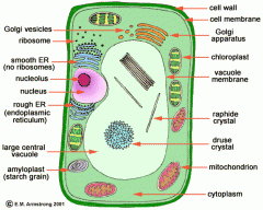
Label and draw a plant cell
|
|
|
|
, How are proteins produced?
|
proteins made at the ribosomes on rough endoplasmic rreticulum proteins are folded and processed transported to Golgi apparatus in vesicles proteins undergo further processing enter more vesicles to be transported around the cell leave by exocytosis |
|
|
Describe the cytoskeleton
|
protein threads arranged in microfilaments and microtubules has four functions -support cell's organelles -strengthen cell and maintain its shape -transport of organelles and materials |
|
|
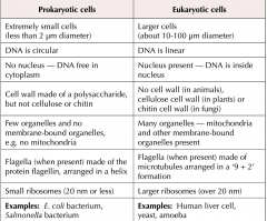
Compare prokaryotes and eukaryotes
|
|
|
|
What is the formula for magnification? |
magnification= image size/ objeect size |
|
|
Define magnification |
how many times bigger the image is than the specimen
|
|
|
What is resolution? |
How well a microscope distinguishes between two points that are close together |
|
|
How do you convert between millimetres, micrometres and nanometres |
mm x1000 micrometre x1000 nm |
|
|
Describe a light microscope |
use light have a lower resolution than electron microscopes maximum resolution of 0.2 micro metres maximum magnification x1500 look at whole cells or tissues |
|
|
Describe a laser scanning confocal microscope |
laser beams can a specimen tagged with fluorescent dye beam focused through a lens aimed at a beam splitter beam is split some, some of light hits dyes on specimen causing them to fluoresce focused through pinhole onto detector produces clearer image |
|
|
Describe transmission electron microscope |
Transmission electron microscope electromagnets focus a beam of electrons transmitted through a specimen 2D images denser parts of specimen absorb more electrons so appear darker max res 0.0002 microm max mag x1000000 |
|
|
Describe a scanning electron miceoscope |
scan a beam of electrons across the specimen
knocks off electrons from specimen gathered in cathode ray tube to form an image can be 3D max res 0.002 microm max mag x 500,000 |
|
|
How can you prepare a microscope slide? |
Dry mount- hairs, pollen, flowers Wet mount- living samples |