![]()
![]()
![]()
Use LEFT and RIGHT arrow keys to navigate between flashcards;
Use UP and DOWN arrow keys to flip the card;
H to show hint;
A reads text to speech;
321 Cards in this Set
- Front
- Back

Observations about Axons of Retinal Ganglion Cells |
They are radially symmetric in two places (fovea and optic nerve). There is little or no overlap (they are very organized). |
|
|
How do axons of the retinal ganglion know where to go? How do they connect with their targets? |
Axon growth and guidance. |
|
|
How many bytes of information does DNA provide for wiring? |
750 megabytes. |
|
|
Does it matter if axon wiring is correct? |
To a point, no. Plasticity and experience can allow us to correct wiring when done wrong. However, there still is a way that most wiring is done. |
|
|
Retinotopy |
A map of the visual field is preserved in the brain. |
|
|
What happens when you mess with the retinotopy? |
When you mess with it, it disrupts your ability to see the image. Plasticity cannot fix this. |
|
|
2 Mechanisms for Axon Growth |
1. Predetermined 2. Functional Selection of Exuberant (extra) Connections |
|
|
Predetermined Axon Growth |
There is a clear plan for what connections are needed. |
|
|
Functional Selection of Exuberant |
Make many connections and prune away the less useful ones. |
|
|
Which axon growth mechanisms are used? |
Both; mechanisms are in play in us most of the time. |
|
|
Why are newts unique? |
At the optic chasm, their axons all cross instead of half crosses and half remaining on the same side. This give newts very strong peripheral vision. |
|
|
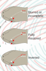
Roger Sperry's Experiment (Action, 3 possible results, actual result, and conclusion) |
Action: Cut optic nerve in newts and turned the left eyeball 180 degrees. 3 Possible Results: incomplete degeneration of vision, axons follow paths straight back and vision is fine, or axons cross back to their original connections and vision is inverted. Actual Result: Inverted vision Conclusion: Even with inverted vision, newts could start to be trained that everything is switched. This shows that axon connections are prewired, but also that plasticity can allow for adaptation in the case of (in this case intentional) mistakes. |
|
|
Point to point connections are like... |
seats on an airplane; particular neurons go particular places. |
|
|
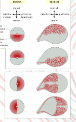
Retina to Tectum Experiment |
Played around with removing sections of RGC. Axons left on bottom of retina = axons on top of tectum Axons left on top of retina = axons on bottom of tectum Axons left on left side of retina = axons on right side of tectum Axons left on right side of retina = axons on left side of tectum Conclusion: Certain neurons are predetermined to go to certain places. There is clearly a specific map. |
|
|
How are axon maps achieved? |
Different gradients of chemicals is what axons look for when growing. Axon guidance molecules. |
|
|
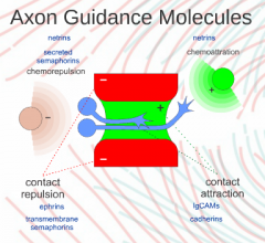
4 Kinds of Axon Guidance Molecules |
1. Contact Repulsion 2. Contact Attraction 3. Chemoattraction 4. Chemorepulsion |
|
|
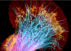
Structure of a Growing Neuron |

Cell body, axon, and growth cones that extend looking for synapses. |
|
|
Contact Repulsion Molecules |
Growth cone touches one of these and then will go the other way. Imagine setting up a channel that the axon must remain inside of. Note: contact MUST occur for this to work |
|
|
Example of Contact Repulsion Molecules at Work |
Dendrites; they want to spread out, so if they touch they want to go the other direction. |
|
|
Contact Attraction Molecules |
Growth cone touches one of these and will continue to grow often along it |
|
|
What commonly has contact attraction? |
Other axons. |
|
|
What kind of molecules are contact attraction and repulsion molecules? |
Short range molecules that are membrane tethered. |
|
|
What kind of molecules are chemoattraction and chemorepulsion molecules? |
Long range secreted molecules. |
|
|
Chemoattraction |
A factor that when released causes axons to grow towards it. Axons sense the gradient and seek out the higher gradient. |
|
|
Chemorepulsion |
A factor that when released causes axons to grow away from it; they sense the gradient and seek away from higher gradient. |
|
|
How are images projected on the retina and then to the tectum? |
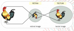
|
|
|
Describe the Visual of the Two Kinds of Axon Growth Mechanisms |
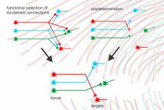
|
|
|
Summary Diagram of Axon Guidance Molecules |
|
|
|
Newt Experiment: Shows axon connections are prewired. |
|
|
Retina to Tectum Experiment: Shows certain neurons are predetermined to go to certain places. There is clearly a specific map. |
|
|
How does ~Gigabyte of info in the DNA specify 1014 connections in our brain? |
General, overarching question. |
|
|
Transection |
To cut across (?) |
|
|
Regeneration in Amphibians |
Amphibians can regenerate nerve connections after damage by re-extending truncated axons back t their targets. |
|
|
Chemoaffinity Hypothesis |
"It seems a necessary conclusion from these results that cells and fibers of the brain and (spinal) cord must carry some kind of individual identification tags, presumably cytochemical in nature, by which they are distinguished one from another almost, in many regions, to the level of the single neuron; and further, that the growing fibers are extremely particular when it comes to establishing synaptic connections, each axon linking only with certain neurons to which it becomes selectively attached by specific chemical affinity." |
|
|
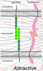
Ig CAMs |
Cell adhesion molecules for contact mediated attraction. |
|
|
Cadherins |
Cell adhesion molecules for contact mediated attraction. |
|
|
Semaphorins |
Evolutionarily conserved and widely used axon guidance cues, they consist of secreted and transmembrane variants and mostly act as repellants. Some transmembrane variants can also act as receptors. |
|
|
Netrins |
A long distance axon guidance molecule. |
|
|
Epherents |
The most common contact repulsion molecules. |
|
|
How do gradients work? |
Chemoattractants: target secretes a protein. The protein is at the highest concentration nearest the target neuron, so an axon grows towards high concentration and away from low concentration. Chemorepulsion can also be released; grows towards low concentration and away from high concentrations. |
|
|
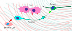
Where is an example of a place a chemorepulsive protein would be released from? |
Stromal cell. |
|
|
2 Uses of Gradients |
1. Steer Growth Cone 2. Establish Point to Point Connection |
|
|
How do gradients establish point to point connections? |
Short range; allows neurons to info the exact spot along a gradient. When they hit a specific point in the gradient, they stop. This is their point, thus point to point connection. |
|
|
Early Experiment 1 on Point to Point Connections |
Take anterior tectum, heated anterior tectum (to denature proteins), posterior tectum, and heated posterior tectum and see what grows. Results: all four options showed similar growth because there is no "choice" for where to grow (whole plate is the same part of tectum). |
|
|
Retina to Tectum Connection |
Nasal retina connects to posterior tectum.
Temples retina connection to anterior tectum. |
|
Label the Kinds of Axon Guidance Molecules |
Stomal cell - chemorepulsion Glial cell - contact repulsion Target cell - chemoattraction Not pictured - chemoattraction |
|
|
Results from Early Experiment 2 on Point to Point Connections |
PAP with temporal axons - only grew on anterior stripe PAP heated with temporal axons - grows all over APA with nasal axons - grows all over APA heated with nasal axons - grows all over. |
|
|
Possible Explanations for Results from Early Experiment 2 on Point to Point Connections |
1. Anterior is attracted to temporal axons, but this attraction denatures when whole thing is heated (temporal axons need to have the receptor for an anterior chemoattraction if this is the case). 2. Maybe the temporal axons have a receptor for a repulsive agent in the posterior tectum. (nasal axons don't care because they don't have |
|
|
Determining the Correct Hypothesis for Results from Early Experiment 2 on Point to Point Connections |
TEST: only heat or denature either P or A in the PAP dish. Results: When you only heat or denature A then you get results like if you hadn't heated it at all (only growth on A). When you only heat denature P then there is growth all over in. This shows us that P has the repellent and when we heat denature P it stops working resulting in total, not point to point growth. |
|
|
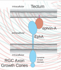
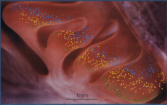
What does a retina stained for eph receptors look like? |
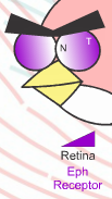
Nasal is white and temporal is stained dark. |
|
|
What does a stain of the tectum for ephrin (the ligand) look like? |
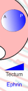
Similar gradient as retina; dark in posterior and none in anterior. |
|
|
Explanation of the Retina/Tectum Gradient |
Ephrin is repellant and the tectum has the repellant; repellent is in the posterior area. Eph is a trans-membrane receptor; retina RGC axon growth cone shave these receptors to sense the gradient of ephrin. Nasal to posterior Temples to anterior; lots of eph receptors prevent temple axons from ending up in the posterior. |
|
|
Eph Facts |
Eph is/has a kinase, so when it binds to ephrin it signals the cell causing the axon to do things like turn away, slow down, or even stop growing altogether. |
|
|
Forward Signaling |
When an eph receptor binds an ephrin and the cell responds; really both cells respond! |
|
|
Axon Growth Competition Experiment |
Gradients of eph receptors and ligands are known. Set up: change the gradient of eph receptors so that now nasal axons have a ton of eph receptors (when they usually have none and now they have more than temple axons). Prediction: even the smallest amount of ephrin will repel them so they should grow far to the anterior side. Actual Result: everything remains evenly spread just switched around a bit. This shows that competition between axons can cause different sorting rather than just the whole group moving to the anterior. |
|
|
What does eph/ephrin look like? |
|
|
|
Axon Growth Competition Experiment Visual |
|
|
|
Two Gradient Experiment (?) |
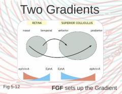
There must also be a nasal axon gradient that goes in the opposite direction. This allows for precise axon to tectum matching. |
|
|
Spinal Cord Injuries and How to potentially resolve them! |
Lesion cuts axon in spinal cord. If we could put the right chemoattractants in the spinal cord then we should be able to get the brain neurons to extend into the spinal cord which would allow the body to work around the lesions. If we knew the correct receptors then we could potentially do this. We need more brains to be donated to science! |
|
|
IgCAMS |
Cell adhesion molecules; important chemoattractants |
|
|
Cadherins |
Calcium dependent cell adhesion molecules that bind when calcium is present; important chemoattractants |
|
|
What happens when you remove IgCAMs and cadherins? |
Neurons do not grow at all. |
|
|
Are axon paths predetermined? |
Some are
|
|
|
Axon Guidance Summary (8) |
Some axon paths are predetermined. Point to point connections. Proteins mediate and some are membrane bound. Ligand/receptor gradients. Relative (not absolute). Competition. Attraction, cell adhesion. Functional selection. |
|
|
GPI |
Liquid anchor that attaches an extracellular protein to the cell membrane; can be released. |
|
|
Forward Signaling |
Normal signaling. |
|
|
Reverse Signaling |
The process by which a protein that "normally" functions as a ligand functions as a receptor and a protein that "normally" functions as a receptor functions as a ligand. |
|
|
FGF |
Growth factors in signaling pathways (?) |
|
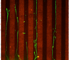
Hypotheses for why axons grow as pictured: |
Attractive Hypothesis: There are cell adhesion molecules all over the black area. If you start to leave the black area, you want to go back because you like the black. Repulsive Hypothesis: Ephrin is on the red and the neurons growth cone has eph receptors, so as it grows it is repulsed by the ephrin keeping it in the center. Note that repulsion is caused by CONTACT, so axon veers towards red but it then repulsed by the red once contact is made. |
|
|
What does attractive molecule bonding look like? |
Cell adhesions molecules from separate neurons bind. |
|
|
What does repulsive molecule bonding look like? |
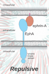
Ephrin from one neuron binds to eph receptor on another neuron. |
|
|
Why do growth cones/axons not get stuck when they bind to adhesion molecules? |
The binding does stop the neuron. However, the neuron keeps growing so new axon continues to grow and bind. Growth cone is what reaches out in front, but what is left behind (axon) remains attached. This method is slightly more common than repulsive, which is why repulsive is more common. |
|
|
Which kind of molecule bonding is more common (attractive or repulsive)? |
Repulsive. |
|

What experiment could you do to test between the attractive and repulsive hypotheses for axon growth as pictured? |
Really consider one.... |
|
|
Growth Cone Structure Parts |
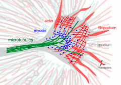
Lamellipodium, filopodium, receptors, myosin, actin, and microtubules. |
|
|
Lamellipodium |
The veil out in front of the growth cone; very thin and dynamic. Connects everything together. Made of actin in a lattice structure. |
|
|
Filopodium |
Thread like appendages; fingers that explore what is far ahead of growth cone and axon. They move around a lot and are made of bundles of actin. |
|
|
Receptors |
On the ends of filopodium; they are the first structure that explores out far ahead. |
|
|
Myosin |
Helps actin move around. |
|
|
Microtubules |
Go all the way down the axon and make up the main structure of the growth cone. They come out slightly into the growth cone. As it builds, more microtubules need to be built to lengthen it out. |
|
What is each color represent? |
Purple - actin Yellow - myosin Green - tubulin |
|
|
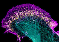
What do you see in growth cone video? |
Filopodium are all over the place; they sort of wave back and forth. Lamellipodium look like they are trying to go backwards - retrograde flow. |
|
|
Red = actin Yellow = myosin Blue = tubulin |
|
|
Growth Cone Turning Experiment |
Label individual filopodium so we can tell them apart. Hypothesis: when we put in long range attractant, after some time growth cones will turn towards bottom while still growing. Result: If there is a repellant or no attractant than growth cone filopodium on opposite side from attractant collapse because of retrograde flow. Filopodium on the side of the attractant turn and get locked in, so additionally we build new filopodium to allow us to turn more. In the diagram, the blue filopodium is the leading filopodium because it is most locked in, so not tubulin gets built in its direction (the direction of the leading filopodium) and everything else gets built around it. |
|
|
What is growth cone turning really? |
It is really just rebuilding in a new direction (the direction of the leading filopodium). |
|
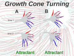
Which more accurately represents growth cone turning? |
B - far side retracts and new filopodium are built on the near side because the other ones get locked in. |
|
|
How do filopodium release from adhesion with attractant? |
They have to go up, out, and away. Growth cones can get away from adhesion (even if it is hard), but along the axon is where adhesion molecules are truly stuck and bound together. |
|
|
What are the filaments of actin constantly doing? |
Pulling back towards the growth cones' central domain, so just to remain the same length we have to continuously repolymerize the leading edge. |
|
|
How do we repolymerize the leading edge in growth cones? |
G actin is sent out there as growth. |
|
|
How do we extend growth cones? |
When extending, myosin's get inhibited and polymerization increases.
|
|
|
Where is the central domain vs. peripheral domain of the growth cone? |
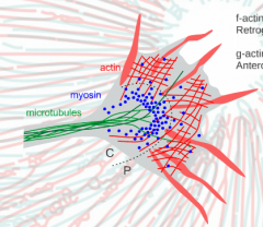
C = central P = peripheral |
|
|
How do we retract growth cones? |
When retracting, myosin's pull back and we have less polymerization. |
|
|
Actins and Their Flows |
F Actin = retrograde flow G Actin = anterograde flow |
|
|
When you have a stable f-actin, myosin pulls it back (left). When you take apart back of f-actin where myosin is pulling on then you don't need that part anymore and g-actin is sent to the front. The top shows growth and the bottom shows collapse. Get this diagram explained. |
|
|
Functional Selection |
When you don't use neurons, they are pruned away. |
|
|
Polymerization |
Attaching G actin to the front of actin filaments to preventing shrinking of filaments or to stretch them out. f |
|
|
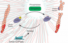
RhoA |
Inhibits Cofilin, stabilizing F actin so filaments get pulled back into main body of growth cone. |
|
|
Ocular Dominance |
Preference for receiving and/or representing visual input from one eye over the other eye. |
|
|
Ocular Dominance Columns |
When cells in the same vertical columns share the same ocular dominance ocular dominance columns are produced. |
|
|
Turning Growth Cones |
One filament extends, others shrink, and new ones grow. |
|
|
Cofilin |
Increases the amount of severing of f actin, which makes g actin more available and allows extending to occur in filaments. |
|
|
Slingshot |
Activated when receptor at edge of filament touches a contact attractant. Activates cofilin. |
|
|
Anosmia |
Lacking the ability to smell. |
|
|
Phantosmia |
Smelling phantom smells that aren't actually there. |
|
|
Who is phantosmia common in? |
People who have damage to their olfactory epithelium. |
|
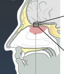
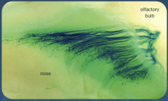
Olfactory Bulb |
Purple spoon shaped item right above the pink near the box. |
|

Olfactory Epithelium |
Pink blob |
|
|
Olfactory Epithelium |
Way up behind the eyes; contains mucus with neurons whose sensors stick right out through the mucus into the air to detect volatile chemicals. |
|

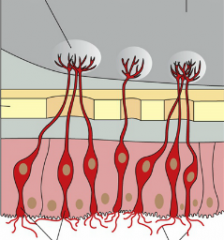
Glomerulus |
Bulbs at top of neurons. |
|
|
Glomerulus |
A cluster of nerve endings, spores, or small blood vessels, in particular. |
|

Olfactory Receptor Neurons |
The neurons in the image. |
|
|
Olfactory Receptor Neurons |
Neurons that go directly into the cortex. |
|
|
Why do olfactory receptor neurons connect directly into the brain? |
It is an ancient system and the wiring remains simple because it developed at a simpler time in our evolution. |
|
|
What could the direct connection of olfactory receptor neurons into the brain be responsible for? |
Consider how smells are the best sense for evoking different scenes or memories; this could have something to do with the direct connection. |
|

Olfactory Cilia |
Cilia at bottom of neurons. |
|
|
How good are dogs noses? |
They are so good at smelling that they can smell a change in R group on a chemical. They can differentiate smells the way we can differentiate notes on a piano. |
|
Bone |
The yellow across the center. |
|

Where is the top of this image? |
The olfactory bulb. |
|

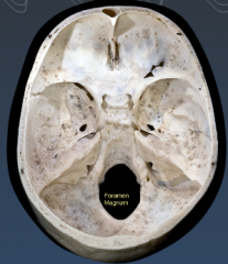
Describe what we are looking at. |
We are looking down at the skull. The front is at the top. |
|

What is the hole? |
Foramen Magnum - where the spinal cord goes. |
|

What is the depression on the right? |
Temporal lobe. |
|

What is the front depression? |
Frontal lobe.
|
|
|
How many holes are there into the brain? |
Not that many; we only want a few holes in and out of the brain because we do not want a lot coming in and out. |
|
|
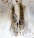
Cribiform plate |
Lots of little holes with nose and nasal epithelium right underneath; bundles of olfactory receptor axons come right into the brain through these holes. |
|
|
Cribiform Plate |
|
Where is the cribiform plate? |
Front center. |
|
|
Which sensory system is the fastest? |
Auditory. |
|
|
How fast is smell in comparison to other sensory systems? |
Smell is about as fast as vision. |
|
|
What molecules did scientists predict would be involved in the olfactory transduction pathway and which molecules actually were? |
Calcium - obviously has an effect, but not specific enough to really help us study the pathway. cGMP - key component in the visual system, so why not? They tried it, but it turns out cAMP is actually the molecule involved in this system. Note: they actually had to use dBcGMP and dBcAMP so that the molecule could enter the cell. Once in the cell it turned into just regular cAMP or cGMP. |
|
|
Molecules of the Olfactory Transduction Pathway Experiment |
Dissect out olfactory receptors. Sprinkle on dBcGMP. See what happens: if cGMP is important then there should be a change. Results: no change. However, when they tried it again with dBcAMP there was a very strong response! |
|
|
What happens when you sprinkle cAMP on a cell? |
Nothing. It has to be dBcAMP to get into the cell and cause a cAMP response. |
|
|
What is the summary of the olfactory transduction pathway? |
GPRC to GaOlf to ACIII to CNG |
|
|
GPRC in the Olfactory Transduction Pathway |
On the surface of neurons; senses molecules |
|
|
GaOlf in the Olfactory Transduction Pathway |
Activates ACIII |
|
|
ACIII in the Olfactory Transduction Pathway |
Makes cAMP |
|
|
CNG in the Olfactory Transduction Pathway |
CNG channels open allowing cations to flow; the result is depolarization! |
|
|
Are olfactory receptors depolarized or hyper polarized by an odor? |
Depolarized. |
|
|
How is the olfactory system similar to the visual system?
|
Both have sensory neurons, both have CNG channels, both have cA/GMP. |
|
|
What is the main difference between the olfactory and visual systems? |
In visual systems when activated the cGMP is converted to GMP causing channels to close and thus resulting in hyper polarization. In olfactory systems when activated cAMP is made and opens channels resulting in depolarization. |
|
|
Why is turning off the olfactory system so important? |
We forget about smells pretty quickly because we are good at adapting and recovering from them. |
|
|
What molecule turns off the olfactory system? |
Calcium. |
|
|
How is the olfactory system turned off? |
When the CNG channels open, the cell is depolarized but also calcium is entering. Once there is a large enough volume of calcium, calmodulin is activated. Calmodulin activation results in the closing of CNG channels, turning off of ACIII, and turning on of phosphodiesterase which then gets rid of cAMP. |
|
|
What are the three things calmodulin does when activated? |
1. Closes CNG channels (no more depolarization). 2. Turns off ACIII (so not more cAMP is being made). 3. Turns on phosphodiesterase (gets rid of remaining cAMP). |
|
|
Turning off the Olfactory System Experiment Set Up |
Put mutations in parts of calmodulin and take extraceullar recording (this means depolarization inside cell appears as hyper polarization).
|
|

Turning off the Olfactory System Experiment Graph Explaination |
Black line: scientists control scent puffer in the rodents nose; the square bumps in the line are when a puff of scent was released. Blue line: normal response. Red line: response amplitude remains normal, so adaptation abilities are fine, but recovery ability is diminished. |
|

Describe the Normal Response |
Blue line = normal response. First puff evokes a strong response. Response almost returns to normal, but not quite. Second puff evokes a less strong response. Every subsequent response will continue to be less and less; this is adaptation. |
|
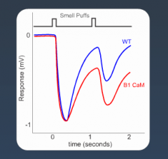
Describe the Response wen Calmodulin is Mutated |
Red line = mutated response. Amplitudes of responses remain identical to normal response. After first response though we never even make it back to the level that a normal response makes it to. This shows that recovery is slower. Only recovery is being affected because only part of calmodulin has been mutated. Adaptation remains effective. |
|
|
What causes slower recovery in the olfactory system? |
Less active calmodulin means CNG channels stay open longer resulting in less recovery. |
|
|
How many passes are in GPCR? |
7 |
|
|
Where is GPCR? |
Transmembrane. |
|
|
What does GPCR stand for? |
G-protein coupled receptor. |
|
|
What kind of receptors are odorant receptors? |
GPCR |
|
|
What is a flaw of GPCRs? |
They are so complex that there are many parts that can get mutated. |
|
|
How many odorant receptors do we have? |
~400 |
|
|
How many different genes do we have for odorant receptors? |
Since each odorant receptor is different, each one has a different gene. Therefore, we have ~400 different genes for odorant receptors. Note: this is 400 out of only 20,000! |
|
|
Pseudogenes |
Like the archeological remains of GPCRs. |
|
|
Why do we have pseudogenes? |
Our ancestors probably had a much better sense of smell than we did, but since we haven't needed a keen sense of smell for so long it would seem since it was no longer selected for that these genes have "decayed." |
|
|
How many pseudogenes do we have? |
~400 |
|
|
How do amounts of genes/pseudogenes for olfactory receptors compare between species? |
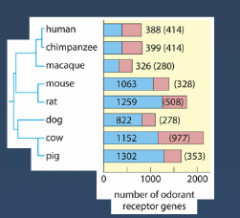
Humans Chimps Macaque Mouse Rat Dog Cow Pig Blue = genes Red = pseudo |
|
|
How many olfactory receptors does a single neuron express? |
One! Each only has one GPCR that detects only one thing. This is good for coding because when a neuron fires you know exactly what it is detecting. |
|
|
Labeled Line |
Every neuron's line (or axon) detects only one thing (a specific smell). |
|
|
Combinations and Smells |
Everything we smell is really a combination of many molecules. |
|
|
What do smells activate? |
Many GPCRs and neurons together! Smells are combinations of many molecules each of which activates a separate GPCR. |
|
|
How does the brain interpret smells? |
The brain interprets patterns of neuronal activity and knows that certain patterns indicate certain smells. |
|
|
Why do dogs and rodents have a better sense of smell than we do? |
Their brains can interpret more patterns and more distinctly than our brains can. |
|
|
Summary of Olfactory Pathway: Bacon Example. |
Fry Bacon. Chemicals release into air (volatile). Chemicals diffuse into nose. Chemicals hit epithelium where olfactory neurons protrude. Chemicals bind with GPCR on neurons. Binding activates G(alpha)OR. G(alpha)OR activates ACIII. ACIII makes cAMP. cAMP starts a cascade to open CNG channels. Open CNG channels = depolarization. Since several chemicals are all doing this simultaneously in separate neurons, several neurons are now active. This varied activity results in patterns of activation in the brain that get interpreted as smells. Soon enough calcium enters through CNG channels to turn on calmodulin. Calmodulin closes CNG channels, turns off ACIII, and turns on phosphodiesterases to get rid of cAMP. This results in the end of smelling the bacon. |
|
What is this? |
An example of the patterns of neuronal activity that indicate smells to the brain. |
|
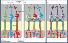
Which correctly indicates the wiring of the olfactory system neurons? How do we know this? |
B because when observing Glomeruli we can see there is distribution of different GPCR among the olfactory epithelium so that excludes A. Then when we trace a single GPCR we can see that even though the different kinds are distributed, they all lead to specific Glomeruli. This implies B. |
|
|
Varying colors shows that different GPCR are spread throughout the olfactory epithelium. |
|
|
Trace of a single GPCR shows us that even though different GPCR may be spread throughout the olfactory epithelium, they all Glomeruli. |
|
|
4 Qualities of Sound |
Amplitude, frequency, timbre, localize. |
|
|
Amplitude |
Height of wave; how loud a sound is. |
|
|
Frequency
|
How frequent waves are. AKA pitch or tone. |
|
|
Localize |
Where the sound is coming from. |
|
|
Timbre
|
What kind of sound it is. |
|
|
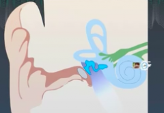
3 Main Parts of the Ear |
Outer, middle, inner. |
|
|
How Affects the Inner Ear |
1. Vibrates cochlea
2. Two membrane - Baslar and Tectorial Plate - move up and down from vibration. 3. As the plates move up and down they shear against each other which moves the hair on the hair cells either one way or the other. |
|
|
What displaces hair cells more? Amplitude or frequency? |
Amplitude. Talking quietly results in little displacement whereas talking lousy results in a lot of displacement. Frequency affects the rate of displacement. |
|
|
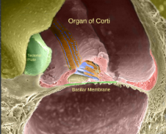
Baslar Membrane |
Lower membrane; everything rests on it. In the inner ear. |
|
|
Tectorial Plate |
In the inner ear. Anchored to the tips of the hair cells. |
|
|
What is the speed of a visual reaction vs. a hearing reaction? |
100 ms vs. 100 to 200 microseconds. |
|
|
What molecules work quickly enough for a hearing reaction? |
Not CNG channels. Sodium stretch channels (basically). "Ion channels at the base of the ropes that connect hairs, so when ropes are displaced y hairs, the trap doors get pulled open allowing for a reaction to occur." - TMC1 Channels |
|
|
How can we observe mutations in hearing? |
Congenital deafness. |
|
|
What mutations have we seen in congenital deafness? |
Mutations in cadherin 23 and protocadherin. These two genes each make up half of the ropes that connect the hairs in the cochlea, so when they are mutated you are deaf. |
|
|
Have we identified TMC1 channels? |
Yes, but are still not positive we understand how they work. |
|
|
How did we show what mutations cause deafness? |
Electron microscopy - stain and observe specific areas. Other methods are discussed in the book. |
|
|
Frequency and the Cochlea |
Only one part of the cochlea responds to one frequency, so which frequency the sound is can be coded by which neurons activate. |
|
|
How is intensity of sound coded? |
By rate of firing. The more firing, the louder the noise must have been. |
|
|
Where does information from the cochlea travel? (2 steps) |
To the spinal ganglion and then to the cochlear nucleus. |
|
|
Middle Ear Bones |
Push on cochlea to activate; more it pushes, the more ripples are sent into cochlea. |
|
|
Organ of Corti |
Organ of corgi moves up and down when cochlea vibrates and this causes the tectoral plate to shear up against the top of it. |
|
|
Spiral Ganglion Neurons |
Send representation of sound from cochlea to brain. |
|
|
Mechanotransduction |
The process in sensory cells by which mechanical stimuli are converted into electrical signals. |
|
|
Tip Link |
Single protein fiber that connects hairs on hair cell. |
|
|
Mechanotransduction Channels |
An ion channel that is gated by mechanical force. |
|
|
Auditory Nerve |
A bundle of axons from spiral ganglion neurons that transmits auditory information to the brainstem. Also contains efferents from brainstem to outer hair cells. 3 |
|
|
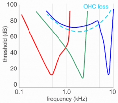
Frequency Tuning |
The property whereby a cell in the auditory system is best activated by sounds of a particular frequency. Represented by a v-shaped curve on a frequency-intensity plot. |
|
|
Tonotopic Map |
The ordered arrangement of cells in the auditory system in physical space according to their frequency tuning. Cochlea and multiple brain regions contain tonotopic maps. |
|
|
Electromotility |
A property of the cochlear outer hairs whereby hyper polarization causes the cells to lengthen, and depolarization for them to shorten along their long axis. |
|
|
Prestin |
Motor molecule that makes outer hair cells move. |
|
|
Frequency Tuning Chart |
|
|
What is required to make the cochlea not just work, but work extremely well? |
Outer hair cells. |
|
|
What do outer hair cells do?
|
Receive information from the brain (efferent). |
|
|
Do outer hair cells send information to the brain? |
No; they are not even connected to the spinal ganglion. |
|
|
What happens to outer hair cells when you are hearing?
|
They contract and expand to instantly change the mechanical properties at the local area.
They instantly adapt to sound by changing local properties so that no single part of the cochlea responds. |
|
|
Outer hair cells are responsible for... |
adaptation. |
|
|
Outer hair cells give the auditory system what kind of properties?
|
Active mechanical. |
|
|
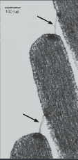
Which plate is which in the auditory complex? |
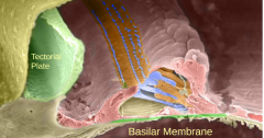
Tectorial - green Basilar - bottom red |
|

Where is the organ of corti? |
Up top! |
|
What are these called? |
Stereocilia. |
|
Where are the stereo cilia? |
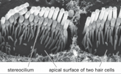
Left arrow. |
|

Where is the apical surface of the hair cells? |

Right arrow. |
|
|
How do you code frequency of sound? |
Labeled line; different axons along the cochlea indicated different frequencies. |
|
|
How do you code amplitude of sound? |
By how many spiral ganglion axons are activate (labeled line) and the RATE of firing of those neurons! |
|
What are these? |
Tip links. |
|
|
Auditory Pathway Summary |
Inner hair cell enervated by spinal ganglion neurons. Axons from spinal ganglion cells go to cochlear nucleus. Information then goes to olivary nuclei. Then to inferior colliculus. Then to thalamus and the medial geniculate nucleus. Finally, auditory cortex. |
|
|
Olivary Nuclei |
Olive structures in a region in the hindbrain. |
|
|
2 Auditory Pathways |
1. Fast route 2. To both sides of the brain |
|
|
Where does information from the left cochlea end up? |
Right side of the brain. |
|
|
Fast Auditory Pathway |
Left cochlea to left cochlear nuclei towards right olivary nuclei but skips it and goes straight to right inferior colliculi, to right medial geniculate nuclei, and the right auditory cortex. |
|
|
Auditory Pathway to Both Sides of the Brain |
Left cochlea to left cochlear nuclei to both superior olivary nuclei. From here the information goes equally to both sides of the brain. (inferior colliculi, thalamus at medial GN, and auditory cortex). |
|
|
Where in the auditory pathway is the sound source determined? |
Superior Olivary Nuclei - most of localization has to do with informationfrom one side or the other. This is the first place information comes togetherfrom both sides, so this is where localization occurs. |
|
|
Where is everything integrated in the auditory pathway? |
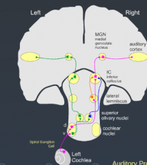
The inferior colliculus where all information compiles into one signal. |
|
|
What "map" may be present in the nuclei in the auditory pathway? |
Spinal ganglion axons encode information about different frequencies, so this is the "map" we preserve as the information travels on. |
|
|
Tonotopy |
You could color each area of the auditory pathway with a gradient of frequencies to make a map. |
|
|
Consider what lesions in different areas of the auditory pathway would do. |
Consider! |
|
|
Auditory Precedence |
Sound comes directly from the source but also echoes around the walls. Our hearing automatically inhibits us hearing all the echoes; the first sound you hear is what your auditory system focuses on. |
|
|
Masking |
You can hide noises in other regions; lateral inhibition in the hearing system. |
|
|
Where are we good at localizing sound? |
The horizontal plane. |
|
|
Where are we not as good at localizing sound? |
From above and below. |
|
|
What animals are really good passive listeners? |
Owls are really good at localization from above and below vis passive listening. |
|
|
What kind of animal is a good active listener and what does this mean?
|
Bats are good active listeners; they make a sound and locate things from where it bounces off of. |
|
|
What other animals are good at localization but not at determining frequencies?
|
Fish/marine animals. |
|
|
Why are owls so good at passive listening localization? |
Owls: "Their whole face is like a big ear." Their feathers channel sound information to their ears which are conveniently placed closer together than our ears (200 microseconds vs our 600 microseconds). |
|
|
Interaural Time Difference |
Time it takes from sound in one ear to get to the other ear.
|
|
|
Describe how neurons come together for us to localize sounds. |
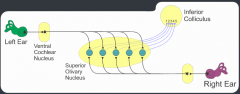
Left ear to ventral cochlear nucleus to olive - 5 branches. Same from right ear, so ten total branched synapsing onto 5 neurons that then go back to the inferior colliculus. |
|
|
Which neurons for localizing sound will be activated if you hear a sound that comes equally into both ears (occurs from directly in front of you)? |
The 3rd neuron. All others receive inputs, but not simultaneously like 3 receives. |
|
|
Coincidence Detector |
How neurons come together for us to localize sound is called a coincidence detector. |
|
|
What does a coincidence detector tell us?
|
It tells us about timing differences between the right and left ears. You need convergence of two ears to do this, so thats why it occurs in the olive. |
|
|
There are also the equivalent of coincidence detectors for detecting the _________ of noises. |
difference in intensities |
|
|
What factor makes coincidence detecting easier? |
When ears are further apart it is easier to differentiate! |
|
|
What is the key factor about how information gets to the superior olives?
|
Axons that go from ear to olive have to have a very large diameter and be well myelinated so information can get to the olive ASAP. |
|
|
How does information travel to the olive vs. past the olive? |
Information has to travel very fast to the olive, but once it gets there from there on axons can be smaller and less myelinated to slow things down. |
|
|
Horizontal Localization in Humans and Frequenceis |
Timing differences for localization (ITDs) onlywork for us at lower frequencies. At higher frequencies the loudness differencetakes over; we localize based on intensity differences at those frequencies. |
|
|
Vertical Localization in Humans: How do we localize sound from above and below us? |
We should receive the same information from each side whether from above or below so how do we distinguish? Shape of the ear! Depending on if the sound comes from above or below, the shape of the ear changes the frequency so we can detect which way it came from. |
|
|
Vocal Learning |
(video of Elise singing) You don't have to see someone walking to be able to walk, but you can't talk without hearing sound. |
|
|
How come deaf people can talk? |
Nowadays they can usually hear something that can help them to speak, but they still aren't perfect because they cannot compare what they are hearing to what they are saying. |
|
|
Wernicke's Area |
Language Comprehension |
|
|
Broca's Area |
Language Production |
|
|
Inferior Colliculus |
A midbrain nucleus that integrates auditory signals from brainstem nuclei and sends signal to thalamus. |
|
|
Precedence Effect |
The ability of a first arriving sound to suppress the perception of later arriving sounds. |
|
|
Coincidence Detectors |
A cell that is maximally activated by simultaneous auditory signals from left and right ears. |
|
|
Delay Lines |
A thin axon fiber that carries auditory signals to target neurons at different locations along the axon with different time delays. |
|
|
Somatosensation
|
Touch
|
|
|
Where does touch information go in the brain?
|
Front of parietal lobe; somatosensory cortex.
|
|
|
Proprioception
|
How reflexes work; how you know when your body is moving.
|
|
|
Thermosensation
|
Temperature; very relative
|
|
|
Nociception
|
Pain receptors
|
|
|
Pruroception
|
itch; not completely understood. People can have chronic itch.
|
|
|
What do we think itching has to do with?
|
Chemicals more so than mechanics; our own immune molecules may be releasing the chemicals.
|
|
|
Interoception
|
Internal organs sensing.
|
|
|
How do skin and spinal cord correlate and why is this important?
|
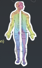
Certain regions of the skin lead to certain places in the spinal cord.
This is important clinically because lesions in certain areas of the skin can indicate issues in different areas of the spinal cord. |
|
|
Where are our sense receptors/organs?
|
The tips of the dendrites in our skin.
|
|
|
Where to processes for sensory neurons go?
|
All the way back to the dorsal route ganglion (right outside the spinal cord) and then into the spine for reflexes and/or into the brain.
|
|
|
What are A alpha Neurons?
|
Proprioceptors; fastest because biggests and well myelinated.
|
|
|
Where do A alpha neurons go?
|
Specific places in the spinal cord.
|
|
|
What is the con of A alpha neurons (why are all neurons not this type)?
|
They take up a lot of room.
|
|
|
B Alpha
|
Middle level proprioceptor neurons.
|
|
|
C Alpha
|
Slowest and smallest proprioceptor neurons.
|
|
|
3 Questions to ask about any Sensor
|
What does it detect?
What type of adaptation? What is its speed of conduction? |
|
|
Common Answers to What Sensors Detect
|
Pressure, indentation of skin, vibration on skin, temperature, pain, etc.
|
|
|
Common Answers to Type of Adaptation for Sensors
|
Not at all, slowly, or rapidly.
|
|
|
Common Answers to Speed of Conduction for Sensors
|
A, B, or C alpha.
What is it conducted on or by? |
|
|
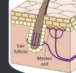
Where is a Merkel cell?
|
|
|
3 Kinds of Neurons
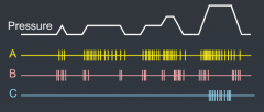
|
A – slow adapting, low threshold, pressure detectorB – rapidly adapting, low threshold, pressure detectorC – not adapting, high threshold, pressure detector
|
|
|
Do more sensitive neurons have bigger or smaller receptive fields?
|
Smaller
|
|
|
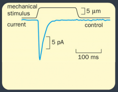
How does touch work molecularly?
(hypothesis, test, results) |
Hypothesis: Stretch channels (mechanically gated ion channels).
Test: Push on some mechanically sensitive cells and see what they do via patch pipette recording (so you are only looking at one channel with no chance of breaking the cell). Results: As you stimulate you see an inward current. |
|
|
How does touch work molecularly: Follow Up
|
We want to take out the channel and see what occurs, so we perform a knockout screen.
We use RNAi to do this, but we need to determine the right RNAi for the protein to knockout the channel. How do we know what to knockout? Tried all sodium channels - nothing. Tried all transmembrane channels with no known function - 2 responses. When we knocked out the channels the cell lost its ability to detect pressure. |
|
How does touch work molecularly?
Interpreting the Results Chart |
Fast adapting cell. You can tell because it only responds right away and then the response fades.
|
|
|
RNAi
|
RNA interference; when added to RNA, the product of that RNA is interfered with and goes away.
|
|
|
What channel protein is responsible for cell pressure detection? |
Piezo |
|
|
When was Piezo discovered?
|
2010 |
|
|
What was Piezo's reversal potential and what does this tell us?
|
0; tells us it is a nonselective ion channel. |
|
|
Piezo |
|
|
What is the structure of Piezo? |
3 blades; when the blades are touched they move opening the receptor site. |
|
|
What 3 Pieces of Evidence are Critical to Prove Piezo is the channel protein? |
1. When you remove it from mechanically sensitive cells they are no longer mechanically sensitive. 2. When you add it to non-mechanically sensitive cells they now are mechanically sensitive. 3. They exist in sensory parts of the body. All 3 sources of evidence have been found. |
|
|
What is thermosensation detected by? |
TRP channels. |
|
|
What do TRP channels look like?
|
Ion channels |
|
|
Capsaicin |
Molecule in peppers that makes you feel like you are hot; not water soluble, which is why drinking water doesn't help you get rid of the heat. |
|
|
Menthol |
In peppermints; activates cool receptors. |
|
|
If you add capsaicin to regular cells what happens? |
Nothing because they don't have TRP channels and are not thermosensitive.
|
|
|
What happens if you add TRPV1 (TRP channel protein) and then capsaicin? |
Cells respond to the heat. |
|
|
DRG |
Dorsal route ganglion |
|
|
Trigeminal Ganglia |
Clusters of somatosensorty neurons near the brainstem involved in sensation of the face. |
|
|
Dermatomes |
An area of skin that is mainly supplied by a single spinal nerve |
|
|
Mechanosensors |
Sensors neurons activated by mechanical force. |
|
|
Merkel |
A specialized epithelial cell at the junction of the dermis and epidermis. |
|
|
Meissner |
They are a type of nerve ending in the skin that is responsible for sensitivity to light touch. |
|
|
TRPV1 |
Capsaicin |
|
|
TRPM8 |
Menthol |
|
|
Expression Cloning |
A strategy for cloning a gene by transferring cells with pools of cDNA and using a functional assay to identify the pool that contains the cDNA of interest. Continue to do this until a single cDNA is identified. |
|
|
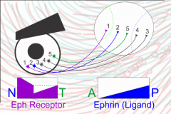
Two Gradient Experiment |
Ephrin = repellant Eph A = receptor High repellant for nasal and posterior. High receptor for temporal and anterior. |
|
|
Practice Exam again |
DO THIS |
|
|
C for Newt Practice Problem |
There can’t just be a chemoreceptor/attractantpair between left tectum that right eye has a receptor for because if there wasthe right eye would have gone to the left tectum to begin with Instead, it is probably that each eye goes toboth tectum, but functional selection sorts out which tectum they stay with.This is actually the result you get (but not actually with functionalselectivity, it is still a good hypothesis though). Otherwise, for this to be true the environmentthe eye is in has to change the receptor that the axons has. o Real answer is that there is a midline repellentthat gets turned on when axons cross, which force them to stay crossed. |
|
|
Outermost hair cells hear _____ frequencies. |
high. |
|
|
Innermost hair cells hear ______ frequencies.
|
low. |
|
|
What happens if you cut the outer hair cells?
|
Inner hair cells hear worse and a bigger variety of frequencies. Outer hair cells help us finely tune frequencies. |
|
|
3 Possible Explanations for any Stripe Assay |
Attractant in 1 Repellant in other Both of these |
|
|
Never code with... |
height of an AP. |
|
|
When considering the newt experiment always consider... |
contact repellants patterning all over. |
|
|
Where does frequency separation occur? |
Right after the cochlea. |
|
|
Where is mammalian hearing coded labeled line? |
Between the R/L ear. |
|
|
Where is the primary motor cortex? |
In front of the somatosensory cortex. |
|
Name 6 Components |
Outer ear - pinna (filters sound) Ear canal Tympanic membrane Inner ear bones Cochlea Auditory nerve |
|
|
2 Parts of the Cochlea |
Semicircular canals and the cochlea. |
|
|
How do the middle ear bones interact with the cochlea? |
They push on them creating vibrations through the cochlea that the cochlea splits up based on frequency. |
|
|
Important Fluid Filled Tube in Cochlea |
Media - high K+ |
|
|
Where are tectorial and basilar membranes? |
Around the organ of corti. |
|
|
When the cochlea vibrates what happens? |
The organ of corti moves up and down resulting in tectorial membrane shearing against it. |
|
|
What is the hair cell ratio?
|
1 inner to 3 outer. |
|
|
What do hair cells connect to?
|
Tectorial Plate |