![]()
![]()
![]()
Use LEFT and RIGHT arrow keys to navigate between flashcards;
Use UP and DOWN arrow keys to flip the card;
H to show hint;
A reads text to speech;
317 Cards in this Set
- Front
- Back
|
When do we see a prospective nervous system in humans? |
13 to 14 days. |
|
|
Gastrulation is a... |
massive cell movement leading the rearrangement/formation of the germ layers. |
|
|
What do you have at the end of gastrulation? |
Dorsal/Ventral and Anterior/Posterior
You have the formation of a body plan. |
|
|
When is the induction of the nervous system? |
Gastrulation. |
|
|
There are no _________ determined to be ________ pre-gastrulation. |
cells; neural cells |
|
|
Xenopis |
An Amphibian Model System |
|
|
What is the mammal equivalent of the dorsal lip? |
Primitive streak. |
|
|
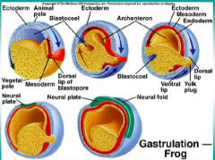
Describe the results of the movement of layers during gastrulation. |
Mesoderm underlying the neural ectoderm on the dorsal side and the endoderm ends in the middle of the whole thing. |
|
|
What does mesoderm give rise to? |
Muscle, blood, notochord. |
|
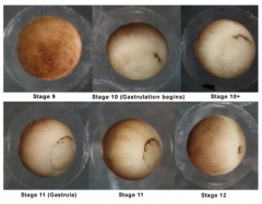
Describe what is happening at stages 9&10. (top row) |
9 - ectoderm, mesoderm, and endoderm are all in a row on the outside. 10 - dorsal lip of blastopore forms; this is where the mesoderm and endoderm first start going into the ball of cells. |
|
What layer does each color represent? |
Ectoderm - blue Mesoderm - pink/red Endoderm - yellow |
|
|
How does the mesoderm get in the right place? |
It has to move along the inside of the embryo; now two different cell types come int contact with each other. |
|
|
Special Factors Development Experiment (set up, results, conclusion) |
Bisected embryo at two cell stage. Took cells and divided them and let them grow individually. (This happens in humans all the time resulting in identical twins). Result: Doesn't harm the embryo at all except if the gray crescent on the dorsal side wasn't evenly distributed between the two daughter cells then you get one normal embryo and one belly piece (ball with no neural ectoderm). Conclusion: the gray crescent contains "special factors" that induce the nervous system. |
|
|
Timing of Neural Tissue Development Experiment (set up, results, conclusion) |
Took embryos at early and late gastrula stages. Took a piece of tissue from the area that would give rise to neural ectoderm (but hadn't yet), and transplanted it to the ventral side of the embryo. Result: In the early embryo the tissue became epidermis, but in the late embryo the neural plate formed on the ventral side. Conclusion: Ectoderm becomes determined to neural tissue between early and late gastrula stages. |
|
|
Organizer Experiment (set up, results, conclusion) |
Took toy bit of dorsal lip from one embryo and put it in the ventral region into the ventral region of another embryo. Result: After 24 hours there was a secondary nervous system growing; the transplanted tissue was organizing cells on the ventral side to give rise to this resulting in two conjoined twin embryos if they let the second nervous system grow. Conclusion: the dorsal lip can induce ventral tissue to adopt a neural fate and pattern (organize) it. |
|
|
Regional Identity and Development Experiment (set up, results, conclusion) Not super important... |
Took mesoderm from a late gastrula embryo and stuffed it into an early gastrula embryo. Result: Structure reflected the anterior/posterior quality of that mesoderm (where it was put determined where it grew). Conclusion: The underlying mesoderm imparts a regionalidentity to neural ectoderm as it induces it. |
|
|
Gastrula |
An embryo at the stage following the blastula, when it is a hollow cup-shaped structure having three layers of cells. |
|
|
The dorsal lip is the... |
organizer. |
|
|
Noggin Experiment |
Screen to isolate specific proteins in dorsal lip that were being secreted as the dorsal mesoderm came into the mesoderm. Found Noggin. Long version: cDNAplasmid library constructed from lithiumtreated dorsalizedembryos;RNA was synthesized from this library and injected into ventralizedembryos; those sets of plasmids whose RNA rescued the dorsalaxis of the ventralizedembryos were narrowed down until a single cDNA was isolated - this wasnoggin. |
|
|
Why is Noggin name as such? |
Its over expression results in a HUGE head (completely dorsal-ized embryo). |
|
|
What is cool about noggin? |
It can rescue UV irradiated embryos where UV usually destroys too much for the embryo ti survive. |
|
|
What kind of protein did noggin compare to? |
None; it was completely novel! |
|
|
How does noggin function? |
Binds to another growth factor (BMP), so that the growth factor cannot bind to its receptor; it is inhibitory. |
|
|
What does BMP signaling result in? |
Skin |
|
|
What happens when you inhibit BMP signaling? |
Neurons |
|
|
What happens when you overexpress Wnts? |
Anterior genes are down regulated. |
|
|
What happens when Wnt inhibitor is overexpressed? |
Anterior genes are upregulated; it anteriorizes the nervous system. |
|
|
What does Wnt do?
|
Regionalizes the nervous system. |
|
|
High BMP = |
skin |
|
|
High Wnt and no BMP = |
posterior neural tissue (trunk and spinal cord) |
|
|
No Wnt and no BMP = |
head/brain tissue |
|
|
Describe Wnt and BMP Gradient |
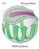
Wnt = purple BMP = green |
|
|
How does tissue begin to adopt a neural fate/express neural genes? |
Transcription Factor Cascade: 1. BMP signaling (lack thereof) activates TFs 2. TFs lead to transcription of Neurogenin (another TF) 3. Neurogenin activates NeuroD 4. NeuroD activates neural specific genes |
|
|
Why study early development? (2 reasons) |
1. To help understand complex neurological diseases (autism, schizophrenia). 2. To understand environmental factors causing disease. |
|
|
Blastula |
An animal embryo at the early stage of development when it is a hollow ball of cells. |
|
|
Neurulation |
The stage of development where the construction of the nervous system occurs (post gastrula). |
|
|
What happens during neurulation? 3 Main Events |
Dorsal dise, ectodermal cells overlying mesoderm structure calle notochord thicken to form the neural plate. Center of the neural plate drops towards interior of embryo while two edges move towards neural fold and eventually fuse at the center creating the neural tube. Neural tube is covered by layers of ectoderm derived epidermal cells. |
|
|
Notochord |
A midline mesodermal structure in vertebrate embryos ventral to the spinal cord that produces secreted cues for patterning of the spinal cord. |
|
|
Neural Plate |
The layer of ectodermal cells overlaying the notochord that invaginates and gives rise to the neural tube during neurulation. |
|
|
Neural Tube |
A hollow tube surrounded by layers of neuralectodermal cells; embryonic precursor to the vertebrate CNS. |
|
|
What does the lumen of the neural tube develop into? |
Ventricles that house the cerebrospinal fluid. |
|
|
4 Steps during Development |
1. Egg 2. Blastula 3. Gastrula 4. Neurula |
|
|
3 Processes during Development |
1. Fertilization/Cleavage 2. Gastrulation 3. Neurulation |
|
|
Egg to Blastula = |
fertilization/cleavage |
|
|
Blastula to Gastrula = |
gastrulation |
|
|
Gastrula to Neurula = |
neurulation |
|
|
Where is the future gut in a gastrula? |
It is the big hole in the middle. |
|
|
Where is the future notochord in the gastrula? |
Above the future gut; mesoderm area. |
|
|
Where are the neural folds in the neurula? |
The folds at the top.
|
|
|
Where is anterior vs. posterior in the neurula?
|
Anterior = top Posterior = bottom |
|
|
Describe Cell Movement during Neurulation |
Start with neural plate over notochord. Neural plate folds in at bottom to form a crater and then sides pull together at top creating the neural fold. Neural fold closes forming the neural tube covered by a layer of epidermal cells. Neural crest forms above neural tube. |
|
|
Where is dorsal vs. ventral during neurulation? |
Dorsal is the top (neural plate) and ventral is the bottom (notochord). |
|
|
What do morphogens do? |
Turn cells with an undetermined fate into cells with a specific fate by enhancing or inhibiting transcription. |
|
|
What does different amounts of morphogens do? |
Different amounts can specify very general or very specific features. |
|
|
Progenitor |
A cell pre-differentiation. |
|
|
Steps for a Neural Progenitor (5)
|
1. Get in the right place 2. Neural birth/migration 3. Axon guidance 4. Building of dendritic arbor 5. Synapses form and are modified through life |
|
|
Transcription Factor |
A protein that binds to a promotor/enhancer and causes it to be transcribed. |
|
|
What do we need Tf to turn on to get neurons? |
Neuronal genes. |
|
|
How many genes does on TF turn on? |
Up to thousands. |
|
|
What controls TF? |
Morphogens. |
|
|
Where are most TF located? |
Inside the cell. |
|
|
Where are most morphogens located? |
Outside the cell. |
|
|
How do morphogens work? |
They diffuse into the cell and cause TF to react. |
|
|
Morphogen |
A diffusable protein that enhances or inhibits transcription. |
|
|
Why are they called morphogens? |
If you mess with them during development then you become very morphed (survival rate is limited). |
|
|
Human Example of Morphogens: Pax and Emx |
Pax6 and Emx2 are TF. Pax6 is highly common in front of brain and less in the back; Emx2 is the reverse gradient. High concentrations of one inhibits the other! |
|
|
Examples of Morphogens |
FGF and BMPs/Wnts |
|
|
How do FGFs and BMPs/Wnts affect Pax6 and Emx2? |
FGF causes Pax6 to be expressed through Emx2 inhibition. BMPs and Wnts inhibit Pax6 by inducing Emx2. |
|
|
How do morphogens (FGF and BMPs/Wnts) interact with gradients (Pax6 and Emx2)? |
Morphogens set up the gradients inside the cells and the gradients cause the brain to organize itself. |
|
Mutations to cause a more anterior based brain? (3 options) |
1. Increased Pax6 2. Increased FGF inhibition which pushes Emx2 gradient further to the right and Pax6 fills in the rest (ends up causing increased Pax6). 3. Knockout BMPs or Wnts because they help posterior development. |
|
Mutations to cause a more posterior based brain? (3 options) |
1. Increase Emx2 2. Knockout FGFs (which inhibit Emx2) 3. Increase BMPs and Wnts |
|
|
What steps of neural development occur early postnatally? |
Dendrite and synapse formation. |
|
|
Neuronal Birth Experiment (set up, results, conclusion) |
Take a pregnant mouse and inject embryos with radioactive thymidine (that will go into DNA and remain there) at different stages of development.
Results: observed mice 8 days after birth. Black dots show neurons. 13 day injection - neurons near center 15 day injection - neurons nearer edge 17 day injection - neurons on edge Conclusion: newest neurons travel the longest distance, while older neurons remain where they are born. |
|
|
Why isn't the entire image black with cells injected with thymidine in the neuronal birth experiment? |
Only neurons born at injection retain the thymidine. Other cells born at this time continue dividing, so lose a high enough dosage to be seen. Cells not born at this time don't really get the injection I believe. |
|
|
How do neurons organize after birth? |
Older neurons DO NOT just get pushed to the outside. Newer neurons actually migrate past existing neurons into a more peripheral position; they travel the longest distance while older neurons remain near where they are born. |
|
|
Post-Mitotic |
No longer performing mitosis after birth. |
|
|
What cells are post-mitotic? |
Neurons. |
|
|
Where do progenitors live?
. |
Near (inside) ventricles. |
|
|
Where are neurons born? |
Right next to ventricles. |
|
|
What are neurons born from? |
Stem cells. |
|
|
Steps of Neuronal Differentiation (4 steps) |
1. Progenitor makes a copy of itself. 2. First neurons are born when progenitors are no longer proliferating; its resulting cell splits into itself + a baby neuron (asymmetrical division). 3. Progenitor is now called a radial glia and baby neuron crawls up its extensions to the top of the brain. 4. Notch and delta result in differentiation. |
|
|
What does it mean to Proliferate? |
To make a symmetrical copy of itself. |
|
|
Asymmetrical Division |
When a progenitor splits into a copy of itself and a baby neuron. |
|
|
What are radial glia? |
Mature neural progenitors. |
|
|
What do radial glia do?
|
They make processes that extend to the top of the brain and then baby neurons crawl up this process to get to the outside of the brain. The more new neurons we get, the longer the processes become and the further out the baby neurons travel. |
|
|
Intermediate Progenitors |
Progenitors that split into two baby neurons and that is it. |
|
|
Can one radial glia create multiple neurons? |
Look in Book/Ask Around |
|
|
4 Parts of Fly Sense Organ |
1. Hair (cell) 2. Socket 3. Neuron that senses hair touch 4. Sheath cell that protects it |
|
|
Where do the four parts of a fly sense organ come from? |
The same single progenitor. |
|
|
How does a progenitor result in 4 cells? |
Sensory organ progenitor > two progeny > four cells. |
|
|
Fly Sense Organ Mutation Experiment |
Fly mutant screen: found a mutation that makes the flies numb; called the mutation "numb." |
|
|
What happened if you expressed no numb in the flies? |
All socket cells. |
|
|
What happened if you over expressed numb in the flies? |
All neuron cells. |
|
|
Did over or under expression of numb result in a functioning fly sense organ? |
No. |
|
|
What happened when we stained cells for the numb protein? |
SOP showed numb on the right, so when it divided only some of the progeny got it. (?) |
|
|
What does numb actually do? |
Inhibits notch for notch-delta. |
|
|
Notch |
Transmembrane receptor. |
|
|
Delta |
Ligand for notch. |
|
|
How does notch and delta work? |
Notch and delta are on the surface of two cells. Notch on ones gets activated by binding delta. This cell starts expressing more notch and less delta. No notch activation results in just delta. This results in different kinds of cells near each other. |
|
|
What do notch and delta do? |
Enhance the differences of cells near each other. |
|
|
Notch and delta only work when they are on... |
opposite cells. Notch and delta cannot communicate within one cell. |
|
|
Stochastic
|
A random start where once it is started a pattern emerges. |
|
|
The result of notch and delta is a... |
checkerboard. |
|
|
How does a cell binding notch affect its neighbors? |
If one cell has notch, its neighbors get delta. Then the neighbors of delta get notch and so on and so on. |
|
|
How do many systems use notch and delta? |
Lots of different systems use notch and delta at different TIMES resulting in organization of different structures. |
|
|
SHH gene is a ________. |
Morphogen |
|
|
What is the receptor of SHH? |
Patch |
|
|
What does patch do?
|
Causes intracellular signaling that allows or represses the transcription of different things in the cell. |
|
|
Where is SHH? |
It is a diffusant outside of the cell. |
|
|
3 Parts of a Slice of Spinal Cord in Early Development |
Notochord (bottom circle) Floor Plate (just above notochord; bottom of upper piece) Spinal Cord (upper piece) |
|
|
What shape is the spinal cord in early development? |
Tube |
|
|
Where do motor neurons live?
|
The ventral part of the spinal cord (when developed will be the ventral horn). |
|
|
Where do motor neurons live in early development? |
The floor plate is the ventral location the motor neurons live for now. |
|
|
Where is SHH expressed? |
Floor plate and notochord; highly on the ventral side! |
|
|
Where is ventral/dorsal in the spinal cord slice? |
Ventral = down Dorsal = up |
|
|
Where are interneurons located in early development? |
Upper, dorsal portion of the spinal cord. |
|
|
How do we specify different types of neurons? |
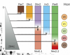
Different gradients can specify. |
|

What inhibits Pax7? |
Any amount of SHH. |
|
|
What do different amounts of SHH do? |
Different amounts of SHH inhibit different TF and there is a pattern of these TF that leads to a particular neuronal fate. |
|
|
How do you get motor neurons? |
You need a middle amount of SHH to get Pax6 and Nkx6.1 but you can't have too much or too little or else Irx3 or Nkx2.2 kick in and inhibit the formation of motor neurons. |
|
|
What do these gradients determining neuron type result in? |
Very nice edges on what neurons will become what; there can be no "in between" a motor or sensory neuron because of up and down regulation. |
|
|
Homeodomain |
Really early TF are homeodomain TF (hox genes) |
|
|
What does SHH prevent? |
It prevents you from becoming a cyclops. |
|
|
What does cyclopamine cause? |
It causes you to become a cyclops!
|
|
|
How was cyclopamine discovered? |
Sheep were eating lilies with them in it and birthing cycloptic baby sheep. (they don't live very long, but they do get born) |
|
|
What does cyclopamine inhibit? |
SHH! |
|
|
Guidelines for Getting the Right kinds of Neurons in the Right Place (4) |
1. Morphogen gradients 2. Homedomain TF 3. Combinations of up and down regulation that specify the fates of NPs 4. Notch and delta to differentiate neighbors/progenies |
|
|
Telencephalon |
The most highly developed and anterior part of the forebrain, consisting chiefly of the cerebral hemispheres. |
|
|
Ventricular Zone |
The ventricular zone is the germinal zone located at the walls surrounding the ventricles where the majority of neurones of higher brain areas are generated. From there, neurones migrate to their final target regions using intrinsic or extrinsic cues and/or radial glia. Neural Development Neurons. Where neurons are born. |
|
|
Intrinsic vs. Extrinsic Factors Experiment in Baby Neurons (set up, results, conclusion) |
See if baby neurons can grow on a dish where they cannot be influenced by extrinsic factors. Results: Stage 1 - little ball; little veils form and turn into minor processes Stage 2 - minor processes Stage 3 - one of minor processes takes off as axon Stage 4 - axon is very long; dendritic outgrowth occurs Stage 5 - maturation of processes Conclusion: Neurons can grow with only intrinsic factors. |
|
|
Axon Growth Experiment in Baby Neurons
(set up, results in early or late in development, conclusion) |
Take baby neuron's axon and cut it.
Remainder of original axon degenerates; neuron is basically back to stage 2. If you cut the axon early in development, another process becomes the axon and extends (can be the original process, but chances are low). If you cut the axon later in development (closer to maturation stage) the same process extends out again as the axon. Conclusion: There must be an intrinsic process determining which process becomes the axon. |
|
|
Do adult neurons regenerate if cut? |
Not in humans. |
|
|
LKB1 |
Partitioning protein that specifies the axon. |
|
|
What does a stain for LKB1 look like in a baby neuron? |
Processes that look similar to the naked eye look different under an LKB1 stain. One process clearly has a build up of LKB1 and this is the process that clearly extends. |
|
|
Process of Forming an Axon |
External cues > LKB1 > affects cytoskeleton (forms growth cone) > resulting axon |
|
|
What kind of feedback/inhibition is necessary for transformation from baby neuron to full axon and dendritic arbor? |
Positive feedback within the axon for LKB1 (once one process has it, you want that process to keep getting more of it). Other processes need to be inhibited for LKB1 so that they do not become axons, but rather become the dendritic arbor. |
|
|
In vitro |
Whole cell is in a dish |
|
|
In vivo |
The cell is in the animal |
|
|
Experiment to Discoer Proteins Required for Dendrites (set up, results, conclusion of protein) |
Performed an unbiased genetic screen in drosophila. Results: K1 - no affect K2 - shortens dendrites AND axons K3 - only shortens dendrites K3 is our protein: Dar3 |
|
|
What does Dar3 do? |
It is involved in vesicle traffic to the Golgi. |
|
|
Why is Dar3 important for dendrite building? 2 Hypotheses |
H1: Minus ends of microtubules are only out in dendrites and have to sit on golgi outposts
H2: In dendrites there is so much communication that the golgi outposts are just necessary |
|
|
What microtubule end is out in axons? |
Plus |
|
|
What microtubule end is out in dendrites?
|
Plus and Minus ends |
|
|
Cadherins |
Hold synapse parts together |
|
|
Where is neurexin? |
Presynaptic terminal |
|
|
Where is neuroligand? |
Postsynaptic terminal |
|
|
What do neurexin and neuroligand do? |
Hold synapse in the right place (hold cells together). |
|
|
Functional Selection |
Make too many connections and use functional selection to get rid of the ones you don't need. |
|
|
What is the NT released at a neuromuscular junction? |
Acetylcholine |
|
|
Discovery of Aggrin Experiment |
Took chick muscle precursors, plated them (in vitro), and stain for acetylcholine receptor. Saw the receptors were randomly dispersed (no clusters like you need for a synapse). Added random chemicals. Found aggrin caused synapse-like clusters of the receptors. |
|
|
What causes a growth cone to go to a specific part of a muscle? |
Before avon comes in contact there is some aggregation and the growth cone goes to this part of the muscle that has the aggregates. |
|
|
What happens once a growth cone comes in contact with muscle? |
Aggregation causing the presynaptic cell to turn into a presynaptic terminal. |
|
|
What is the main aggregation signal? |
Aggrin |
|
|
What is the main aggregation receptor? |
MuSK |
|
|
What is aggregation fighting against? |
Dispersal |
|
|
How does a cell combat dispersal?
|
Contact with a growth cone helps counteract dispersal as does the MuSK receptor. |
|
|
What does LRP4 do? |
Tells the presynaptic cell to turn into a synapse. |
|
|
Summary of Aggregation (5) |
1. Aggregation of some acetylcholine receptors on the target cell 2. Axon attracts to this aggregation 3. Aggregation forces vs. dispersal forces (aggrin binds to MuSK causing more clustering and specialization of the postsynaptic side and inhibiting dispersal forces). 4. LRP4 causes specialization of presynaptic side. 5. We have a synapse. |
|
|
What happens when Aggrin binds to MuSK? (2) |
1. Causes more clustering of receptors 2. Causes specialization of the postsynaptic side. |
|
|
Parts of a Synapse (7) |
Snares, VG Ca channels, neurexin, neuroligand, receptors, PSD, vesicles |
|
|
What are snares? |
They attach vesicles to membrane in the presynaptic terminal. |
|
|
Neurexin Experiment (set up, result) |
Take a non neuron and over express neurexin in it. Results: Makes cell (?) into synapse. |
|
|
Neuroligand Experiment |
Take a non neuron and over express neuroligand. Results: Only affects the neuron it is interacting with. |
|
|
Are neurexin and neuroligand necessary or sufficient for synaptic formation. |
Sufficient, but not necessary. |
|
|
What happens if you knock out neurexin and neuroligand in a mouse? |
Synapses still form; there is a large set of additional molecules that allow this to occur. |
|
|
What can result from too many synapses? |
Autism |
|
|
What can result from too few synapses? |
Schizophrenia |
|
|
Is more synapses better? |
No! You want just the right amount. |
|
|
How many synapses do we start with? |
Too many and we get rid of tons of them to allow us to function well. |
|
|
Why can we make many synapses and then get rid of them? |
They are relatively easy to make excess of. |
|
|
Hebb's Rule |
Fire together, wire together. |
|
|
Stereotyped Axon Pruning |
Looked at mice brains: Primary motor neuron needs to go to spinal cord and neurons from visual cortex need to go to the superior colliculus, but both sets of neurons grow both places and then destroy the unnecessary branch. |
|
|
Why does stereotyped axon pruning occur (mouse brain example)? 2 Hypotheses |
H1: Maybe it is like how frogs start as tadpoles; maybe at one point the connection was needed but now it is not. H2: Maybe the neurons follow a leading axon there and it is easier to follow to both locations that to determine preference. |
|
|
Characteristics of Wallerian Degeneration |
When one neuron is cut there is a rapid, synchronous apoptosis like program that occurs. This program is injury based. |
|
|
Do babies or adults have more neurons? |
Babies |
|
|
NGF |
Nerve Growth Factor |
|
|
Development Reveiew (9) |
1. Egg to precursor 2. Precursor to neurons 3. Neuronal migration 4. Neuronal polarity 5. Axon guidance 6. Synaptogenesis 7. Synapse elimination 8. Axon pruning 9. Neuronal apoptosis |
|
|
What are myofibrils made up of? |
Sarcomeres. |
|
|
What are sarcomeres made up of? (3) |
1. Z lines on the ends 2. Thin filaments (actin) 3. Thick filaments (myosin) |
|
|
Is actin thin or thick? |
Thin |
|
|
Is myosin thin or thick?
|
Thick |
|
|
How do muscles work (brief overview)? |
Myosin pulls on actin bringing Z lines closer. |
|
|
Which direction does myosin want to go and how do we prevent it from going that way? |
The spring wants to pull myosin to the left, but we use energy to hold it out to the right. |
|
|
How do we use energy to hold myosin out to the right? |
ATP is hydrolized to ADP in order to hold it out to the right? |
|
|
What role does phosphate play in the actin/myosin relation? |
There is a phosphate molecule in the kink of myosin to hold the structure open; when the phosphate is removed the spring releases and the structure pulls to the left (power stroke). |
|
|
What is it called when myosin pulls to the left?
|
Power Stroke |
|
|
What does myosin do when there is no ATP? |
It remains tightly bound to actin in the rigor state. |
|
|
Brief Overview of ATP/myosin/action (4) |
ATP allows myosin head to release When you hydrolyze ATP you pull it out allowing it to grab onto new actin When phosphate moves out of the way, myosin power strokes pulling the actin towards it Myosin and actin remain bound until another ATP releases it back to the cocked, open position |
|
|
What happens as cross-bridge cycles continue? |
The muscle gets shorter and shorter (contracts). |
|
|
What regulates muscle contraction? |
Troponin and Tropomyosin. |
|
|
Where does tropomyosin sit? |
On actin. |
|
|
What does Troponin bind? |
Calcium |
|
|
What happens when there is no calcium for troponin to bind?
|
The troponin causes all of tropomyosin to cover actin's binding sites. This prevents myosin from binding even when there is plenty of ATP. |
|
|
What happens when toponin binds calcium? |
When there is calcium triggered from AP in muscle, calcium binds to troponin which shifts tropomyosin out of the way resulting in myosin binding and power stroking until calcium is removed (which happens relatively quickly). |
|
|
What does ATP power? |
Cross-bridge cycles. |
|
|
When does power stroke occur? |
When ATP is present and phosphate is released from myosin allowing it to spring forward and stroke. |
|
|
What is the most common state in any of your muscles right now? |
Actin and myosin unbound. Plenty of ATP, but Tropomyosin is blocking actin binding sites because troponin has no calcium bound. Myosin is in a resting intermediated state. |
|
|
How do cross-bridges get started? Where and 4 Steps |
Neuromuscular junction - action potential propagates down the T tubules which increases calcium levels in the cytoplasm of the muscle resulting in cross-bridge cycles. |
|
|
Twitch |
The smallest contraction a muscle can make. |
|
|
What is a twitch the result of?
|
A single AP from an alpha motor neuonr |
|
|
Summation |
Temporal integration within the muscle. |
|
|
What is summation the result of? |
Multiple AP down alpha motor neurons result in twitches summing into a bigger contraction. |
|
|
Tetanus |
Maximum muscle contraction |
|
|
What is tetanus the result of? |
When a train of AP at a certain frequency maxes out. |
|
|
What kinds of APs cause stronger muscle contractions? |
Higher frequency of APs. |
|
|
Why does a twitch result in such a small action? |
A single twitch does not give us enough time to stretch out all the elastic components in a muscle. |
|
|
How much myosin is active during a twitch? |
All of it. |
|
|
How do you get a solid movement? |
Keep calcium level high over time then you can move all the elastin and get a solid movement. |
|
|
What happens when calcium is taken away from a muscle contraction? |
Muscle returns to limp position. |
|
|
Multiple twitches build up _____ and ______. |
Power; contraction |
|
|
If you only have one muscle how to you hold a limb still? (reason and 2 false hypotheses) |
Motor units; not lack of ATP or low frequency stimulation. |
|
|
Motor Unit |
The number of muscle fibers that a single alpha motor neuron enervates. |
|
|
How many fibers does each muscle have? |
Thousands. |
|
|
To hold muscles straight we... |
fully contract one or a couple of motor units. (not twitches! full contraction) |
|
|
How many muscle fibers does each motor unit have?
|
big or small; 1 to 5 fibers usually
|
|
|
Motor unit are _________. |
Non overlapping! Each is only enervated by one motor neuron. |
|
|
How are motor units arranged in the muscle? |
They are distributed throughout the muscle with the smallest towards the middle and the larger ones expanding outwards. |
|
|
Motor Pool |
Set of all motor units for a single muscle. |
|
|
A single upper motor neuron can enervate a ______________. |
motor pool |
|
|
What does it mean that a single upper motor neuron can enervate a motor pool?
|
It means that a single upper motor neuron can enervate all the alpha motor neurons that go to motor units in the muscle. |
|
|
As you pick up lighter to heavier objects, you turn on ______ to ______ motor units. |
smaller to larger feather = smallest motor unit phone = smallest and middle weight lifting = smallest, medium, and large |
|
|
Why do we have such good motor precision? |
Our muscles are basically made up of a thousand different motors that can all be activated differently or all at the same time allowing for precise motor movements. |
|
|
Motor Units Experiment |
Cause an animals reflexes to react by pushing on a spindle organ or tendon. Harder and harder you push/stimulate, the more and larger motor units are activated. |
|
|
Is there a correlation between the size of a muscle and the size of the motor pool?
|
No; big muscles do not necessarily need a bigger motor pool and small muscles might need a big motor pool. It depends on what you need the muscles for. |
|
|
What does a larger motor pool result in? |
More resolution |
|
|
How big of a motor pool would you expect for a muscle that only needs to contract or not contract? |
Small motor pool; probably only one motor unit. |
|
|
How big of a motor pool do motions that need to vary from fine to strong require? |
A large motor pool; example - fingers |
|
|
Is there a correlation between the size of the muscle and the size of the motor unit? |
It depends but yes. There are bigger motor units in bigger muscles because big muscles needs tons of motor units and so some of them are going to be bigger. |
|
|
How do we build muscle? 2 ways |
Either make individual muscle fibers bigger or add more muscle fibers. Both ways make muscles stronger. |
|
|
Can you add neurons to a muscle? |
NO! |
|
|
Can you add branches to existing neurons in a muscle? |
YES! |
|
|
Why do we add branches to existing neurons in muscles? |
First muscle fibers get bigger and then you can add more muscle fibers which require more branches from existing neurons. |
|
|
Where do we add branches to existing neurons in muscles? |
Smaller muscle pools do not get fibers added to maintain precision, but larger ones get fibers added to increase strength; this is where neurons branch to accommodate the increased number of muscle fibers. |
|
|
How can you increase precision? |
Take away muscle fibers from bigger motor units thereby increasing your number of small motor units. |
|
|
Descending Order of Muscle Make Up (6)
|
Muscles > Fascicles > Muscle Cells > Myofibrils > Sarcomeres > Actin and Myosin |
|
|
How do you keep your forearm still, while still flexing your biceps? |
Antagonist Muscle! Triceps. |
|
|
Can you expand a muscle?
|
No, you can only contract it. |
|
|
How do different patterns for walking/muscle coordination occur? |
Pacemaker Neuron |
|
|
How do we know that the brain or the spinal cord don't control muscle coordination? |
Chicken can run without its head and we can still walk with destroyed DRGs in the spine. |
|
|
CPG |
Central Pattern Generator |
|
|
What does the CPG do? |
Fires without input and not continuously. |
|
|
How does the CPG work?
|
They have both de and hyper polarization gated ion channels resulting in cyclical pattern generation.
|
|
|
Motor Pathway Overview (5) |
Primary motor cortex > upper motor neuron > cortical spinal tract > alpha motor neuron (one motor unit) > muscle |
|
|
How many muscles are required to touch your finger to your nose? |
So many more than we think; arms, forearms, wrists, fingers, spine, core, ect. |
|
|
What initiates complex motor programs to allow us to do simple tasks? |
Basal Ganglia |
|
|
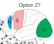
Where is the basal ganglia? |
Purple region |
|
|
Crust of the Pie |
Pataman |
|
|
Filling of the Pie |
Pallidus |
|
|
What part of the brain worries about movement PLANNING? |
Prefrontal/Frontal Cortex |
|
|
Prefrontal cortex and sensory information go to the... |
basal ganglia (patamen) along with dopamine input from the substantial nigra. |
|
|
What happens in the Patamen? |
There is the spiny neurons that inhibits neurons in the Pallidus. |
|
|
What do neurons in the Pallidus do?
|
Inhibit neurons in the thalamus, so when Pallidus neurons are inhibited by the spiny neurons in the Patamen, the thalamus becomes active. |
|
|
What kind of neurons are in the Pallidus? |
Tonic inhibitors; at rest they stop anything that would be occurring in the thalamus. |
|
|
What happens when the thalamus becomes active? |
It talks to motor neurons in the motor cortex and causes movements. |
|
|
What does the BG prevent us from doing |
Extra movements; babies don't have a fully functioning BG which is why they do lots of extra movements. |
|
|
How does the BG terminate movement? |
Disinhibition |
|
|
What is the equivalent of the Pataman in rats? |
Striatum |
|
|
What happens if you excite the striatum? |
Enables movement
|
|
|
What happens if you excited the pallidus?
|
Stops movement; rigid
|
|
|
Optogenetics |
Instead of sticking an electrode into the BG (deep into the brain), we can use ChR2, a light gated channel. |
|
|
ChR2 |
A light gated channel. |
|
|
How does ChR2 work?
|
When we shine light of a specific wavelength very brightly (think like a laser) then it will open the channels and let in ions thereby exciting the cell exactly how an electrode would have. |
|
|
Where can ChR2 be expressed?
|
Any cell! |
|
|
ChR2 Expression in the Spiny Neuron Experiment (set up, result, explanation) |
Spiny neuron expresses D1 (dopamine) receptor, so use the D1 promote in from of ChR2 DNA. When you turn the light on (open the channel) the rat goes from slowly walking to running around like crazy. You have excited the neuron with light which inhibits the inhibitor resulting in motion. |
|
|
How do you express ChR2 in specific cells? |
Find a receptor already expressed in that cell and use that promotor to express the ChR2 channel. |
|
|
What does the cerebellum control? |
Coordination, balance, muscle memory, and basically anything we do automatically (dance, speak, drive). |
|
|
What does damage to the cerebellum do?
|
Makes it very hard to keep your balance because then you have to think about doing this. |
|
|
Where does the cerebellum get information from and give it to? |
Receives input from sensory system and then corrects motor cortex through the thalamus to make better movements happen. |
|
|
How are perkinje neurons organized? |
Tightly stacked.
|
|
|
Where are axons of parallel fibers located? |
They run through the perkinje cell's dendritic arbor. |
|
|
Where does input to parallel fibers come from? |
Mossy fibers |
|
|
How do parallel fibers code? |
Labeled line; this is why there are so many. |
|
|
How do perkinje cells read parallel cells? |
They interpret the patterns and decide if they are appropriate or not. If not appropriate then the cells that fired then becomes less likely to fire again. |
|
|
Where do perkinje cells output? |
Deep Cerebellar Nuclei and Thalamus |
|
|
How do we correct movement? |
Through error detection. |
|
|
Cerebellum Basic Path (3) |
Sensory > Cerebellum > Motor |
|
|
Where does the climbing fiber synapse? |
Along the dendritic tree.
|
|
|
What information does the climbing fiber carry? |
It gets information saying we made a mistake. |
|
|
What does the climbing fiber do? |
Trains perkinje neurons to know what patterns are bad. |
|
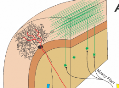
Where is the climbing fiber?
|
Red fiber |
|
|
Ataxia |
The loss of full control of body movements. |
|
|
What does noggin technically do? |
Inhibit BMP
|
|
|
BMP results in... |
body/skin. |
|
|
WNT + BMP = |
Ectoderm |
|
|
WNT = |
Spinal cord |
|
|
No WNT or BMP = |
Head and brain (noggin inhibits them to get this result). |
|
|
Neurexin and Neuroligand Cell Experiments (set up, result, conclusion) |
If you take a neuron that has not yet been polarized (early in development) and place a normal cell expressing either neurexin or neuroligand next to it then it will start expressing the opposite.
This shows us that the neuron must already be expressing both neuroligand and neurexin. |
|
|
If a cell is expressing neuroligand, the neuron will become... |
presynaptic.
|
|
|
If a cell is expressing neurexin, the neuron will become... |
postsynaptic. |
|
|
Why might a cell not express notch and delta (not show patterning)? 2 Hypotheses |
1. If it has a certain level or above of factor X. 2. If the cells are not actually touching (the astrocytes have divided them). |
|
|
Why might cells not be "playing" the notch and delta game correctly? 3 Hypotheses |
1. They might not be touching.
2. The process is just stochastic (random). 3. Consider that a protein may just be on one side of the cell before division resulting in uneven notch/delta distribution. |
|
|
What are the yellow things in between the baby neurons in the practice test question 2?
|
Astrocytes; this is what radial glia become.
|
|
|
Can astrocytes divide?
|
Yes they can divide into more astrocytes. |
|
|
What does cyclopanine do? |
Inhibit Shh |
|
|
What happens when we apply cyclopanine? |
There is slightly less Shh causing the eyes to get pushed to the middle into one eye. |
|
|
Where is there the most Shh? |
The center of the face. |
|
|
Neurotropic Hypothesis |
The target of an axon (target neuron) gives off some growth factor and without this growth factor the neuron dies. |
|
|
NGF |
The nerve growth factor neurons need to receive from their target neurons in order to not die. |
|
|
Trk |
The receptor for nerve growth factor. |
|
|
What happens if you over express Trk? |
Your neurons become more sensitive to NGF and need more of it to continue surviving. |
|
|
Total Motor Pathway Overview |
Pre/Frontal Lobe > Pre-motor area > BG turns on motor program (spiny neuron inhibits inhibitor in the Pallatus turning on motor program in thalamus) > Thalamus > Back to Motor Cortex > Upper motor neuron > Alpha motor neuron > Motor units > Muscles contract > sensors in tendons sense this > DRG and spinal cord > somatosensory cortex > cerebellum > BG for inhibition |
|
|
How do you train the error detection system in the cerebellum to work? |
If perkinje cells fire and make a change that is bad then the inferior olive tells us it is bad and the climbing fibers tell those synapses from perkinje to parallel cells to weaken resulting in less likely chance of firing next time (depression). |
|
|
What happens if you have no climbing fibers?
|
No learning. |
|
|
What does the cerebellum not know? |
What the "right" motion is; it just knows when we do something wrong, but this means the changes it makes to that motion will not necessarily be for the best. |
|
|
What can optogenetics be used for? |
Hyper or depolarizing the cell or just monitoring it. |
|
|
What is useful about optogenetics?
(2) |
Less invasive and allows you to target a specific gene. |
|
|
Optogenetics: How can you put the gene into a cell? (2) |
Trangenic animal (make the animal express it) or a virus/infection. |
|
|
How did we discover the motor pathway/cortical spinal tract? |
Modified rabies virus; tag the virus and mutate it so it can only cross one synapse and then watch as it creates a pathway one synapse at a time from the muscle up (retrograde). |
|
|
Neurotropic Virus |
A virus that can travel within neurons. |
|
|
How does the rabies virus travel? |
Retrograde. |
|
|
Monosynaptic Retrograde |
How the modified rabies virus travels; one synapse at a time in the retrograde direction. |
|
|
Results of the Modified Rabies Virus Experiment (2 set ups, results, and conclusions) |
All injected rabies viruses ended up in the motor cortex when injected to distal limbs. This supports the idea of one long axon as the cortical spinal tract. When we injected rabies virus into proximal limbs though we saw accumulation of rabies virus in the brainstem. This shows that there are several motor pathways. |
|
|
Broad Tuning within M1 Experiment with a Monkey |
Put an electrode in the monkey's brain in a neuron in the finger region. Stimulate > finger movements Ask monkey to move a specific finger > Silent with no movement. Movement of any finger evokes a response. Stronger APs when the preferred finger is moved. Conclusion: upper motor neurons are broadly tuned (will react to many things but react the best to a specific thing). |
|
|
What can you use an electrode array to do? |
Use a monkey's brainwaves to move a robotic arm. |
|
|
Population Vector |
The sum of the preferred direction vectors of a population of neurons weighted by the firing rate of each neuron. The preferred direction of a neuron is a vector in a 3-D space pointing in the direction towards which movement elicits the highest firing rate of the neuron. |
|
|
Agonist |
Causes movement; prime mover. |
|
|
Antagonist |
Opposes movement. |