Reading...
![]()
Play button
![]()
Play button
![]()
Use LEFT and RIGHT arrow keys to navigate between flashcards;
Use UP and DOWN arrow keys to flip the card;
H to show hint;
A reads text to speech;
14 Cards in this Set
- Front
- Back
|
Optokinetic Reflex:
• has involuntary/voluntary movements • goal of its movements • Its afferent limb • It's efferent limb |
Optokinetic Reflex:
• has involuntary movements • goal is to keep the eyes focused on an object and keep it in the center of the visual field • The parieto-occipital eyefield as it receives info about motion and depth • Again, the parieto occipital eyefield which after receiving input, triggers eye movements |
|
|
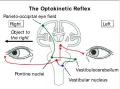
Pathway of the Optokinetic reflex:
• Efferent limb - The number of nuclei involved in this pathway - efferent limb fibers originate here - the trajectory of them descending and where they synapse - where the 2ndry neurons project - where the tertiary neurons project |

Pathway of the Optokinetic reflex:
• Efferent limb - Four (Pontine, Vestibular, Abducens and Oculomotor) - Parieto-occipital eye field - Fibers descend through the internal capsule and ipsilaterally into the pons. They synapse at the Pontine nuclei. - 2ndry neurons: Fibers from the pontine nuclei cross the midline and synapse at the contralateral vestibulocerebellum (flocculo-nodular lobe) - tertiary neurons: Fibers from the VC descend medially and synapse on the ipsilateral medial vestibular nuclei - Quaternary neurons: Fibers from the medial VN ascend, cross the midline and synapse on the contralateral Abducens nucleus - From the Abducens nucleus, The Common Final Pathway for horizontal eye movements is followed > A 1st set of fibers from the abducens nucleus iNN the lateral rectus > A 2nd set of fibers immediately crosses the midline and ascends up the contralateral MLF to synapse at the oculomotor nucleus. Fibers from this nucleus iNN the medial rectus |
|
|
For the Optokinetic Reflex, an object moving to the right activates which side (left/right) of the following structures:
• Parieto-occipital eyefield • Pontine nuclei • Vestibulocerebellum • Vestibular nucleus • Abducens nucleus • Lateral rectus • Oculomotor nucleus • Medial rectus |
For the Optokinetic Reflex, an object moving to the right activates which side (left/right) of the following structures:
• RIGHT - Parieto-occipital eyefield • RIGHT - Pontine nuclei • LEFT - Vestibulocerebellum • LEFT - Vestibular nucleus • RIGHT - Abducens nucleus • RIGHT - Lateral rectus • LEFT - Oculomotor nucleus • LEFT - Medial rectus |
|
|
Opticokinetc Nystagmus:
• When it naturally occurs • its two components |
Opticokinetic Nystagmus:
• Occurs at the end of an opticokinetic reflex movement when the object in focus reaches the limit of eye movements to one side. The fixation on the target is broken and the eyes move quickly in the opposite direction, re-setting the focus of vision onto a new target • Its two components are rhythmical and alternating - slow phase driven by the opticokinetic reflex - fast phase which is a reset phase driven be saccades in the opposite direction |
|
|
The Rotating Chair Experiment
(For clockwise rotation) • The Three phases • The reflex each phase tests and how |
The Rotating Chair Experiment
(For clockwise rotation) • Start of the rotation, VOR tested - Endolymph flows within the canals CCW, and eye movement is also CCW, opposite to the direction of head movement - Once the eyes have reached the limits of conjugate movement, they reset and exhibit horizontal nystagmus in the CW direction • Transition period, Optokinetic reflex tested - Relative to the head, visual targets are moving left inducing the OK reflex CCW with nystagmus movements CW • The abrupt stop, VOR tested - The endolymph continues to flow CCW, the eyes are driven by VOR to move CCW with nystagmus occurring CW. |
|
|
The Pupillary Light Reflex:
• Its fxn • Afferent limb of eye experiencing intense light - The cells that are initially activated - Where the fibers travel and synapse - The 2nd set fibers produced and where they synapse - The third set of fibers produced and where they synapse - The muscle iNN |
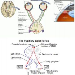
The Pupillary Light Reflex:
• To limit the amount of light falling on the retina and prevent potential damage to the retina by excessive light intensity • Afferent limb of eye experiencing intense light - Retinal ganglion cells are initially activated - Both nasal and temporal fibers form the optic nerve behind the eyeball > 50% of the fibers form the optic tract stay ipsilateral > 50% cross at the optic chiasm and travel w/ the contralateral optic nerve - They synapse at the pre-tectal nucleus of the midbrain - The Pre-tectal midbrain fibers bilaterally synapse at the EW nucleus - Fibers arising from the EW nucleus synapse at L and R Ciliary ganaglion - Short ciliary nerves iNN the constrictor pupilae muscle |
|
|
Corneal Reflex
• Its fxn • Neural Circuitry - Afferent limb > Type of receptors > CN involved > Region of the brain entered, its trajectory and where it synapses - Efferent Limb > Source of its fibers > Where the fibers will synapse |
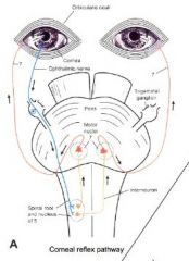
Cornea Reflex
• Protect the eye and cornea from damage • Neural Circuitry - Afferent limb > Nociceptors > opthalmic division of CN V > Enters the pons and descends with in the pons and medulla before synapsing at the facial nuclei - Efferent Limb > Facial nuclei > The obicularis oculi muscles of both eyes |
|
|
The nuclei/ganglion and the muscles responsible for opening the eye (2)
|
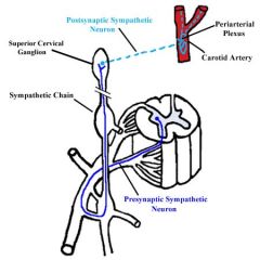
• Muscle: Tarsal
> Pre-ganglionic fiber: Hypothalamic T1-T3 of the spinal cord > Post ganglionic fiber: From Superior cervical ganglion which travels through the carotid plexus |
|
|
Describe testing for Opticokinetic nystagmus and a possible site of damage
|
Testing: Moving a series of stripes in front of the patient and watching for the saccadic eye movements
Possible site of damage: Parieto-occipital eyefield which would interrupt the initiation of the reflex |
|
|
Describe testing for the Pupillary light reflex
|
In a darkened room, ask the patient to look straight ahead while shining light alternatingly into each pupil
Observe for the direct and consenual response |
|
|
Describe testing for the Corneal light reflex
|
The patient is asked to look up and away while the examiner touches the cornea with a fine wisp of cotton, approaching away from the patient's line of vision
Closing of both eyes (direct and consensual) response is monitored |
|
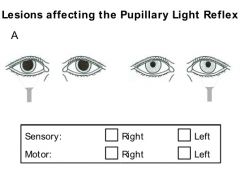
Which limbs of the pupillary light reflex are affected?
|

Right sensory limb is defective since neither a direct or consensual response is observed when the right side is stimulated
|
|

Which limbs of the pupillary light reflex are affected?
|
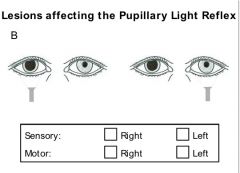
The Right Motor limb is defective
- When the affected eye is directly stimulated, there is no consensual response in the contralateral eye - When the normal eye is directly stimulate, there is no contralateral response in the affected eye. So over all, no parasympathetic response. |
|
|
Bell's Palsy:
• Cause • Clinical manifestation (2) |
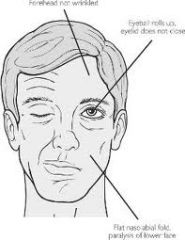
Bell's Palsy:
• A peripheral lesion of the facial nerve • Clinical manifestations - loss of the corneal reflex (Obicularis oculi iNN) - flattened nasolabial fold - Patient cannot raise the eyebrow or wrinkle the forehead on the affected side |