Reading...
![]()
Play button
![]()
Play button
![]()
Use LEFT and RIGHT arrow keys to navigate between flashcards;
Use UP and DOWN arrow keys to flip the card;
H to show hint;
A reads text to speech;
17 Cards in this Set
- Front
- Back
|
The two types of synapses btwn neurons
|
Chemical and Electrical
|
|
|
The Electrical synapse:
• Type of channel it contains • The composition of the channel • fxn of the channel • an example |
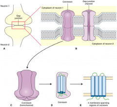
The Electrical synapse:
• Gap Junction • Two hemichannels, called Connexons, each having 6 subunits of 4 membrane spanning regions (connexins) • Fxns to help synchronize the activity of neighboring cells ex. astrocytes and spatial bufering |
|
|
Name & define 5 types of chemical synapses and how they fxn (excitatory/inhibitory)
|
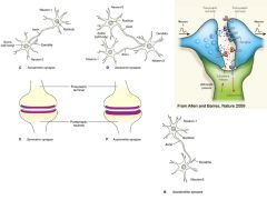
• Axomatic - a postsynaptic membrane is on the cell body of another neuron; Inhibitory
• Axodendritic - a postsynaptic membrane is on the dendrite of another neuron; Excitatory • Axoaxonic - the postsynaptic membrane is on the axon of another neuron; Inhibitory • Symmetric - The pre- and post- synaptic membrane are similar in thickness; Inhibitory • Asymmetrical - The Post-synaptic membrane is thicker than the pre-synaptic membrane; Excitatory |
|
|
Two types of receptors at the at chemical synapses, how they mediate changes in the post-synaptic cell after ligand binding and examples of each
|
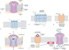
• Ionotropic receptors
- after a ligand binds, they undergo a conformational change to either open/close, thus changing post-synaptic membrane potential ex. Nicotinic AchR, GABA and NMDA • Metabotropic receptors - After a ligand binds, they modify the post-synaptic cell through a G-protein mediated cascade ex. Muscarinic AChR and NorE receptors |
|
|
There are 3 modes of rapid termination of the action of neurotransmitters at the synaptic cleft. Name them and the types of neurotransmitters predominantly using that termination mode.
|
• Diffusion - Neurotransmitters predominantly use this mode though all neurotransmitters can
• Extracellular enzymatic degradation: Predominantly used by acetylcholine • Uptake into nerve endings or glia - Very important mode used for catecholamines, glutamate, seratonin, GABA and glycine |
|
|
Ionotropic glutamate receptors:
• The transmitter that binds them • The 3 most important types • The types of channels they open • These ions are involved in influx/efflux • The net result of ion flux |
Ionotropic glutamate receptors:
• Glutamate • NMDA, AMPA, kainate • cation channels • Influx: Na+; Efflux: K+ • Net Influx of Na+ leads to a small depolarization ---> EPSP |
|
|
Compare the depolarizing and re-polarizing phases of the EPSP in relation to time and what is happening to the receptors
|
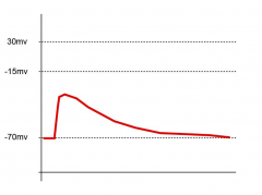
The depolarization phase is fast due to:
- the rapid binding of neurotransmitters to the receptors - and the opening of channels to allow cations to enter The repolarization phase is slow and rate controlled due to: - transmitters unbinding from the receptors, - cation channels closing - and positive charge re-distributing itself across the membrane |
|
|
What is an amplitude of an EPSP from an ionotropic receptor dependent on?
And what does this tell us about the type of potentials that can be elicited? |

The amplitude is dependent on how much glutamate is released by exocytosis in response to a presynaptic impulse
This tells us the depolarizations at the post-synaptic membrane are graded with respect to the [glutamate] applied |
|
|
Describe the ionic basis of Glutamate-induced EPSPs
What causes the depolarization? What is on the post-synaptic membrane that leads to a change in the membrane potential? What is the size of the EPSP dependent on? |
It is the number of cation channels opened in the post-synaptic membrane that determines the value of depolarization for the membrane. Because the channels will only open when bound with Glutamate, a short pre-synaptic impulse will only release a small amount of glutamate into the synaptic cleft to bind to channels.
|
|
|
Definition of the reversal potential
|
It is the value at which there is no net flow of an ion at the synapse (no synaptic current).
|
|
|
Describing the ionic basis of Glycine & GABA induced IPSPs:
• What is E(anion) value predominantly dependent on? • What do - & + driving forces describe? • Regardless of the -/+ sign of the driving force, what does the potential change of the membrane move towards and ultimately what does it tells us about the actions of Glycine and GABA? |
Describing the ionic basis of Glycine & GABA induced IPSPs:
• It is dependent on the equilibrium potential of chloride, E(Cl) • - DF: anions (mainly chloride) will leave the cell + DF: anions will enter the cell • The potential change of the membrane move towards E(Cl) and the actions of Glycine and GABA are inhibitory. |
|
|
The initiation zone:
• Its location in multipolar and sensory neurons • Two points about its excitation threshold and channels |
The initiation zone:
• Multipolar neurons - located at the axon hillock Sensory neurons - located at the sensory endings 1) It is the site where the excitation threshold is lowest 2) and the concentration of v-gated Na+ channels is highest/unit area |
|
|
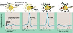
Considering that EPSPs spread with decrement,
these are two means many EPSPs can be combined to attain excitation threshold and generate an AP. What is the main idea of each |
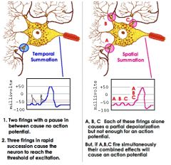
Spatial summation:
Due to many neurons synapsing on the post-synaptic cell, an additive effect is created, allowing the sum of the additive currents to yield larger EPSPs than separate EPSPs by themselves Temporal Summation: Several EPSPs will reach the post-synaptic neuron sequential, in a timeframe where the individual EPSPs of the 1st potentials have yet to relax back to the resting potential. The series of impulses are additive, creating larger EPSPs. |
|
|
What dictates the firing rate of a neuron?
|
The combined effect of the spatial and temporal summation of EPSPs & IPSPs occurring at many sites in its soma and dendrites
|
|
|
Two modes of action for Cocaine
|
1) It blocks Na+ channels from inside the cell (local anesthetic)
2) It blocks the transport roteins responsible for the re-uptake of the following neurotransmitters, thereby prolonging their existence in the synaptic cleft - Dopamine - NorAdrenaline - Seratonin (5-OH tryptamine) |
|
|
Morphine:
• Acts as an agonist for this molecule • Its receptor and location • Its competing transmitters (2) • the outcome of it binding to the receptor |
Morphine:
• μ-enkephalin • nocioreceptors located on C-fibers in the dorsal horn (substantia gelatinosa) • Substance P and Glu (They are excitatory) • It inhibits the relay of pain information to 2nd order neurons in the spinal cord • Its competing transmitters • the outcome of it binding to the receptor |
|
|
Psychiatric, neurological and neurodegenerative diseases are associated with these kinds of activities from these receptors
|
Excessive activation or inactivity of ionotropic and metabotropic receptors
|